INTRODUCTION
The middle cerebral artery (MCA) is the most commonly af-fected artery in ischemic stroke.1-3 The MCA territory can be
di-vided into the superficial MCA territory and lenticulostriate ar-tery (LSA) territory.4 Patients with superficial MCA territory
infarction often develop devastating or fatal stroke.5,6
Patients with superficial MCA territory infarction may show concomitant involvement of the LSA territory. The LSA com-monly originates from the proximal segment (M1) of the MCA trunk or the superior division of the MCA.7 Thus, the presence
of concomitant LSA territory involvement in patients with su-perficial MCA territory infarction provides information on the relevant artery of infarction, useful for the determination of stroke mechanisms.8,9 In addition, co-existing LSA involvement
in patients with superficial MCA territory infarction may sug-gest 1) occlusion of the MCA by larger thrombi, which may re-sult in larger MCA territorial infarctions; 2) more frequent in-volvement of the adjacent corticospinal tract, which can result in more severe or frequent motor weakness; and 3) a higher risk of brain herniation, because of deeply located sizable infarc-tions. All these factors may affect patient outcomes. Further-more, stroke mechanisms may differ in patients with superficial
Lenticulostriate Artery Involvement is Predictive
of Poor Outcomes in Superficial Middle
Cerebral Artery Territory Infarction
Kijeong Lee
1, Eun Hye Kim
1, Dongbeom Song
1, Young Dae Kim
1, Hyo Suk Nam
1, Hye Sun Lee
2, and Ji Hoe Heo
11Department of Neurology, Severance Stroke Center, Yonsei University College of Medicine, Seoul; 2Department of Biostatistics, Yonsei University College of Medicine, Seoul, Korea.
Purpose: Patients with superficial middle cerebral artery (MCA) territory infarction may have concomitant lenticulostriate artery (LSA) territory infarction. We investigated the mechanisms thereof and the outcomes of patients with superficial MCA territory infarction according to the presence or absence of LSA involvement.
Materials and Methods: Consecutive patients with first-ever infarction in the unilateral superficial MCA territory were included in this study. They were divided into the superficial MCA only (SM) group and the superficial MCA plus LSA (SM+L) group.
Results: Of the 398 patients, 84 patients (21.1%) had LSA involvement (SM+L group). The SM+L group more frequently had sig-nificant stenosis of the proximal MCA or carotid artery and high-risk cardioembolic sources. Stroke severity and outcomes were remarkably different between the groups. The SM+L group showed more severe neurologic deficits (National Institute of Health Stroke Scale score 10.8±7.1 vs. 4.0±5.0, p<0.001) and larger infarct in the superficial MCA territory (40.8±62.6 cm3 vs. 10.8±21.8
cm3, p<0.001) than the SM group. A poor functional outcome (mRS >2) at 3 months was more common in the SM+L group (64.3%
vs. 15.9%, p<0.001). During a mean follow-up of 26 months, 67 patients died. All-cause (hazard ratio, 2.246) and stroke (hazard ra-tio, 9.193) mortalities were higher in the SM+L group than the SM group. In multivariate analyses, LSA involvement was an inde-pendent predictor of poor functional outcomes and stroke mortality.
Conclusion: LSA territory involvement is predictive of poor long-term outcomes in patients with superficial MCA territory infarction.
Key Words: Lenticulostriate artery, middle cerebral artery, cerebral infarction, prognosis, mortality
Yonsei Med J 2017 Jan;58(1):123-130
https://doi.org/10.3349/ymj.2017.58.1.123 pISSN: 0513-5796 · eISSN: 1976-2437
Received: December 17, 2015 Revised: May 13, 2016 Accepted: June 7, 2016
Corresponding author: Dr. Ji Hoe Heo, Department of Neurology, Severance
Stroke Center, Yonsei University College of Medicine, 50-1 Yonsei-ro, Seodaemoon-gu, Seoul 03722, Korea.
Tel: 82-2-2228-1605, Fax: 82-2-393-0705, E-mail: [email protected] •The authors have no financial conflicts of interest.
© Copyright: Yonsei University College of Medicine 2017
This is an Open Access article distributed under the terms of the Creative Com-mons Attribution Non-Commercial License (http://creativecomCom-mons.org/licenses/ by-nc/3.0) which permits unrestricted non-commercial use, distribution, and repro-duction in any medium, provided the original work is properly cited.
MCA territory infarction, depending on the presence or ab-sence of co-existing LSA territory infarction.
In the present study, we compared the mechanisms thereof, severities of stroke, and long-term outcomes in patients with the superficial MCA territory infarction in relation to the pres-ence or abspres-ence of LSA involvement.
MATERIRALS AND METHODS
Study population and groupThis was a retrospective observational study using a prospec-tively registered cohort data.10 The present study included
pa-tients admitted between January 2010 and December 2012 with first-ever infarction involving the unilateral MCA superficial territory within 7 days of symptom onset. Patients who had co-exiting infarctions in other territories were excluded. The pa-tients were evaluated using standardized protocols, including brain computed tomography (CT), magnetic resonance imag-ing with diffusion-weighted imagimag-ing (DWI), and cerebral angi-ographic studies, such as MR angiography, CT angiography, and digital subtraction angiography. Cardiac evaluation in-cluded transesophageal echocardiography (TEE), transtho-racic echocardiography (TTE), heart CT scan, and Holter or con-tinuous electrocardiography monitoring during patient stay in the stroke unit. The patients were managed using standardized protocols of our stroke center based on given guidelines.11,12
We divided the patients with superficial MCA territory in-farction into two groups according to the presence of concomi-tant LSA territory infarction: the superficial MCA only (SM) group and the superficial MCA plus LSA (SM+L) group. The Institutional Review Board of Severance Hospital, Yonsei Uni-versity Health System approved this study and waived the need for informed consent, owing to the retrospective and ob-servational nature of the study.
Definition of risk factors
Hypertension was defined as blood pressure recordings of systolic ≥140 mm Hg or diastolic ≥90 mm Hg on repeated mea-surements during hospitalization or being on antihypertensive medication. Diabetes mellitus was diagnosed based on a fast-ing plasma glucose level ≥7 mmol/L, a glycated hemoglobin value ≥6.5%, or when patients had been treated with oral hy-poglycemic agents or insulin. Hypercholesterolemia was de-fined as a fasting serum total cholesterol level ≥6.2 mmol/L, low-density lipoprotein-cholesterol >4.1 mmol/L, or being treated with lipid lowering agents after a diagnosis of hypercholesterol-emia. Patients were regarded as current smokers if they smoked within 1 year prior to admission.
Mechanism of stroke
The mechanism of stroke was determined based on the Trial of Org 10172 in the Acute Stroke Treatment (TOAST)
classifi-cation.13 This study included patients with cardioembolism,
large artery atherosclerosis, or stroke of undetermined etiology due to two or more identified causes or due to negative evalu-ation. Patients with other determined etiologies were exclud-ed from the study. Significant stenosis of the MCA or internal carotid artery (ICA) was defined as stenosis >50% or occlusion of the artery relevant to the infarction. We identified high-risk potential cardiac source of embolism (PCSE) based on the TOAST classification. The stroke subtype and the degree of ar-terial stenosis were discussed during weekly stroke conferences and determined in consensus among stroke specialists. These data were registered in the prospective registry.
Infarction volume
The volume of infarction was measured on DWI in a semi-au-tomatic manner using Xelis software (Infinitt, Seoul, Korea). An investigator (EHK) who was blinded to the clinical informa-tion measured the infarcinforma-tion volumes for the superficial MCA territory and those for the LSA territory. The infarction volume of the superficial MCA territory was compared between the groups.
Stroke severity and long-term outcomes
Stroke severity was determined on the basis of National Insti-tute of Health Stroke Scale (NIHSS) at admission. Significant brain swelling was defined as brain edema that required os-motherapy or hemicraniectomy. Poor functional outcome was defined as a modified Rankin Scale (mRS) score >2 at 3 months after stroke onset. Long-term mortality and causes of death were identified by using death certificates from the Korean National Statistical Office, by reviewing medical records, and by conducting face-to-face or telephone interviews with a re-search nurse or stroke specialist. Mortality data from the Kore-an National Statistical Office are considered reliable, since they are collected on the basis of a unique 13-digit identification code assigned to subjects at birth and on the death certificate.14
The cause of death was coded according to the International Classification of Disease (ICD), 10th revision. Stroke mortality was defined by the ICD code as I60–I69. Patients who were alive on August 31, 2014 were censored.
Statistical analyses
All statistical analyses were performed using SPSS software for Windows (version 20.0; SPSS Inc., Chicago, IL, USA). The Pear-son χ2 test was used to compare the frequency of categorical
variables. The independent t-test was used to compare contin-uous variables. The variables achieving p-values less than 0.05 in the univariate analyses were entered for multivariate analy-ses using the logistic regression model to determine the factors associated with poor functional outcome. With regard to long-term mortality, Kaplan-Meier analysis was used to estimate sur-vival, and the log-rank test was used to compare rate estimates. We tested the proportional-hazards assumption by including
an interaction term between variables and natural log-trans-formed follow-up time. Also, we checked log-minus-log sur-vival plots. All variables met a proportional-hazards assump-tion. Cox proportional hazard regression analysis was performed to calculate crude and adjusted hazard ratios (HR) with 95% confidence intervals (CI). All statistical analyses were two-tailed, and p<0.05 was considered statistically significant.
RESULTS
Demographic and clinical characteristics of the patients
During the study period, 1754 consecutive patients were reg-istered to the prospective cohort. At first, 885 patients were ex-cluded because they did not have any infarction in the superfi-cial MCA territory. After excluding 180 patients with co-existing infarctions in other territories or bilateral superficial MCA
ter-ritories, 689 patients had unilateral infarction involving the superficial MCA territory. Of the 689 patients, we excluded 291 patients who received thrombolytic treatment, had a his-tory of previous cerebral infarction, had other determined etiol-ogy based on the TOAST classification, or did not undergo DWI or cerebral angiography. Finally, 398 patients were included in this study (Supplementary Fig. 1, only online).
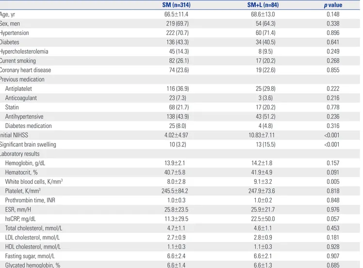
Of the 398 patients with superficial MCA territory infarc-tion, the mean age was 66.9±11.7 years and 273 (68.6%) were men. Of the 398 patients, 369 (92.7%) underwent at least one cardiac evaluation (heart CT in 278, TEE in 245, and TTE in 197), and 295 patients (74.1%) were evaluated using continuous electrocardiography monitoring for at least 24 hours. Eighty-four patients (21.1%) had concomitant lesions in the LSA ter-ritory (SM+L group). The SM+L group showed higher levels of white blood cells than the SM group. The other demographic characteristics were not different between the groups (Table 1).
Table 1. Comparison of Baseline Characteristics
SM (n=314) SM+L (n=84) p value Age, yr 66.5±11.4 68.6±13.0 0.148 Sex, men 219 (69.7) 54 (64.3) 0.338 Hypertension 222 (70.7) 60 (71.4) 0.896 Diabetes 136 (43.3) 34 (40.5) 0.641 Hypercholesterolemia 45 (14.3) 8 (9.5) 0.249 Current smoking 82 (26.1) 17 (20.2) 0.268
Coronary heart disease 74 (23.6) 19 (22.6) 0.855
Previous medication Antiplatelet 116 (36.9) 25 (29.8) 0.222 Anticoagulant 23 (7.3) 3 (3.6) 0.216 Statin 68 (21.7) 17 (20.2) 0.778 Antihypertensive 138 (43.9) 43 (51.2) 0.236 Diabetes medication 25 (8.0) 4 (4.8) 0.316 Initial NIHSS 4.02±4.97 10.83±7.11 <0.001
Significant brain swelling 10 (3.2) 13 (15.5) <0.001
Laboratory results
Hemoglobin, g/dL 13.9±2.1 14.2±1.8 0.157
Hematocrit, % 40.7±5.8 41.9±4.9 0.091
White blood cells, K/mm3 8.0±2.8 9.1±3.2 0.005
Platelet, K/mm3 245.5±84.2 247.9±73.6 0.818
Prothrombin time, INR 1.0±0.3 1.0±0.2 0.848
ESR, mm/H 25.8±23.5 25.9±21.7 0.976
hsCRP, mg/dL 11.3±29.5 22.5±50.0 0.057
Total cholesterol, mmol/L 4.7±1.1 4.6±1.1 0.453
LDL cholesterol, mmol/L 2.7±0.9 2.8±0.9 0.181
HDL cholesterol, mmol/L 1.1±0.3 1.1±0.3 0.928
Fasting sugar, mmol/L 6.6±2.4 6.6±2.1 0.907
Glycated hemoglobin, % 6.6±1.4 6.6±1.3 0.685
SM, superficial middle cerebral artery only; SM+L, superficial middle cerebral artery plus lenticulostriate artery; NIHSS, National Institutes of Health Stroke Scale; INR, international normalized ratio; ESR, erythrocyte sedimentation rate; hsCRP, high sensitivity C-reactive protein; LDL, low-density lipoprotein; HDL, high-density lipoprotein.
Mechanism of infarction
Stroke subtypes were different between the groups. Large ar-tery atherosclerosis and two or more causes were more com-mon in the SM+L group. Negative evaluation was more frequent in the SM group (20.1%) than in the SM+L group (4.8%).
Com-pared with the SM group, the SM+L group more commonly had significant stenosis of the MCA or ICA relevant to an infarc-tion and high-risk PCSE (Table 2).
Table 2. Subtype and Stroke Mechanisms
SM (n=314) SM+L (n=84) p value
Stroke subtype 0.003
Large artery atherosclerosis* 76 (24.2) 31 (36.9)
Cardioembolism 109 (34.7) 27 (32.1)
Two or more causes* 66 (21.0) 22 (26.2)
Negative evaluation 63 (20.1) 4 (4.8)
Stroke mechanism
Significant MCA stenosis 43 (13.7) 33 (39.3) <0.001
Significant ICA stenosis 45 (14.3) 28 (33.3) <0.001
High-risk PCSE 84 (26.8) 35 (41.7) 0.008
SM, superficial MCA only; SM+L, superficial MCA plus lenticulostriate artery; MCA, middle cerebral artery; ICA, internal carotid artery, PCSE, potential cardiac source of embolism.
Values are numbers (%).
*p<0.005; comparison with negative evaluation using Bonferroni post-hoc analysis.
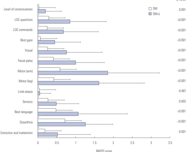
Fig. 1. Comparison of mean NIHSS scores for each item between the groups. Scores for all NIHSS items except that for limb ataxia are significantly higher in the SM+L group. NIHSS, National Institute of Health Stroke Scale; SM, superficial middle cerebral artery only; SM+L, superficial middle ce-rebral artery plus lenticulostriate artery; LOC, loss of consciousness.
Extinction and inattention Dysarthria Best language Sensory Limb ataxia Motor (leg) Motor (arm) Facial palsy Visual Best gaze LOC commands LOC questions Level of consciousness 0 0.5 1 1.5 NIHSS score 2 0.001 <0.001 <0.001 0.002 0.487 <0.001 <0.001 <0.001 <0.001 <0.001 <0.001 <0.001 0.001 p-value 2.5 3 3.5 SM SM+L
Initial stroke severity and infarct volumes
Initial neurologic deficits were more severe in the SM+L group than in the SM group (mean NIHSS, 10.83±7.11 vs. 4.02±4.97,
p<0.001) (Table 1). We compared the scores of each NIHSS
item between the groups to determine whether the higher NI-HSS score in the SM+L group was due to symptoms related with LSA lesions, such as weakness, dysarthria, and sensory changes. In doing so, scores for all NIHSS items, except those for ataxia, were significantly higher in the SM+L group (Fig. 1). The infarct volume of the superficial MCA territory was larger
in the SM+L group than in the SM group (40.8±62.6 cm3 vs.
10.8±21.8 cm3, p<0.001) (Supplementary Fig. 2, only online).
Significant brain swelling was also more frequent in the SM+L group (15.5% vs. 3.2%, p<0.001) (Table 1).
Long-term outcomes
Poor functional outcome (mRS>2) at 3 months was more com-mon in the SM+L group than in the SM group (64.3% vs. 15.9%,
p<0.001) (Fig. 2). Univariate analysis showed that the patients
with poor functional outcome were less commonly men, were
Fig. 2. Modified Rankin Scale at 3 months of symptom onset. Numbers are percentages (%). SM, superficial middle cerebral artery only; SM+L, super-ficial middle cerebral artery plus lenticulostriate artery.
SM SM+L 39 8 34 16 12 12 5 16 6 27 1 13 3 8 0 1 2 3 4 5 6
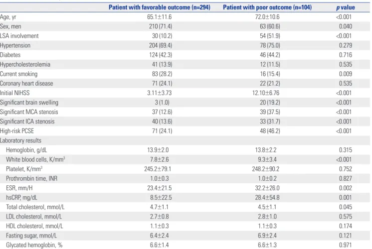
Table 3. Univariate Analysis of Factors Associated with a Poor Outcome at 3 Months
Patient with favorable outcome (n=294) Patient with poor outcome (n=104) p value
Age, yr 65.1±11.6 72.0±10.6 <0.001 Sex, men 210 (71.4) 63 (60.6) 0.040 LSA involvement 30 (10.2) 54 (51.9) <0.001 Hypertension 204 (69.4) 78 (75.0) 0.279 Diabetes 124 (42.3) 46 (44.2) 0.716 Hypercholesterolemia 41 (13.9) 12 (11.5) 0.535 Current smoking 83 (28.2) 16 (15.4) 0.009
Coronary heart disease 71 (24.1) 22 (21.2) 0.535
Initial NIHSS 3.11±3.73 12.10±6.76 <0.001
Significant brain swelling 3 (1.0) 20 (19.2) <0.001
Significant MCA stenosis 37 (12.6) 39 (37.5) <0.001
Significant ICA stenosis 40 (13.6) 33 (31.7) <0.001
High-risk PCSE 71 (24.1) 48 (46.2) <0.001
Laboratory results
Hemoglobin, g/dL 13.9±2.0 13.8±2.2 0.315
White blood cells, K/mm3 7.8±2.6 9.3±3.4 <0.001
Platelet, K/mm3 245.2±79.1 248.2±90.2 0.752
Prothrombin time, INR 1.0±0.3 1.0±0.2 0.827
ESR, mm/H 23.4±21.5 32.2±26.0 0.002
hsCRP, mg/dL 8.5±22.5 28.4±54.8 0.001
Total cholesterol, mmol/L 4.7±1.1 4.5±1.1 0.045
LDL cholesterol, mmol/L 2.7±0.8 2.8±1.0 0.575
HDL cholesterol, mmol/L 1.1±0.3 1.1±0.3 0.174
Fasting sugar, mmol/L 6.4±2.4 6.9±2.4 0.121
Glycated hemoglobin, % 6.6±1.4 6.6±1.3 0.971
LSA, lenticulostriate artery; NIHSS, National Institutes of Health Stroke Scale; MCA, middle cerebra artery; ICA, internal carotid artery; PCSE, potential cardiac source of embolism; INR, international normalized ratio; ESR, erythrocyte sedimentation rate; hsCRP, high sensitivity C-reactive protein; LDL, low-density lipopro-tein; HDL, high-density lipoprotein.
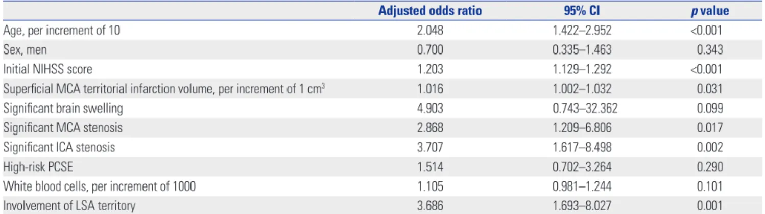
more commonly older age, and had more commonly LSA in-volvement, current smoking history, higher initial NIHSS score, significant brain swelling, significant MCA stenosis, significant ICA stenosis, high-risk PCSE, and higher white blood cell count, ESR, hsCRP and total cholesterol levels (Table 3). Multivariate logistic regression analysis showed that LSA involvement was an independent predictor of poor functional outcome (ad-justed odds ratio 3.686, 95% CI 1.693–8.027, p= 0.001), along with age, initial NIHSS, significant stenosis of the MCA or ICA relevant to an infarction, and the infarct volume of the super-ficial MCA territory (Table 4).
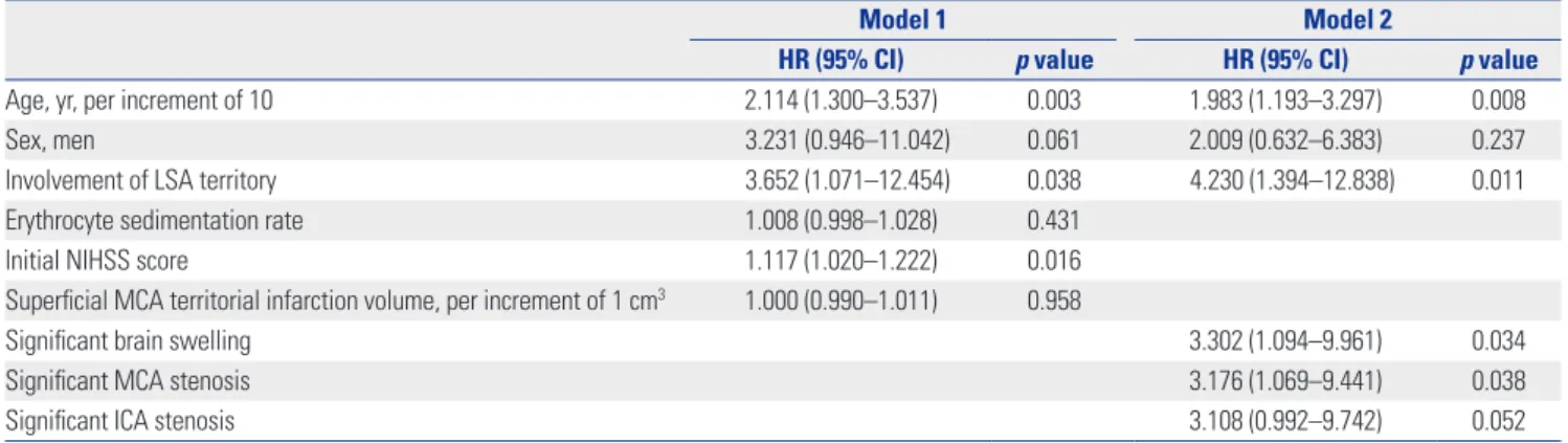
During a follow-up of mean 26±15 months, 67 patients (16.8%) died. Eighteen patients (4.5%) died of stroke. The SM+L group showed significantly higher all-cause mortality (HR, 2.246; 95% CI, 1.339–3.768) (Fig. 3A) and stroke mortality (HR, 9.193; 95% CI, 3.442–24.556) (Fig. 3B) than the SM group. Multivariate Cox regression analysis, after adjusting factors that were significant on univariate Cox regression analysis (Supplementary Table 1, only online), showed that LSA involvement was independent-ly associated with long-term stroke mortality along with age,
higher initial NIHSS, significant brain swelling, and significant stenosis of the MCA trunk (Table 5). Meanwhile, however, mul-tivariate Cox regression analysis (Supplementary Table 2, only online), revealed that LSA involvement was not independent-ly associated with all-cause mortality.
DISCUSSION
In the present study, we investigated whether initial stroke se-verity, stroke mechanism, and long-term outcomes differed among patients with superficial MCA territory infarction ac-cording to the presence or absence of co-existing LSA territory infarction. We found that initial neurologic deficits were more severe in patients with SM+L group than in those with SM alone. LSA involvement can cause weakness, sensory changes, or dysarthria,15,16 and these symptoms might have contributed to
the severe neurologic deficits in the SM+L group. However, when baseline NIHSS scores for each item were compared between the groups, all of the items except for ataxia showed higher Table 4. Multivariate Analysis of Factors Associated with a Poor Outcome at 3 Months
Adjusted odds ratio 95% CI p value
Age, per increment of 10 2.048 1.422–2.952 <0.001
Sex, men 0.700 0.335–1.463 0.343
Initial NIHSS score 1.203 1.129–1.292 <0.001
Superficial MCA territorial infarction volume, per increment of 1 cm3 1.016 1.002–1.032 0.031
Significant brain swelling 4.903 0.743–32.362 0.099
Significant MCA stenosis 2.868 1.209–6.806 0.017
Significant ICA stenosis 3.707 1.617–8.498 0.002
High-risk PCSE 1.514 0.702–3.264 0.290
White blood cells, per increment of 1000 1.105 0.981–1.244 0.101
Involvement of LSA territory 3.686 1.693–8.027 0.001
NIHSS, National Institute of Health Stroke Scale; MCA, middle cerebral artery; ICA, internal carotid artery; PCSE, potential cardiac source of embolism; LSA, len-ticulostriate artery; CI, confidence interval.
Fig. 3. Kaplan-Meier curve for all-cause mortality (A) and stroke mortality (B). SM, superficial middle cerebral artery only; SM+L, superficial middle cerebral artery plus lenticulostriate artery.
1.00 0.75 0.50 0.25 0.00 0 1 2 3 4 (years) A SM SM+L 1.00 0.75 0.50 0.25 0.00 0 1 2 3 4 (years) B SM SM+L
scores in the SM+L group. These results suggest that the severe neurologic deficits in SM+L patients may be attributed to the wider hemispheric dysfunction, not simply the consequence of the additional LSA territory infarction. Consistent with this hypothesis, the infarction volume of the MCA superficial terri-tory excluding that in the LSA territerri-tory was much greater in SM+L patients than in SM patients.
The initially severe stroke in the SM+L group could be, in part, ascribed to differences in the stroke mechanisms between SM and SM+L. In the present study, the SM+L group more fre-quently had significant stenosis or occlusion in the MCA or ICA and high-risk PCSE than the SM group. The larger infarc-tion volume in the SM+L group might be associated with those etiologies. The possible mechanisms of superficial MCA terri-tory infarction are embolism from the heart, artery-to-artery embolism from the proximal arterial trees, such as the aorta, carotid artery and MCA, and in-situ thrombosis of the MCA,17,18
To produce the concomitant LSA lesion, thrombi from the heart or proximal arteries should be large enough to occlude the MCA trunk or multiple to involve both the superficial MCA and LSA territories.19,20 In patients with significant stenosis of
the MCA, thromboemboli from the heart or proximal arteries might have been more easily trapped at the MCA trunk to completely occlude the MCA.21 In addition, cardioembolism is
known to produce larger infarction and more severe stroke.22,23
Finally, the local MCA trunk atheroma could also be the source of the emboli to both the LSA and the superficial MCA branches.
In this study, the presence of LSA territory infarction was an independent predictor of poor long-term outcomes and in-creased risk of mortality, even after adjustment for the initial NIHSS score and infarction volume. Functional outcome mea-sured by mRS, largely depends on the degree of motor weak-ness.24 Motor tracts, which pass as compact bundles through
the internal capsule,25 could be more frequently damaged in
the SM+L group, and this might have caused more severe mo-tor weakness and impeded functional recovery. In patients with large superficial territory infarction, additionally sizable
infarc-tions in the subcortex due to LSA involvement might increase the risk of compressing deep brain structures and brain herni-ation. In a previous study, patients who had both large artery atherosclerosis and cardioembolism showed very high long-term mortality, while those without showed very low long-long-term mortality.26 Consistent with the previous findings, the SM+L
group more frequently had two or more causes (large artery atherosclerosis and cardioembolism), while the SM group more frequently had undetermined etiology due to negative evaluation.
This study has limitations. First, this study is not free from selection bias because this was a retrospective study, although the cohort data were prospectively and consecutively collect-ed. Second, patients who received reperfusion therapy were excluded from this study. Consequently, some patients with hyperacute MCA infarction were not included in this analysis. Third, the mortality rate of this study is relatively low, com-pared to the previous studies.27,28 We excluded patients with
co-existing infarctions in other territories or those with recana-lization therapy. These patients might have initially more se-vere stroke that may be associated with stroke mortality. In addi-tion, cause of death was determined based on medical records or death certificates in some patients. Thus, stroke mortality might not be accurately reflected in some patients. Finally, although the infarction volume in the superficial MCA territory can be influenced by the presence and degree of collateral circula-tions,20,29 they could not be determined because the
angiograph-ic studies were not standardized.
In conclusion, among patients with superficial MCA territo-ry infarction, the presence of co-existing LSA territoterrito-ry infarc-tion was associated with initially severe stroke and larger in-farct volume in the MCA superficial territory. LSA involvement was almost the most powerful predictor of poor functional out-come and stroke mortality. Therefore, greater concern should be given when treating patients with MCA superficial territory infarction and a co-existing LSA lesion.
Table 5. Cox Regression Model of Stroke Mortality in Multivariate Analysis
Model 1 Model 2
HR (95% CI) p value HR (95% CI) p value
Age, yr, per increment of 10 2.114 (1.300–3.537) 0.003 1.983 (1.193–3.297) 0.008
Sex, men 3.231 (0.946–11.042) 0.061 2.009 (0.632–6.383) 0.237
Involvement of LSA territory 3.652 (1.071–12.454) 0.038 4.230 (1.394–12.838) 0.011
Erythrocyte sedimentation rate 1.008 (0.998–1.028) 0.431
Initial NIHSS score 1.117 (1.020–1.222) 0.016
Superficial MCA territorial infarction volume, per increment of 1 cm3 1.000 (0.990–1.011) 0.958
Significant brain swelling 3.302 (1.094–9.961) 0.034
Significant MCA stenosis 3.176 (1.069–9.441) 0.038
Significant ICA stenosis 3.108 (0.992–9.742) 0.052
LSA, lenticulostriate artery; NIHSS, National Institute of Health Stroke Scale; MCA, middle cerebral artery; ICA, internal carotid artery.
Model 1 includes age, sex, erythrocyte sedimentation rate, initial NIHSS, superficial territorial infarction volume and involvement of LSA and model 2 includes age, sex, significant brain swelling, significant MCA stenosis, significant ICA stenosis and involvement of LSA.
ACKNOWLEDGEMENTS
This work was supported by a grant from the Korea Health 21 R&D Project, Ministry of Health & Welfare, Republic of Korea (HI08C2149).
REFERENCES
1. Mohr JP, Lazar RM, Marshall RS. Middle cerebral artery disease. In: Mohr JP, Grotta JC, Wolf PA, Moskowitz MA, Mayberg MR, Von Kummer R, editors. Stroke: pathophysiology, diagnosis, and man-agement. 5th ed. Philadelphia, PA: Elsevier Saunders; 2011. p.384-424.
2. Kang J, Park TH, Lee KB, Park JM, Ko Y, Lee SJ, et al. Symptomatic steno-occlusion in patients with acute cerebral infarction: preva-lence, distribution, and functional outcome. J Stroke 2014;16:36-43. 3. Bang OY, Lee PH, Heo KG, Joo US, Yoon SR, Kim SY. Specific DWI
lesion patterns predict prognosis after acute ischaemic stroke within the MCA territory. J Neurol Neurosurg Psychiatry 2005;76: 1222-8.
4. Berman SA, Hayman LA, Hinck VC. Correlation of CT cerebral vas-cular territories with function: 3. Middle cerebral artery. AJR Am J Roentgenol 1984;142:1035-40.
5. Moulin DE, Lo R, Chiang J, Barnett HJ. Prognosis in middle cere-bral artery occlusion. Stroke 1985;16:282-4.
6. Hacke W, Schwab S, Horn M, Spranger M, De Georgia M, von Kummer R. ‘Malignant’ middle cerebral artery territory infarction: clinical course and prognostic signs. Arch Neurol 1996;53:309-15. 7. Gibo H, Carver CC, Rhoton AL Jr, Lenkey C, Mitchell RJ.
Micro-surgical anatomy of the middle cerebral artery. J Neurosurg 1981; 54:151-69.
8. Lee DK, Kim JS, Kwon SU, Yoo SH, Kang DW. Lesion patterns and stroke mechanism in atherosclerotic middle cerebral artery disease: early diffusion-weighted imaging study. Stroke 2005;36:2583-8. 9. Choi HY, Yang JH, Cho HJ, Kim YD, Nam HS, Heo JH. Systemic
atherosclerosis in patients with perforating artery territorial infarc-tion. Eur J Neurol 2010;17:788-93.
10. Lee BI, Nam HS, Heo JH, Kim DI; Yonsei Stroke Team. Yonsei Stroke Registry. Analysis of 1,000 patients with acute cerebral in-farctions. Cerebrovasc Dis 2001;12:145-51.
11. Adams HP Jr, del Zoppo G, Alberts MJ, Bhatt DL, Brass L, Furlan A, et al. Guidelines for the early management of adults with ischemic stroke: a guideline from the American Heart Association/Ameri-can Stroke Association Stroke Council, Clinical Cardiology Coun-cil, Cardiovascular Radiology and Intervention CounCoun-cil, and the Atherosclerotic Peripheral Vascular Disease and Quality of Care Outcomes in Research Interdisciplinary Working Groups: the American Academy of Neurology affirms the value of this guide-line as an educational tool for neurologists. Stroke 2007;38:1655-711.
12. Furie KL, Kasner SE, Adams RJ, Albers GW, Bush RL, Fagan SC, et al. Guidelines for the prevention of stroke in patients with stroke or transient ischemic attack: a guideline for healthcare professionals from the american heart association/american stroke association.
Stroke 2011;42:227-76.
13. Adams HP Jr, Bendixen BH, Kappelle LJ, Biller J, Love BB, Gordon DL, et al. Classification of subtype of acute ischemic stroke. Defi-nitions for use in a multicenter clinical trial. TOAST. Trial of Org 10172 in Acute Stroke Treatment. Stroke 1993;24:35-41.
14. Kim HC, Choi DP, Ahn SV, Nam CM, Suh I. Six-year survival and causes of death among stroke patients in Korea. Neuroepidemiol-ogy 2009;32:94-100.
15. Weiller C, Ringelstein EB, Reiche W, Thron A, Buell U. The large striatocapsular infarct. A clinical and pathophysiological entity. Arch Neurol 1990;47:1085-91.
16. Russmann H, Vingerhoets F, Ghika J, Maeder P, Bogousslavsky J. Acute infarction limited to the lenticular nucleus: clinical, etiolog-ic, and topographic features. Arch Neurol 2003;60:351-5.
17. Saito I, Segawa H, Shiokawa Y, Taniguchi M, Tsutsumi K. Middle cerebral artery occlusion: correlation of computed tomography and angiography with clinical outcome. Stroke 1987;18:863-8. 18. Ueda S, Fujitsu K, Inomori S, Kuwabara T. Thrombotic occlusion
of the middle cerebral artery. Stroke 1992;23:1761-6.
19. Marinkovic SV, Milisavljevic MM, Kovacevic MS, Stevic ZD. Perfo-rating branches of the middle cerebral artery. Microanatomy and clinical significance of their intracerebral segments. Stroke 1985;16: 1022-9.
20. Cho HJ, Yang JH, Jung YH, Kim YD, Choi HY, Nam HS, et al. Cor-tex-sparing infarctions in patients with occlusion of the middle ce-rebral artery. J Neurol Neurosurg Psychiatry 2010;81:859-63. 21. Wong KS, Gao S, Chan YL, Hansberg T, Lam WW, Droste DW, et al.
Mechanisms of acute cerebral infarctions in patients with middle cerebral artery stenosis: a diffusion-weighted imaging and micro-emboli monitoring study. Ann Neurol 2002;52:74-81.
22. Timsit SG, Sacco RL, Mohr JP, Foulkes MA, Tatemichi TK, Wolf PA, et al. Brain infarction severity differs according to cardiac or arteri-al embolic source. Neurology 1993;43:728-33.
23. Kang DW, Chalela JA, Ezzeddine MA, Warach S. Association of ischemic lesion patterns on early diffusion-weighted imaging with TOAST stroke subtypes. Arch Neurol 2003;60:1730-4.
24. Banks JL, Marotta CA. Outcomes validity and reliability of the modified Rankin scale: implications for stroke clinical trials: a lit-erature review and synthesis. Stroke 2007;38:1091-6.
25. Djulejic´ V, Marinkovic´ S, Georgievski B, Stijak L, Aksic´ M, Puškaš L, et al. Clinical significance of blood supply to the internal cap-sule and basal ganglia. J Clin Neurosci 2016;25:19-26.
26. Kim YD, Cha MJ, Kim J, Lee DH, Lee HS, Nam CM, et al. Long-term mortality in patients with coexisting potential causes of isch-emic stroke. Int J Stroke 2015;10:541-6.
27. Hong KS, Kang DW, Koo JS, Yu KH, Han MK, Cho YJ, et al. Impact of neurological and medical complications on 3-month outcomes in acute ischaemic stroke. Eur J Neurol 2008;15:1324-31.
28. Kim J, Song TJ, Park JH, Lee HS, Nam CM, Nam HS, et al. Different prognostic value of white blood cell subtypes in patients with acute cerebral infarction. Atherosclerosis 2012;222:464-7. 29. Gado M, Marshall J. Clinico-radiological study of collateral
circula-tion after internal carotid and middle cerebral occlusion. J Neurol Neurosurg Psychiatry 1971;34:163-70.