저작자표시 2.0 대한민국 이용자는 아래의 조건을 따르는 경우에 한하여 자유롭게 l 이 저작물을 복제, 배포, 전송, 전시, 공연 및 방송할 수 있습니다. l 이차적 저작물을 작성할 수 있습니다. l 이 저작물을 영리 목적으로 이용할 수 있습니다. 다음과 같은 조건을 따라야 합니다: l 귀하는, 이 저작물의 재이용이나 배포의 경우, 이 저작물에 적용된 이용허락조건 을 명확하게 나타내어야 합니다. l 저작권자로부터 별도의 허가를 받으면 이러한 조건들은 적용되지 않습니다. 저작권법에 따른 이용자의 권리는 위의 내용에 의하여 영향을 받지 않습니다. 이것은 이용허락규약(Legal Code)을 이해하기 쉽게 요약한 것입니다. Disclaimer 저작자표시. 귀하는 원저작자를 표시하여야 합니다.
Feasibility Study of Low Dose Chest CT for
Initial Evaluation of Blunt Chest Trauma
Patient
by
Sung Jung Kim
Major in Medicine
Department of Medical Sciences
The Graduate School, Ajou University
Feasibility Study of Low Dose Chest CT for
Initial Evaluation of Blunt Chest Trauma
Patient
by
Sung Jung Kim
A Dissertation Submitted to The Graduate School of
Ajou University in Partial Fulfillment of the Requirements
for the Degree of Master of Medicine
Supervised by
Joo Sung Sun, M.D.
Major in Medicine
Department of Medical Sciences
The Graduate School, Ajou University
This certifies that the dissertation
of Sung Jung Kim is approved.
SUPREVISORY COMMITTEE
Joo Sung Sun
Tae Hee Kim
Seung Soo Shin
The Graduate School, Ajou University
June, 23rd, 2014
i
- ABSTRACT-
Feasibility study of Low dose chest CT for initial evaluation of
blunt chest trauma
Introduction: Multi-detector computed tomography (MDCT) is now modality of choice for
evaluation of the polytrauma patients including thoracic injury. However, the routine use of
CT results in rapid increase of the CT radiation dose in blunt chest trauma evaluation.
Therefore, according to the ALARA (as low as reasonable achievable) principle, radiation
dose adjustment in the patients with blunt chest trauma is inevitable. We aimed to evaluate
the diagnostic performance and inter-observer consistency of low-dose chest CT (LDCT) in
the patients with blunt chest trauma.
Materials and Methods: A total of 69 patients who met criteria indicative of blunt chest
trauma (77% of male; age range, 16-85) were prospectively included. All patients underwent
LDCT without intravenous (IV) contrast and standard-dose chest CT (SDCT) with IV
contrast using parameters as followings: LDCT, 40mAs with automatic tube current
modulation (ATCM) and 100kVp (body mass index (BMI) <25, n=51) or 120kVp (BMI>25,
n=18); SDCT, 180mAs with ATCM and 120kVp. Transverse, coronal, saggital images were
reconstructed with 3-mm slice thickness without gap for investigators. Reference standard
images (transverse, coronal, saggital) were reconstructed using standard chest CT data with
1-mm slice thickness without gap. Reference standard was established by 2 experienced
thoracic radiologist by consensus. Three investigators independently evaluated blunt chest
ii
wall injury, diaphragmatic injury, aortic injury and upper abdominal injury). Investigators
recorded results with 4 confidence scale (0-3 point). Comparison of radiation dose was done
by vendor providing CT dose index volume (CTDIvol).
Results: Radiation dose of LDCT (average CTDIvol = 2.72mGy) was significantly lower
than those of SDCT (average CTDIvol = 13.4mGy) (79.4% dose reduction). Multiple-reader
receiver operating characteristic (ROC) analysis for comparing areas under the ROC curves
(AUC) demonstrated that LDCT was comparable to SDCT for evaluation of blunt chest
injury (AUC in LDCT : 0.823-1.000, AUC in SDCT : 0.781-1.000, p>0.05). ROC
comparison analysis revealed no significant difference of diagnostic performance between
LDCT and SDCT for the diagnosis of pulmonary injury, skeletal trauma, mediastinal injury
and chest wall injury (p>0.05). Intraclass correlation coefficient (ICC) was measured for
inter-observer consistency and revealed that there was good inter-observer consistency in
each examination of LDCT and SDCT for evaluation of blunt chest injury (0.8601~1.000).
Aortic and upper abdominal injury could not be appropriately compared due to LDCT
underwent without using contrast materials and this was limitation of this study.
Conclusion: Our conclusion is that there is a great potential benefit to use of LDCT for initial evaluation of blunt chest trauma because LDCT could maintain diagnostic image quality as SDCT and provide significant radiation dose reduction. Further study of LDCT with IV contrast for evaluation of aortic and upper abdominal injury is needed.
Keywords: Low dose chest CT, Blunt chest trauma, Diagnostic performance, Radiation
iii
TABLE OF CONTENTS
ABSTRACT ··· i
TABLE OF CONTENTS ··· iii
LIST OF FIGURES ··· iv
LIST OF TABLES ··· v
I. INTRODUCTION ··· 1
II. MATERIALS AND METHODS ··· 3
A. STUDY POPULATION ··· 3 B. CT EXAMINATION ··· 5 C. IMAGE EVALUATION ··· 5 D. REFERENCE STANDARD ··· 7 E. STATISTICAL ANALYSIS ··· 7 III. RESULTS ··· 8 IV. DISCUSSION ··· 19 V. CONCLUSION ··· 22 REFERENCES ··· 23 국문요약 ··· 26
iv
LIST OF FIGURES
▶ Figure 1. A 54-year-old woman after driver accident. ··· 16
▶ Figure 2. A 57-year-old man after driver accident. ··· 17
v
LIST OF TABLES
▶ Table 1. Table of Inclusion Criteria ··· 4
▶ Table 2. Injuries of Blunt Chest Trauma ··· 9
▶ Table 3. AUC Comparison in Each Observer between LDCT vs. SDCT for Pulmonary and Tracheobronchial Injuries ··· 11
▶ Table 4. AUC Comparison in Each Observer between LDCT vs. SDCT for Skeletal trauma ··· 12
▶ Table 5. AUC Comparison in Each Observer between LDCT vs. SDCT for Mediastinal, Chest wall and Diaphragmatic Injuries ··· 13
▶ Table 6. Intraclass Correlation Coefficient for Inter-observer Consistency of LDCT and SDCT ··· 15
1
I. INTRODUCTION
Multi-detector computed tomography (MDCT) is now modality of choice for evaluation
of the patients with multiple traumas including thoracic injury, overwhelming routine chest
x-ray, which could frequently miss significant thoracic injury (Trupka et al., 1997). The
routine use of CT was encouraged in blunt chest trauma, because it could increase diagnostic
yield of the blunt chest trauma patients, whose injuries were clinically and radiographically
silent (Brink et al., 2008; Deunk et al., 2009). Routine MDCT of the chest in trauma patients
could provide substantial information and it also enhanced clinical yield of the multi-trauma
patients with high-risk mechanisms (Plurad et al., 2007; Brink et al., 2008; Deunk et al.,
2009). However, the routine use of CT results in rapid increase of the CT examination for
trauma evaluation. One study revealed that the chest CT taken to the adult emergency
department increased by 226% from 2000 to 2005, while the number of patients increased by
13% (Broder and Warshauer, 2006).
The trauma patients were initially investigated with the extensive diagnostic studies due to
their urgency. Naturally, the radiation exposures in acute period of the trauma patients were
significant. One quantitative study demonstrated that median effective radiation dose
received by adult trauma patient during their first 24 hours assessment was 40.2 millisievert
(mSv) (Winslow et al., 2008). Radiation exposures were also concerned with the patients
with major trauma, who were repeatedly performed CT examinations during admission (Kim
2
organs (e.g. thyroid gland and breast) and the majority of the trauma patients were young.
Therefore, it is very important to adjust radiation dose according to specific indication of the
examination according to the ALARA (as low as reasonable achievable) principle (1991;
Slovis, 2003).
Low dose chest CT (LDCT) has been used for the detection of pulmonary nodule,
especially, lung cancer surveillance, because of the high given contrast in the chest and low
attenuation of radiation. Although radiation reduction from LDCT could limit proper
evaluation of CT image data, we recently have accumulated experience of LDCT and
empirically we could deduce that LDCT could be useful to primary survey of blunt chest
trauma patients. One recent literature reported that the subjectively scored diagnostic
interpretability of low dose CT for total body trauma was comparable (Grupp et al., 2013).
However, to date, the direct comparison of the diagnostic performance of LDCT with
standard dose CT (SDCT) in patients with blunt chest trauma has not yet been reported.
Therefore, this prospective study aimed to evaluate the diagnostic performance and
3
II. MATERIALS AND METHODS
A. Study Population
This prospective study was approved by our institutional review board and written
informed consent was obtained from all the patients. The current study was performed in
emergency department of tertiary educational hospital, the Ajou University Medical Centre.
From August 2012 through December 2013, we enrolled a total of 69 patients who visited in
emergency department with blunt chest trauma. All patients were included according to
hereby inclusion criteria (Table 1). The major exclusion criteria were instability of
hemodynamics or pregnancy, and any conditions needed prompt surgical intervention. Initial
clinical evaluation was performed and recorded by emergency medicine residents who were
supervised by emergency medicine physician and simultaneous chest anteroposterior chest
4 Table 1. Table of Inclusion Criteria
Inclusion criteria
Any signs of thoracic injury on initial CXR - Pulmonary contusion - Hemothorax
- Subcutaneous emphysema
- Suspicious aortic injury including a widened mediastinum and loss of aortic contour - Spinal fracture
- Rib fracture - Scapular fracture - Clavicular fracture - Diaphragmatic rupture Any abnormal findings on physical examination of the
chest wall or lungs
- Decreased breathing sounds at auscultation - Subcutaneous emphysema at palpation - Tenderness to palpation of the chest wall - Lacerations or hematoma of the chest wall
Respiratory insufficiency - SaO2 < 95%
- Respiratory rate < 10 or > 29/min High-energy mechanism of injury and high-energy crush
injury impact to the chest wall
- Fall from height >3 m
- Motor vehicle accident >50 km/h - Ejection from vehicle
- Car rollover
- Severe impact damage to car - Struck pedestrian >10 km/h - Struck bicyclist >30 km/h
5 B. CT Examination
All examinations were performed using a 16-detector-row CT scanner (Sensation 16; Siemens, Erlangen, Germany) that was located in the emergency department. The CT scans were obtained with end-inspiration state, as far as possible, in a supine position. Chest CT scans were performed from the thoracic inlet to the upper abdomen. CT examination was obtained with LDCT without intravenous contrast agent and standard dose CT (SDCT) after intravenous injection of nonionic contrast media (2.5 mL/s, 100mL Iomeprol—Iomeron 300; Bracco, Milan, Italy) using a power injector. The parameters of LDCT scans were as follows: 0.75 mm-collimation; pitch, 1.0; reference effective tube current time product of 40 mAs with automatic tube current modulation (ATCM); tube voltage of 100kV (BMI<25) or 120kV (BMI≥25). The parameters of SDCT scans were as follows: 0.75 mm-collimation; pitch, 1.0; reference effective tube current time product of 180 mAs with ATCM; tube voltage of 120kV. Coronal, sagittal multi-planar reformatted images were reconstructed in both LDCT and SDCT with 3-mm slice thickness without gap. All CT scans were reconstructed using a soft filter kernel. Vendor providing CT dose index volume (CTDIvol) and dose-length product (DLP) were recorded ad CT radiation dose descriptor for LDCT and SDCT.
C. Image Evaluation
Two board-certified radiologists and 1radiology trainee independently performed retrospective analysis of the images of these prospectively collected data. They were unaware of the any clinical information, except the images were from the patients with blunt chest trauma. LDCT and SDCT image data sets were evaluated separately. The order of the each image sets was randomized using a standard random number generator. Firstly, LDCT image data sets were reviewed by 3 investigators. And then, after one month, the image sets of SDCT were reviewed as same fashion. One month of time interval between the reviews was due to minimize recall bias.
Injuries identified on chest CT scan were categorized as follows: 1) pulmonary and tracheobronchial injury (lung contusion, pneumatocele, pneumothorax, hemothorax and tracheobronchial injury); 2) skeletal trauma (fractures of the ribs, clavicle, scapula, sternum and vertebra); 3) mediastinal injury (esophageal injury, pneumomediastinum, hemomediastinum, pneumopericardium, hemopericardium); 4) chest wall injury (chest wall
6
hematoma, chest wall free air); 5) diaphragmatic injury; 6) aortic injury; 7) upper abdominal injury (liver injury, splenic injury, pneumoperitoneum and hemoperitoneum). The each injuries were defined as follows. Lung contusion was defined as geographic ground-glass or nodular opacities or consolidation that not confined with the segmental and lobar boundaries. Pneumatocele was defined as round or oval shape, thin-walled, gas-filled space within the lung. Tracheobronchial tree injury was defined as direct cut-off of the trachea or the bronchus with with extraluminal air collection. Fracture of bone was defined as definitely visible radiolucent line. The reviewers bewared of frequently missed buckle fractures of the rib. Esophageal injury was defined as direct cut-off of the esophagus or peri-esophageal air or fluid leakage. Diaphragmatic injury was defined as the defect or discontinuation of the diaphragm or waist-like stricture of herniated structures (stomach or bowel or fat) or contrast extravasation at diaphragm. Aortic injury was defined as direct signs (intimal flap, pseudoaneurysm, abnormal aortic contour, intraluminal thrombus, contrast extravasation) or indirect sign (periaortic haematoma with periaortic mediastinal fat plane obliteration). Liver and splenic injuries were including laceration, intraparenchymal or subcapsular hematoma or contrast extravasation. Pneumothorax, penumomediastinum, pneumopericardium, chest wall free air, pneumoperitoneum were defined as air density[-1000 Hounsfield units (HU)] within the pleural cavity, mediastinum, pericardium, chest wall and abdominal cavity, respectively. Pneumomediastinum was differentiated from pneumothorax by the presence of the internal septa (Shuman, 1997; Dyer et al., 1999; Steenburg et al., 2008; Oikonomou and Prassopoulos, 2011; Cho et al., 2012). Hemothorax, hemopericardium, hemoperitoneum were defined as blood density (30-100HU) within the pleural cavity, pericardium and abdominal cavity, respectively. Mediastinal hematoma was defined as inhomogenous soft tissue density within the mediastinal fat, with obscuration or obliteration. Chest wall hematoma was defined as area of soft tissue density causing anatomical alteration. We aware of the critical limitation for assessment of aortic injury and upper abdominal injury using non-contrast CT, so LDCT without contrast enhancement of this current study was not appropriate for proper evaluation.
Three investigators made assessment with four-point confidence scale as follows: 0 point, definitely negative; 1 point, probably negative; 2 point, probably positive; 3 point, definitely positive. After evaluation, if multiple lesions were identified in same kind of injury, the highest confidence scale was selected. For example, if multiple rib fractures were identified
7
(e.g. confidence scale for multiple rib fractures range from 1~3), the highest score of the lesions was selected to represent (e.g. rib fracture, confidence scale 3).
D. Reference standard
Axial, coronal, sagittal images were used as reference standard images using standard post-contrast chest CT data with 1-mm slice thickness without gap. The consensus interpretations by two experienced thoracic radiologists were used as reference standard.
E. Statistical Analysis
For the evaluation of diagnostic performance of each injuries of both LDCT and SDCT, a multiple-reader receiver operating characteristic (ROC) analysis was used for comparing areas under the ROC curves (AUC) from thee observers.
To assess the inter-observer agreement, the intraclass correlation coefficient (ICC) was calculated. A correlation greater than 0.9 was considered excellent, 0.7 to 0.9 as good, 0.5 to 07 as fair, and 0.5 or below as poor. 95% level of confidence was used between the AUCs and a P value of .05 was considered as statistically significant difference.
8
III. RESULTS
A. Patient Demographics
Of all patients with blunt chest trauma, a total 69 patients who met inclusion criteria and agree of our study were enrolled. There were 53 men and 16 women (age range, 16-85 years; mean age, 48 years). Fifty one patients (BMI<25) were taken LDCT with the use of 100kVp and 18 patients (BMI≥25) were taken LDCT with the use of 120kVP. The causes of blunt chest trauma were car accidents (42%, 29/69), pedestrian accident (20%, 20/69), fall down (24.6%, 17/69) and motorbike accident (4.3%, 3/69). Most common injuries were rib fracture (n=54), lung contusion (n=34), hemothorax (n=30), and pneumothorax (n=18) (Table 2). There was no case of tracheobronchial tree injury or oesophageal injury in current study.
9 Table 2. Injuries of Blunt Chest Trauma
Pulmonary and tracheobronchial injury
n Mediastinal injury n
Lung contusion 34 Pneumomediastinum 3
Pneumatocele 8 Hemomedistinum 5
Pneumothorax 18 Pneumopericardium 0
Hemothorax 30 Hemopericardium 0
Tracheobronchial tree injury 0
Skeletal injury Chest wall injury
Rib 54 Chest wall hematoma 4
Clavicle 9 Chest wall free air 12
Scapular 7 Diaphragmatic injury 1
Sternum 5 Aortic injury 1
Vertebra 13 Upper abdomen
Liver 16
10
B. Diagnostic performance of LDCT and SDCT for blunt chest trauma
The diagnostic performance for the diagnosis of pulmonary injury using LDCT and SDCT was good to excellent (AUC, from 0.887 to 1.000; from 0.890 to 1.000, respectively). AUC comparison analysis demonstrated no significant difference of diagnostic performance between LDCT and SDCT in all observers (Table 3). Concerning the diagnostic performance for the evaluation of skeletal truam, mediastinal and chest wall injury using LDCT and SDCT was also good to excellent (Table 4, 5). However, evaluation of the mediastinal injuries was limited because absence of oesophageal injury, pneumopericardium, and hemopericardium.
Comparative evaluation of diagnostic performance of LDCT and SDCT to assess injury of aorta, upper abdominal solid organ was limited. Because LDCT was performed without intravenous contrast and SDCT was done with intravenous contrast in current study protocol.
11
Table 3. AUC Comparison in Each Observer between LDCT vs. SDCT for Pulmonary and Tracheobronchial Injuries
Observer 1 Observer 2 Observer 3 LDCT SDCT LDCT SDCT LDCT SDCT Lung contusion AUC (95% CI) 0.979
(0.912-0.999) 0.972 (0.900-0.997) 0.918 (0.827-0.970) 0.890 (0.792-0.953) 1.000 (0.948-1.000) 1.000 (0.948-1.000) P value 0.332 0.306 1.000
Pneumatocele AUC (95% CI) 0.887 (0.788-0.951) 0.903 (0.807-0.961) 0.888 (0.789-0.951) 0.903 (0.807-0.961) 0.923 (0.833-0.974) 0.938 (0.852-0.982) P value 0.833 0.836 0.865
Pneumothorax AUC (95% CI) 0.972 (0.901-0.997) 1.000 (0.948-1.0) 0.944 (0.861-0985) 0.972 (0.901-0.997) 0.917 (0.825-0.970) 0.972 (0.901-0.997) P value 0.317 0.571 0.173
Hemothorax AUC (95% CI) 1.000 (0.948-1.0) 0.999 (0.946-1.0) 0.906 (0.811-0.963) 0.933 (0.847-0.979) 1.000 (0.948-1.000) 0.975 (0.906-0.98) P value 0.412 0.398 0.188
12
Table 4. AUC Comparison in Each Observer between LDCT vs. SDCT for Skeletal trauma
Observer 1 Observer 2 Observer 3
LDCT SDCT LDCT SDCT LDCT SDCT
Rib AUC (95% CI) 0.928 (0.840-0.977) 0.936 (0.851-0.981) 0.833 (0.724-0.912) 0.781 (0.665-0.872) 0.884 (0.784-0.949) 0.936 (0.850-0.981) P value 0.855 0.365 0.324
Clavicle AUC (95% CI) 0.880 (0.779-0.946) 0.885 (0.786-0.949) 0.833 (0.724-0.912) 0.936 (0.850-0.981) 0.885 (0.786-0.949) 0.944 (0.861-0.985) P value 0.352 0.356 0.301
Scapular AUC (95% CI) 0.849 (0.743-0.924) 0.917 (0.825-0.970) 0.786 (0.670-0.875) 0.786 (0.670-0.875) 0.833 (0.724-0.912) 1.000 (0.948-1.0) P value 0.349 1 0.073
Sternum AUC (95% CI) 0.872 (0.770-0.940) 0.959 (0.882-0.992) 0.900 (0.804-0.959) 0.884 (0.785-0.949) 0.898 (0.802-0.958) 0.878 (0.777-0.945) P value 0.323 0.154 0.898
Vertebra AUC (95% CI) 0.953 (0.874-0.989) 0.980 (0.913-0.999) 0.755 (0.636-0.850) 0.828 (0.718-0.908) 0.823 (0.713-0.905) 0.826 (0.715-0.906) P value 0.536 0.343 0.981
13
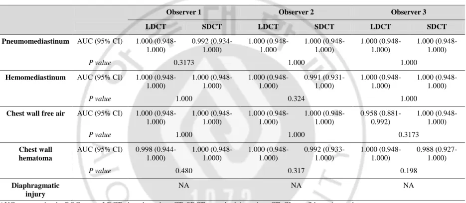
Table 5. AUC Comparison in Each Observer between LDCT vs. SDCT for Mediastinal, Chest wall and Diaphragmatic Injuries
Observer 1 Observer 2 Observer 3 LDCT SDCT LDCT SDCT LDCT SDCT Pneumomediastinum AUC (95% CI) 1.000
(0.948-1.000) 0.992 (0.934-1.000) 1.000 (0.948-1.000 1.000 (0.948-1.000) 1.000 (0.948-1.000) 1.000 (0.948-1.000) P value 0.3173 1.000 1.000
Hemomediastinum AUC (95% CI) 1.000 (0.948-1.000) 1.000 (0.948-1.000) 1.000 (0.948-1.000) 0.991 (0.931-1.000) 1.000 (0.948-1.000) 1.000 (0.948-1.000) P value 1.000 0.324 1.000
Chest wall free air AUC (95% CI) 1.000 (0.948-1.000) 1.000 (0.948-1.000) 1.000 (0.948-1.000) 1.000 (0.948-1.000) 0.958 (0.881-0.992) 1.000 (0.948-1.000) P value 1.000 1.000 0.3173 Chest wall hematoma AUC (95% CI) 0.998 (0.944-1.000) 1.000 (0.948-1.000) 1.000 (0.948-1.000) 0.992 (0.933-1.000) 1.000 (0.948-1.000) 0.988 (0.927-1.000) P value 0.480 0.317 0.198 Diaphragmatic injury NA NA NA
AUC : area under the ROC curve, LDCT : low dose chest CT, SDCT : standard dose chest CT, CI : confidence interval ** NA : not applicable due to all observers identified with confidence 3 (definite positive)
14 C. Radiation Dose Reduction
The use of lower tube voltage and tube current with ATCM in chest MDCT of blunt chest trauma patients could provide substantial dose reduction. The average CTDIvol on LDCT was 2.72 mGy and the average CTDIvol on SDCT was 13.2 mGy. About 79.4 % of dose reduction was achieved in LDCT, compared with SDCT.
D. Intraclass Correlation Coefficient
The ICC values of each injury are summarized in Table 6. ICC analysis revealed that there was an excellent inter-observer consistency for the evaluation of the pulmonary injury (ICC, from 0.8815 to 0.9859), skeletal trauma (ICC, from 0.8601 to 0.9396), mediastinal injury (ICC, from 0.9592 to 1.0000) and chest wall injury (ICC, from 0.8959 to 0.9975) in both LDCT and SDCT. ICC of 3 observers for diagnosis of aortic injury using SDCT was also very good (0.9594).
15
Table 6. Intraclass Correlation Coefficient for Inter-observer Consistency of LDCT and SDCT
LDCT (95% CI) SDCT (95% CI) Pulmonary and tracheobronchial injury
Lung contusion 0.9611 (0.9420-0.9747) 0.9631 (0.9450-0.9760) Pneumatocele 0.8955 (0.8441-0.9320) 0.8815 (0.8232-0.9228) Pneumothorax 0.9576 (0.9368-0.9724) 0.9859 (0.9790-0.9908) Hemothorax 0.9226 (0.8845-0.9456) 0.9210 (0.8821-0.9485) Skeletal injury Rib 0.9074 (0.8619-0.9397) 0.8601 (0.7982-0.9065) Clavicle 0.9077 (0.8669-0.9384) 0.9396 (0.9128-0.9596) Scapula 0.9123 (0.8734-0.9414) 0.9201 (0.8848-0.9467) Sternum 0.8859 (0.8354-0.9238) 0.9071 (0.8660-0.9380) Vertebra 0.8995 (0.8551-0.9329) 0.8918 (0.8439-0.9277) Mediastinal injury Pneumomedistinum 1.0000 0.9592 (0.9390-0.9734) Hemomediastinum 0.9664 (0.9499-0.9781) 0.9682 (0.9526-0.9793)
Chest wall injury
Hematoma 0.9628 (0.9446--0.9758) 0.8959 (0.8446-0.3229)
Free air 0.9843 (0.9766-0.9898) 0.9975 (0.9963-0.9984)
Diaphragmatic injury 1.0000 1.0000
Aortic injury NA 0.9844 (0.9767-0.9898)
Upper abdominal injury
Liver NA 0.9430 (0.9150-0.9629)
Spleen NA 0.9532 (0.9302-0.9695) LDCT : low dose chest CT, SDCT : standard dose chest CT, CI : confidence interval
16 A B
C D
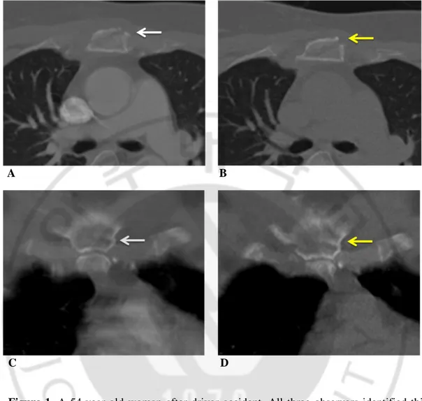
Figure 1. A 54-year-old woman after driver accident. All three observers identified this
sternal fracture in both SDCT (white arrows in A, C) and LDCT (yellow arrows in B, D) with confidence 3 (definite fracture). Axial and coronal 3-mm thickness image of SDCT examination (120kVp and 180mAs with ATCM) and LDCT (100kVp, 40mAs with ATCM) show fracture of sternum clearly.
17 A B
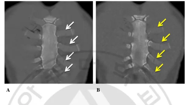
Figure 2. A 57-year-old man after driver accident. All three observers identified this
costal cartilage fractures in both SDCT (white arrows in A) and LDCT (yellow arrows in B) with confidence 2 or 3 (probable or definite fracture). Coronal 3-mm thickness image of SDCT examination (120kVp and 180mAs with ATCM) and LDCT (100kVp, 40mAs with ATCM) show fractures of left costal cartilage.
18
A B
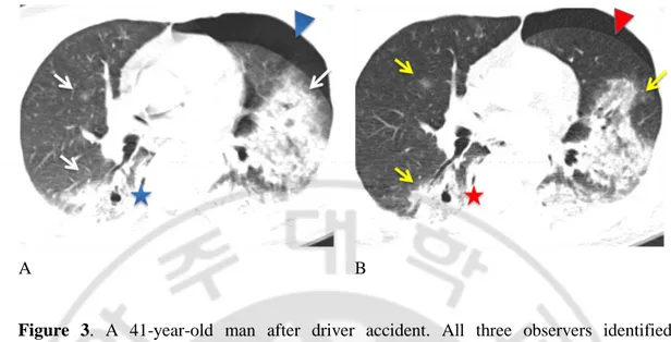
Figure 3. A 41-year-old man after driver accident. All three observers identified
pulmonary contusions (arrows), pneumatocele (star), pneumothorax (arrow head) in both SDCT (white, blue in A) and LDCT (yellow, red in B) with confidence 3 (definitely positive). Axial 3-mm thickness image of SDCT examination (120kVp and 180mAs with ATCM) and LDCT (100kVp, 40mAs with ATCM) show pulmonary lesions clearly.
19
IV. Discussion
Our study demonstrates several key results. First, there was good to excellent inter-observer consistency for the assessment of LDCT image data in patients of blunt chest trauma compared with assessment of SDCT in all observers. Second, there was no significant difference of diagnostic performance between LDCT and SDCT for the evaluation of pulmonary injury, skeletal trauma and mediastinal injury in all observers (except injuries that need post-contrast CT for proper evaluation such as aortic injury). Third, radiation dose reduction could be achieved up to about 79% of SDCT in LDCT.
Although chest radiography plays an important role in the evaluation and management of patients with blunt chest trauma, chest CT has became the most important imaging modality in the evaluation of patients with blunt chest trauma, because of chest CT scan could provide accurate and additional information quickly, especially since the introduction of the MDCT (Brenner and Hall, 2007; Brink et al., 2008; Deunk et al., 2009). In the situation of patients with major blunt trauma, there is a high probability of repeat exposure of radiation from medical imaging. However, radiation hazards of the trauma patients could be easily neglected because of its urgent clinical setting. Moreover, exposure to ionizing radiation from medical imaging is becoming concerned recently due to the fact that increased use of CT scan in recent year (Brenner and Hall, 2007).
Several studies supported a harmful effect of low-dose exposure of radiation. One study reported that radiation exposure induced by medical imaging had significant relevance to cancer risk, causing 0.6-3.2% of malignant tumors in developed countries (Berrington de Gonzalez and Darby, 2004) and Hall et al. estimated that the lifetime cancer risk increased 4% per sievert from low-dose exposure (Hall, 1991).
It is clear that radiologists have to make much effort to reduce unnecessary exposure of ionizing radiation from CT according to the principle of ALARA by the International Commission of Radiological Protection (1991; Slovis, 2003). The radiation dose could be reduced by application of the appropriate criteria for CT examination. Several published guidelines were available and American College of Radiology appropriateness criteria asserted that that in the patients with high-mechanism injury, abnormal chest radiographs, altered mental status, distracting injuries, or clinically suspected thoracic injury, routine use of chest CT should be strongly considered (Jonathan H. Chung, 2013).
20
Another method for CT radiation dose reduction is adjustment of the scanning parameters. Radiation dose could be lowered by reduction of tube current, reduction of tube voltage, using minimum scan length, adjustment of filter size accommodate to patient size, using of automatic exposure and the addition of noise-reducing reconstruction techniques (Kubo et al., 2008). Appropriate adjustment of image parameters ensured CT radiation dose reduction without compromising image. However, it is more important to keep diagnostic quality rather than keep image quality. And our major concern is not to study whether MDCT should be performed routinely or selectively for evaluation of blunt chest trauma patients, but to evaluate feasibility of LDCT instead of SDCT for initial survey of blunt chest trauma patients. Therefore, our study tried to evaluate the diagnostic performance of LDCT in the blunt chest trauma, in the ways of direct comparison of SDCT.
In terms of evaluation of pulmonary and tracheobronchial tree injury, our study revealed that no diagnostic performance for the diagnosis of lung contusion between LDCT and SDCT in all observers with good inter-observer consistency. Lung contusion is most important contributing factors of mortality and morbidity in polytrauma patients with thoracic trauma (Gaillard et al., 1990). It may result in severe respiratory failure, adult respiratory distress syndrome, septic conditions and multiple organs failure. Lung contusion may be frequently missed in chest x-ray especially early phase due to it took with supine position and some contusions is not visible in first hours after trauma. On the other hand, chest CT can accurately visualized lung contusions immediate time after injuries and detect even small pulmonary contusions (Trupka et al., 1997).
Chest CT is superior to chest x-ray for detection of fractured rib, scapula, sternum and vertebra (Traub et al., 2007). Our study also demonstrated that LDCT is comparable with SDCT in detection of the fractures in bony thorax. This result is consistent with previous reports that dose-reduced CT was sufficient for diagnosis of fracture in cervical spine trauma (Mulkens et al., 2007), and in experimental animal model (Moritz et al., 2012). However, subtle rib contour abnormality (buckle fracture) could not be easily detected using low dose CT. Thus, additional coronal and sagittal reformatted images can be helpful for detection of fractures that missed on axial images (Cho et al., 2012).
With regard to pneumothorax and pneumomediastinum, it is not surprising that CT is extremely accurate in detecting of abnormally accumulated of air density (Trupka et al., 1997; Dissanaike et al., 2008; Scaglione et al., 2008; Oikonomou and Prassopoulos, 2011).
21
In this current study, diagnostic performance of MDCT was excellent, AUC of LDCT and SDCT for diagnosing pneumothorax was range from 0.917 to 1.000 and from 0.972 to 1.000, respectively. Furthermore, there was excellent inter-observer consistency in both LDCT (ICC=0.9576) and SDCT (ICC=0.9858) in all observers.
Soft tissue injuries in the chest wall are rarely life-threatening (Collins, 2000). However, chest wall injuries frequently overlooked in daily practice and chest wall hematoma may become life-threatening in the patient with anticoagulant therapy. In low dose CT scan, evaluation of mediastinum and soft tissue of chest wall has limited. It is because inherent limited tissue contrast of these areas, which is intensified in dose reduction techniques (Baker et al., 1974; Prasad et al., 2002). In this study, only four cases of chest wall hematoma were identified and these were well detected in both LDCT (AUC, from 0.998 to 1.000) and SDCT (AUC, from 0.988 to 1.000). Inter-observer consistency for evaluation of chest wall hematoma was also good at LDCT (ICC=0.9628) and SDCT (ICC=0.8959).
There were a few limitations in our prospective study. First, comparative evaluation of aortic and upper abdominal solid organ injury was limited due to non-contrast LDCT. Second, our study did not apply iterative reconstruction (IR) technique, Using IR technique could provide substantial improvement of image quality with reduced radiation dose (Noel et al., 2011).
22
V. CONCLUSION
Our study is first prospective comparison study of LDCT with SDCT in initial evaluation of blunt chest trauma. It has strength of direct intra-observer comparison of diagnostic performance between LDCT and SDCT. This preliminary study suggests that LDCT could maintain diagnostic performance for the initial evaluation of blunt chest trauma patients with significant reduction of radiation dose. Further larger study of low dose CT is needed with intravenous contrast for evaluation of aortic injury and upper abdominal solid organ injury.
23
REFERENCES
1. 1990 Recommendations of the International Commission on Radiological Protection. Ann ICRP 21: 1-201, 1991
2. Baker SP, O'Neill B, Haddon W, Jr., Long WB: The injury severity score: a method for describing patients with multiple injuries and evaluating emergency care. J Trauma 14: 187-196, 1974
3. Berrington de Gonzalez A, Darby S: Risk of cancer from diagnostic X-rays: estimates for the UK and 14 other countries. Lancet 363: 345-351, 2004
4. Brenner DJ, Hall EJ: Computed tomography--an increasing source of radiation exposure. N Engl J Med 357: 2277-2284, 2007
5. Brink M, Deunk J, Dekker HM, Kool DR, Edwards MJ, van Vugt AB, Blickman JG: Added value of routine chest MDCT after blunt trauma: evaluation of additional findings and impact on patient management. AJR Am J Roentgenol 190: 1591-1598, 2008
6. Broder J, Warshauer DM: Increasing utilization of computed tomography in the adult emergency department, 2000-2005. Emerg Radiol 13: 25-30, 2006
7. Cho SH, Sung YM, Kim MS: Missed rib fractures on evaluation of initial chest CT for trauma patients: pattern analysis and diagnostic value of coronal multiplanar reconstruction images with multidetector row CT. Br J Radiol 85: e845-850, 2012
8. Collins J: Chest wall trauma. J Thorac Imaging 15: 112-119, 2000
9. Deunk J, Brink M, Dekker HM, Kool DR, van Kuijk C, Blickman JG, van Vugt AB, Edwards MJ: Routine versus selective computed tomography of the abdomen, pelvis, and lumbar spine in blunt trauma: a prospective evaluation. J Trauma 66: 1108-1117, 2009
10. Dissanaike S, Shalhub S, Jurkovich GJ: The evaluation of pneumomediastinum in blunt trauma patients. J Trauma 65: 1340-1345, 2008
11. Dyer DS, Moore EE, Mestek MF, Bernstein SM, Ikle DN, Durham JD, Heinig MJ, Russ PD, Symonds DL, Kumpe DA, Roe EJ, Honigman B, McIntyre RC, Jr., Eule J, Jr.: Can chest CT be used to exclude aortic injury? Radiology 213: 195-202, 1999
12. Gaillard M, Herve C, Mandin L, Raynaud P: Mortality prognostic factors in chest injury. J Trauma 30: 93-96, 1990
24
Schwabe P, Streitparth F: Reducing Radiation Dose in Emergency CT Scans While Maintaining Equal Image Quality: Just a Promise or Reality for Severely Injured Patients? Emerg Med Int 2013: 984645, 2013
14. Hall E: Scientific view of low-level radiation risks. Radiographics 11: 509-518, 1991
15. Jonathan H. Chung CWC, Tan-Lucien H. Mohammed, Jacobo Kirsch, Kathleen Brown, Debra Sue Dyer, Mark E. Ginsburg, Darel E. Heitkamp, Jeffrey P. Kanne, Ella A. Kazerooni, Loren H. Ketai, James G. Ravenel, Anthony G. Saleh, Rakesh D. Shah (2013). ACR Appropriateness criteria (2013, American College of Radiology ).
16. Kim PK, Gracias VH, Maidment AD, O'Shea M, Reilly PM, Schwab CW: Cumulative radiation dose caused by radiologic studies in critically ill trauma patients. J Trauma 57: 510-514, 2004
17. Kubo T, Lin PJ, Stiller W, Takahashi M, Kauczor HU, Ohno Y, Hatabu H: Radiation dose reduction in chest CT: a review. AJR Am J Roentgenol 190: 335-343, 2008
18. Moritz JD, Hoffmann B, Sehr D, Keil K, Eggerking J, Groth G, Caliebe A, Dischinger J, Heller M, Bolte H: Evaluation of ultra-low dose CT in the diagnosis of pediatric-like fractures using an experimental animal study. Korean J Radiol 13: 165-173, 2012
19. Mulkens TH, Marchal P, Daineffe S, Salgado R, Bellinck P, te Rijdt B, Kegelaers B, Termote JL: Comparison of low-dose with standard-dose multidetector CT in cervical spine trauma. AJNR Am J Neuroradiol 28: 1444-1450, 2007
20. Noel PB, Fingerle AA, Renger B, Munzel D, Rummeny EJ, Dobritz M: Initial performance characterization of a clinical noise-suppressing reconstruction algorithm for MDCT. AJR Am J Roentgenol 197: 1404-1409, 2011
21. Oikonomou A, Prassopoulos P: CT imaging of blunt chest trauma. Insights Imaging 2: 281-295, 2011
22. Plurad D, Green D, Demetriades D, Rhee P: The increasing use of chest computed tomography for trauma: is it being overutilized? J Trauma 62: 631-635, 2007
23. Prasad SR, Wittram C, Shepard JA, McLoud T, Rhea J: Standard-dose and 50%-reduced-dose chest CT: comparing the effect on image quality. AJR Am J Roentgenol 179: 461-465, 2002
25
computed tomography and blunt chest trauma. Eur J Radiol 65: 377-388, 2008
25. Shuman WP: CT of blunt abdominal trauma in adults. Radiology 205: 297-306, 1997
26. Slovis TL: Children, computed tomography radiation dose, and the As Low As Reasonably Achievable (ALARA) concept. Pediatrics 112: 971-972, 2003
27. Steenburg SD, Ravenel JG, Ikonomidis JS, Schonholz C, Reeves S: Acute traumatic aortic injury: imaging evaluation and management. Radiology 248: 748-762, 2008
28. Traub M, Stevenson M, McEvoy S, Briggs G, Lo SK, Leibman S, Joseph T: The use of chest computed tomography versus chest X-ray in patients with major blunt trauma. Injury 38: 43-47, 2007
29. Trupka A, Waydhas C, Hallfeldt KK, Nast-Kolb D, Pfeifer KJ, Schweiberer L: Value of thoracic computed tomography in the first assessment of severely injured patients with blunt chest trauma: results of a prospective study. J Trauma 43: 405-411; discussion 411-402, 1997
30. Winslow JE, Hinshaw JW, Hughes MJ, Williams RC, Bozeman WP: Quantitative assessment of diagnostic radiation doses in adult blunt trauma patients. Ann Emerg Med 52: 93-97, 2008
26 -국문 요약-
흉부 외상 환자의 초기 평가에서의 저선량 흉부
전산화단층촬영의 유용성 검사
아주대학교 대학원 의학과 김성중 (지도교수: 선주성) 서론: 전산화단층촬영 (Computed tomography, 이하 CT)은 흉부 외상을 포함한 중증 외상환자의 진단에 있어 핵심적인 역할을 하고 있다. 하지만 흉부 외상환자에서 일상적인 CT 의 사용으로 인하여 CT 에 의한 방사선량이 급격히 증가하였다. 따라서 ALARA (as low as reasonable achievable)에 따라 흉부 외상환자에서도 방사선량의 조정이 불가피하다. 따라서 이 연구를 통하여 흉부 외상 환자에서 저선량 흉부 CT (Low dose chest CT, 이하 LDCT)의 진단능과 관찰자들 간의 진단 일치도를 알아보고자 한다.연구 방법: 전향적 연구로써 선정 기준에 부합하는 총 69 명의 흉부 외상환자를 연구하였다 (남자 : 77%, 16 세 ~ 85 세). 모든 환자들에게 조영제를 주입하지 않은 LDCT 와 조영제를 주입한 일반선량 흉부 CT (Standard dose chest CT, 이하 SDCT)를 모두 시행하였으며, 다음과 같이 CT 변수를 조정하였다. : LDCT, 40mAs with automatic tube current modulation (ATCM) and 100kVp (body mass index, 이하 BMI<25, n=51) or 120kVp (BMI>25, n=18); SDCT, 180mAs with ATCM and 120kVp. CT 영상은 3mm 절편두께로 횡축, 종축, 시상축으로 재구성하였다. 진단의 참조 표준은 CT 를 1mm 절편두께로 재구성한 영상을 2 명의 흉부 영상의학과 전문의가 일치를 이루어 판독한 결과를 이용하였다. 3 명의 관찰자가 독립적으로 흉부외상 (흉부실질과 기관지 손상, 골격 손상, 종격동 손상, 흉벽 손상, 횡격막 손상, 대동맥 손상, 상복부 손상)에 대해 진단을 하였다. 관찰자들은 진단의 확신 정도를 4 점의 척도를 이용하여
27
평가하였다(0-3 점). 방사선량의 비교는 vendor 가 제시하는 CT dose index volume(CTDIvol) 값을 이용하였다.
연구 결과: LDCT의 방사선량 (average CTDIvol = 2.72mGy)은 SDCT의 방사선량 (average CTDIvol = 13.4mGy)보다 현저하게 낮은 값을 보였다 (약 79.4%의 방사선량 감소). 다중-관찰자 수신자특성곡선 (Multiple-reader receiver operating characteristic, 이하 ROC)의 분석을 통한 area under the ROC curve(AUC)의 값을 분석한 결과 흉부외상을 진단하는데 LDCT와 SDCT 모두 높은 진단능을 보였으며 두 군간의 통계적으로 유의한 차이를 보이지 않았다 (0.823-1.000, 0.781-1.000, p>0.05). 유목내 상관 계수 (Intraclass correlation coefficient, 이하 ICC)를 통하여 관찰자들 간의 진단 일치도를 분석한 결과, LDCT와 SDCT 모두 흉부외상를 진단하는데 있어 좋은 진단 일치도를 보였다 (ICC=0.8601 – 1.000). LDCT에서 조영제를 사용하지 않았기 때문에 대동맥과 상복부 손상을 비교하는데 제한이 있었다. 결론 : LDCT 는 SDCT 와 비교하여 방사선량의 획기적으로 줄이면서 진단능을 유지할 수 있기 때문에 흉부 외상의 초기 평가에 있어서 굉장히 이점을 가지고 있다. 추후에 대동맥과 상복부 손상을 비교하기 위하여 조영제를 이용한 LDCT 를 이용한 연구의 진행이 필요하다. 핵심어: 저선량 흉부 CT, 흉부 외상, 진단능, 방사선량 감소