ABSTRACT
BACKGROUND/OBJECTIVES:Curcuma zedoaria R. (Zingiberaceae) has been used to treat headache, fever, and hypertension-related symptoms in Asian countries, including Korea, China, and Japan. We investigated whether dietary intake of a C. zedoaria extract (CzE) affected atherosclerosis in vivo.
MATERIALS/METHODS: Apolipoprotein E-deficient (ApoE−/−) mice (n = 32) were fed a normal
diet (ND), a high-cholesterol diet (HCD), an HCD containing CzE (100 mg/kg/day), or an HCD containing simvastatin (10 mg/kg/day) for 12 weeks. The anti-atherosclerotic effects were evaluated by observing changes in fatty streak lesions, immunohistochemical analysis, ex vivo fluorescence imaging, lipid profiles, and western blot analysis.
RESULTS: The CzE-fed group showed a 41.6% reduction of atherosclerosis. Furthermore, CzE significantly reduced the levels of serum triglyceride, high-density lipoprotein, the chemokine (C-X3-C-motif ) ligand 1, the adhesion molecules vascular cell adhesion molecule-1, intracellular adhesion molecule-1, and E-selectin; down-regulation of tumor necrosis factor-α, interleukin-6, high mobility group box-1, and cathepsin levels in the aortic sinuses and aortas of ApoE−/− mice were also observed.
CONCLUSIONS: The results suggest that the inclusion of a water extract of C. zedoaria in a HCD is closely correlated with reducing the risk of vascular inflammatory diseases in an ApoE mouse model.
Keywords:Curcuma zedoaria; atherosclerosis; ApoE knockout mice; VCAM-1; chemokine CX3CL1
INTRODUCTION
Atherosclerosis is a sustained inflammatory disease in which the inside of arteries narrows due to the build-up of an atheromatous plaque. Several studies have shown that systemic inflammation and pro-inflammatory cytokines characterize early atherogenesis [1]. Oxidized low-density lipoproteins (OxLDLs) are reported to increase a series of pro-inflammatory reactions via different mediators (tumor necrosis factor [TNF]-α, interleukin [IL]-6, high
Original Research
Received: May 4, 2020 Revised: Jul 3, 2020 Accepted: Dec 20, 2020 §Corresponding Authors: Kung-Woo NamDepartment of Life Science and
Biotechnology, Soonchunhyang University, 22 Soonchunhyang-ro, Asan 31538, Korea. Tel. +82-41-530-4770
Fax. +82-41-530-4883 E-mail. [email protected] Sungwook Chae
Herbal Medicine Research Division, Korea Institute of Oriental Medicine, 1672 Yuseong-daero, Yuseong-gu, Daejeon 34054, Korea. Tel. +82-42-868-9524
Fax. +82-42-868-9301 E-mail. [email protected]
©2021 The Korean Nutrition Society and the Korean Society of Community Nutrition This is an Open Access article distributed under the terms of the Creative Commons Attribution Non-Commercial License (https:// creativecommons.org/licenses/by-nc/4.0/) which permits unrestricted non-commercial use, distribution, and reproduction in any medium, provided the original work is properly cited.
ORCID iDs
Ki Mo Kim
https://orcid.org/0000-0003-4893-4841 Joo Young Lee
https://orcid.org/0000-0001-8761-5187 Byeong Hwa Jeon
https://orcid.org/0000-0002-3347-2739 Khong Trong Quan
https://orcid.org/0000-0003-2630-1031
Ki Mo Kim 1,2, Joo Young Lee 1, Byeong Hwa Jeon 3, Khong Trong Quan 4,
MinKyun Na 4, Kung-Woo Nam 5§, and Sungwook Chae 1,2§
1Herbal Medicine Research Division, Korea Institute of Oriental Medicine, Daejeon 34054, Korea
2 Department of Korean Life Science and Technology, University of Science and Technology, Daejeon 34113, Korea
3Department of Physiology, School of Medicine, Chungnam National University, Daejeon 34134, Korea 4 Department of Pharmacognosy, College of Pharmacy, Chungnam National University, Daejeon 34134,
Korea
5Department of Life Science and Biotechnology, Soonchunhyang University, Asan 31538, Korea
Extract of Curcuma zedoaria
R. prevents atherosclerosis in
apolipoprotein E-deficient mice
MinKyun Na https://orcid.org/0000-0002-4865-6506 Kung-Woo Nam https://orcid.org/0000-0002-1168-5266 Sungwook Chae https://orcid.org/0000-0001-9789-0043 Funding
This work was supported by a grant (KSN2012330) from the Korea Institute of Oriental Medicine (KIOM) and by the Soonchunhyang University Research Fund.
Conflict of Interest
The authors declare no potential conflicts of interests.
Author Contributions
Conceptualization: Nam KW, Chae S; Data curation: Kim KM, Lee JY, Jeon BH; Formal analysis: Lee JY, Quan KT, Na M; Investigation: Kim KM; Writing - original draft: Kim KM; Writing - review & editing: Nam KW, Chae S.
mobility group box [HMGB]-1, etc.) [2]. In the early period of atherogenesis, OxLDL prompts expression of the chemokine (C-X3-C-motif ) ligand 1 (CX3CL1) in vascular smooth muscle and endothelial cells (ECs). CX3CL1 is produced as a membrane-bound long protein, with its chemokine domain being situated on a mucin-like stalk. Cleavage at the base of this stalk generates a TNF-α-converting enzyme, a soluble chemokine with roles as a strong chemoattractant for T cells and monocytes, while the cell-bound chemokine enhances the strong adhesion of leukocytes to activated ECs [3]. Furthermore, high levels of inflammatory factors result in monocyte adhesion and differentiation into macrophages and increase the production of several adhesion molecules, containing vascular cell adhesion molecule-1 (VCAM-1), intracellular adhesion molecule-1 (ICAM-1), and E-selectin [4]. The activated macrophages migrate into the intima, becoming lipid-laden and transforming into foam cells. Some studies have revealed non-traditional roles for cathepsins in extracellular matrix remodeling. Cysteine protease cathepsins have a critical role in extracellular matrix remodeling and have been involved in the development and progression of atherosclerosis-based cardiovascular diseases [5]. The most potent of these cathepsins is cathepsin S, an elastolytic enzyme expressed predominantly in macrophages and inflamed medial smooth muscle cells [6]. Thus, natural products that control vascular inflammatory mediators may be efficacious in atherosclerosis. Curcuma zedoaria R. (Zingiberaceae) is a traditional Korean medicinal plant used to treat symptoms such as fatigue and weakness after illness. The major chemical constituents of C. zedoaria include curcumenol and zedoarofuran [7], curcumenone [8], bisabolone-9-one, turmeronol B, bisdemethoxycurcumin, desmethoxycurcumin, and curcumin [9]. This plant has been reported to exhibit anti-oxidant [10], cancer [11-13], inflammatory [14,15] and fungal effects [16], to reduce drunkenness [17], and to exert analgesic [18] and antitrypanosomal activities [19]. However, the relationship between a C. zedoaria extract (CzE) and atherosclerosis has not been fully elucidated. A previous report showing an anti-inflammatory activity of C. zedoaria encouraged us to study the potential anti-atherogenic effects of CzE. Currently, an apolipoprotein E-deficient (ApoE−/−) mouse model
is the most frequently used animal model in the study of atherosclerosis. The ApoE−/− mouse
model's main advantage is that the rate of atherogenesis can be markedly hastened by a high-fat, high-cholesterol diet (HCD), which results in significant increases in plasma lipid levels. Provision of an HCD is the best method for inducing systematic lesions in ApoE−/− mice.
Thus, we investigated the effects of CzE on atherosclerosis and associated inflammatory mediators in ApoE−/− mice fed an HCD.
MATERIALS AND METHODS
Plant material
Dried C. zedoaria material was obtained from a commercial supplier (Kwangmyung-Dang, Ulsan, Korea). A voucher specimen was deposited in the Herbal Medicine Resources Research Center (Korea Institute of Oriental Medicine). The rhizome (3.0 kg) was extracted by using distilled water under reflux for 3 h. The obtained product was evaporated to dryness under vacuum and finally freeze-dried, producing the CzE (103.3 g). The lyophilized extract used in the animal experiments was stored at −20°C. The chemical components contained in CzE were zedoarondiol, isozedoarondiol, procurcumadiol, procurcumenol, and curcumenol. An high-pressure liquid chromatography analysis was performed using a Shimadzu system with ultraviolet detection at 265 nm and a LC-18 column (150 mm × 4.6 mm, 5 mm) from Phenomenex. The solvent system used was water:acetonitrile (95:5; v/v -> 30:70 -> 1:10) injected at a flow rate of 1 mL/min and an injection volume of 10 µL.
Animals
ApoE−/− mice [20] were acquired from Central Laboratory Animal Inc. (Seoul, Korea) and
housed in cages (3 mice per cage) under specific pathogen-free conditions. All animal protocols were in accord with those of the Committee of Animal Care and Experiments of Chungnam National University, Korea (CNUH-015-A0017). Thirty-two male ApoE−/− mice were randomly
separated into 4 dietary groups: normal diet (ND), n = 6; HCD, n = 10; HCD compensated with CzE, n = 10; and HCD supplemented with simvastatin, n = 6. The HCD included 15% fat (7.5% lard and 7.5% cacao butter), 1.25% cholesterol, and 0.5% cholic acid. The mice were given water and chow ad libitum. Mouse body weight was measured every 3 weeks for 12 weeks. After 12 weeks, parameters were analyzed by western blot analysis, immunohistochemistry, and bioluminescence imaging (PerkinElmer Inc., Waltham, MA, USA).
Histological examinations
The size of the aortic sinus lesions was quantified as described in a previous paper [21], with a slight modification. The heart and aorta were perfused with phosphate-buffered saline for 10 minutes and then with 3.5% paraformaldehyde for 5 minutes. The heart was then rapidly removed, fixed in 10% buffered neutral formalin for 24 h, and washed in tap water to remove the formalin. Atherosclerotic plaque samples were stained with Oil Red O. Six lesion areas were compared by using computer-supported morphometry (Image-Pro Plus; Media Cybernetics, Rockville, MD, USA) at 30-μm intervals, and the average lesion size was estimated (n = 22). To detect the cytokine level, frozen sections were stained individually with 1:200 dilutions of anti-TNF-α, CX3CL1, or HMGB-1 antibodies (Santa Cruz Biotechnology, Santa Cruz, CA, USA). Harris hematoxylin was used as the counterstain.
Serum parameter analysis
Analysis of the serum parameters was performed according to methods described in a previous study [22]. Briefly, serum levels of aspartate aminotransferase (AST), alanine aminotransferase (ALT), triglyceride (TG), total cholesterol (TC), glucose, and high-density lipoproteins (HDLs) were determined using a clinical chemistry analyzer system (Dri-Chem 700i; Fujifilm, Tokyo, Japan). The serum low-density lipoprotein (LDL) content was determined using LDL Assay kits (KOMA Biotech, Seoul, Korea).
Ex vivo fluorescence imaging
Ex vivo fluorescence was evaluated by applying the method reported previously [23]. Briefly, mice were anesthetized with isoflurane/oxygen/nitrogen, and the aorta, liver, and lung were excised and subsequently underwent fluorescent signal analysis via imaging performed using an in vivo imaging system (IVIS Lumina XR; PerkinElmer Inc.). The fluorescent intensity of ProSense 750 (PerkinElmer Inc.) in the selected organ was analyzed.
Western blot analysis
Proteins obtained from mouse aortas were ultrasonically homogenized in lysis buffer that included protease inhibitors (Roche Diagnostics, Indianapolis, IN, USA). Protein concentrations were analyzed by using a Pierce BCA Assay Kit (Thermo Fisher Scientific, Rockford, IL, USA). Western blot analysis was undertaken as previously described [24]. The blotted membranes were developed by using an enhanced chemiluminescence system (Thermo Scientific), and the blots were identified using an LAS-4000 mini luminescent image analyzer (GE Healthcare, London, UK). Quantitative band density was determined by using ImageJ software (National Institutes of Health, Bethesda, MD, USA).
Statistical analysis
All measurements were obtained in triplicate, and the data are presented as mean ± standard deviation values. Analysis of variance in conjunction with Tukey's test was used to analyze group differences, and a P-value < 0.05 was considered to indicate statistical significance.
RESULTS
CzE regulates lipid profile in blood of ApoE
−/−mice
The body weights of ApoE−/− mice fed the ND and those in the experimental diet groups were
recorded periodically to observe changes related to CzE treatment. As listed in Table 1, the average body weights of the ND- and HCD-fed mice were significantly different. A significant reduction in body weight was observed in ApoE−/− mice when treated with HCD and CzE. In
addition, as shown in Table 2, HCD-fed ApoE−/− mice had significantly elevated serum total
TG, TC, glucose, and LDL levels compared to those in ND-fed ApoE−/− mice. Thus, treatment
with CzE remarkably lowered both TG and HDL levels.
Anti-atherogenic effect of CzE on ApoE
−/−mice
After 12 weeks of CzE administration, the effect of CzE on the formation of atherosclerotic lesions in the aortic sinuses of ApoE−/− mice was assessed. Fig. 1 shows sections of the aortic
roots from ApoE−/− mice in the control, HCD, and HCD with CzE groups. Aortic roots of
the control group mice showed no fatty streak lesions, whereas mice fed the HCD for 12 weeks showed considerable fatty streak lesions on the intima of the aortic sinuses (Fig. 1B). However, in the HCD with CzE group mice, the diet-induced aortic injury area was 41.6% smaller than that of the HCD-fed mice (5.50 ± 0.87 [× 106] μm2 vs. 3.21 ± 0.18 [× 106] μm2,
respectively P < 0.01) (Fig. 1C and D).
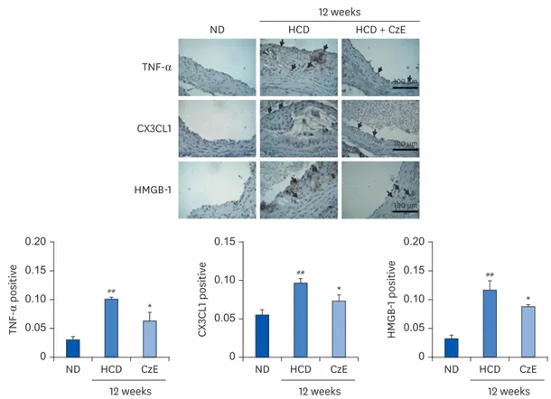
Expressions of TNF-α, CX3CL1, and HMGB-1 induced by CzE in normal intima
and atherosclerotic lesions of ApoE
−/−mice
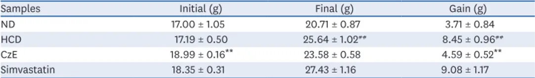
Frozen aortic sinus sections were stained with antibodies for TNF-α, CX3CL1, and HMGB-1 in accordance with the methods described previously [25]. Consistent with our western blot Table 1. Body weight changes in ApoE−/− mice after 12 weeks of CzE administration
Samples Initial (g) Final (g) Gain (g)
ND 17.00 ± 1.05 20.71 ± 0.87 3.71 ± 0.84
HCD 17.19 ± 0.50 25.64 ± 1.02## 8.45 ± 0.96##
CzE 18.99 ± 0.16** 23.58 ± 0.58 4.59 ± 0.52**
Simvastatin 18.35 ± 0.31 27.43 ± 1.16 9.08 ± 1.17
Data are presented as mean ± standard error of mean values (n = 6, each group). ApoE−/− mice treated with
simvastatin were considered positive controls.
ND, normal diet; HCD, high-cholesterol diet; CzE, Curcuma zedoaria extract (HCD with CzE treatment group), Simvastatin, HCD with simvastatin treatment group.
**P < 0.01 vs. HCD group; ##P < 0.01 vs. ND group.
Table 2. Effect of CzE on serum lipid profile levels in ApoE−/− mice
Samples ALT (U/L) AST (U/L) TG (mg/dL) TC (mg/dL) Glucose (mg/dL) HDL (mg/dL) LDL (mg/dL) ND 56.7 ± 1.8 192.7 ± 21.8 75.3 ± 11.8 294.0 ± 14.5 50.0 ± 4.2 63.3 ± 2.4 231.1 ± 11.9 HCD 92.0 ± 15.6 264.0 ± 65.2 190.0 ± 15.3## 874.0 ± 13.0### 135.3 ± 12.7## 180.0 ± 2.9### 777.9 ± 12.7###
CzE 74.0 ± 5.0 181.3 ± 5.9 108.7 ± 1.3** 842.0 ± 23.2 80.0 ± 25.0 92.7 ± 3.5*** 724.3 ± 27.9 Simvastatin 68.7 ± 11.2 177.0 ± 9.3 106.7 ± 11.9* 701.3 ± 17.7** 58.7 ± 3.3** 82.0 ± 7.6*** 562.0 ± 19.8** Data are presented as mean ± standard error of mean values (n = 3, each group). ApoE−/− mice treated with simvastatin were considered positive controls.
ND, normal diet; HCD, high-cholesterol diet; CzE, Curcuma zedoaria extract (HCD with CzE treatment group); Simvastatin, HCD with simvastatin treatment group; ALT, alanine aminotransferase; AST, aspartate aminotransferase; TG, triglyceride; TC, total cholesterol; HDL, high-density lipoprotein; LDL, low-density lipoprotein. *P < 0.05, **P < 0.01, ***P < 0.001 vs. HCD group; #P < 0.01, ##P < 0.001 vs. ND group.
analysis results, ApoE−/− mice demonstrated reduced expression of the inflammatory markers
TNF-α, CX3CL1, and HMGB-1 (P < 0.05) compared with those of HCD-fed ApoE−/− mice. Both
TNF-α and CX3CL1 were highly expressed in the HCD-fed group, but that increased expression was remarkably suppressed by the combined HCD–CzE treatment (Fig. 2). Compared with wild-type mice, the vascular HMGB-1 expression level was notably amplified in ApoE−/− mice
(P < 0.01) (Fig. 2). However, this was reversed by CzE treatment. Additionally, the western blot analysis results showed a pattern similar to that for atherosclerotic lesions (Fig. 4). In addition, TNF-α, and CX3CL1 were expressed not only in ECs, but also in the intima. However, HMGB-1 was detected primarily in the intima of the atherosclerotic lesions (Fig. 2).
CzE treatment decreases arterial cathepsin activity in HCD-fed ApoE
−/−mice
The HCD-fed group exhibited an increased proteolytic fluorescence level in the carotid arteries compared with that in control mice (P < 0.01); however, CzE reduced the cathepsin activity in the aorta and carotid arteries (Fig. 3). The ApoE−/− mice fed the HCD with
simvastatin added showed markedly reduced cathepsin activity in the aorta (Fig. 3). We also detected a significant reduction in ProSense 750 signals in mice fed the HFD with CzE (P < 0.01) compared to that in the HFD-fed group (Fig. 3).
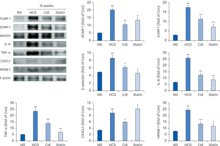
Effect of CzE on vascular inflammatory molecules in HCD-induced ApoE
−/−mice
We also compared the expressions of inflammatory and adhesion molecules in the atherosclerotic aorta of HCD-fed ApoE−/− mice by examining the western blot results. The
expression levels of VCAM-1, ICAM-1, and E-selectin in HCD-fed mice were notably increased compared with those of control mice. In contrast, the CzE- and simvastatin-treated mice
(A) (B) ** 0 100,000 200,000 300,000 400,000 600,000 500,000 700,000 ND ND HCD HCD CzE CzE Lesion ar ea (µm 2) (D) (C)
Fig. 1. Representative images of Oil Red O-stained gross atherosclerotic lesions in HCD-fed and control mice (A-C). Typical cross-sections of aortic valves (× 100) in hearts from control (ND) (A), HCD (B), and CzE-treated HCD (C) mice. Treatment with CzE (100 mg·kg−1·d−1) for 12 weeks affects atherosclerosis in ApoE−/− mice. (D) Mean atherosclerotic
lesion area in the aortic sinus was determined; data are expressed as mean ± SD values (n = 6 for each group). ND, normal diet; HCD, high-cholesterol diet; CzE, Curcuma zedoaria extract; ApoE−/−, apolipoprotein E-deficient. **P < 0.01 vs. HCD group.
## * 0 0.05 0.10 0.15 0.20 ND HCD CzE HMGB -1 positiv e 12 weeks ## * 0 0.05 0.10 0.15 ND HCD CzE CX 3CL1 positiv e 12 weeks ## * 0 0.05 0.10 0.15 0.20 ND HCD CzE TNF -α positiv e 12 weeks ND HCD HCD + CzE TNF-α CX3CL1 HMGB-1 12 weeks 100 µm 100 µm 100 µm
Fig. 2. Immunohistochemical identification of inflammatory molecules in ApoE−/− mouse aortas. ApoE−/− mice were fed an HCD for 12 weeks, after which,
inflammatory molecules in plaques in the aortic sinuses were assessed histologically and immunohistochemically. In the control aorta, TNF-α, CX3CL1, and HMGB-1 showed negligible staining (brown staining). HCD-fed ApoE−/− mice showed over-expression of TNF-α, CX3CL1, and HMGB-1 not only in endothelial cells, including
in some intimal areas, but also in smooth muscle cells (dark-brown staining: black arrows). However, in HCD–CzE-fed ApoE−/− mice, the expressions of these
inflammatory molecules were significantly inhibited. Arrowheads indicate TNF-α-, CX3CL1-, or HMGB-1-positive cells. All results are shown as mean ± SD values. ND, normal diet; HCD, high-cholesterol diet; CzE, Curcuma zedoaria extract; ApoE−/−, apolipoprotein E-deficient; TNF-α, tumor necrosis factor-α; CX3CL1,
chemokine (C-X3-C-motif) ligand 1; HMGB-1, high mobility group box-1. *P < 0.05 vs. HCD group; ##P < 0.01 vs. ND group. Epi-fluorescence (×106) ## ** ** 0 2 4 6 8 10 12 1.6 1.4 1.2 1.0 0.8 ND HCD HCD + CzESimvastatin
ND HCD HCD + CzESimvastatin ND HCD HCD + CzESimvastatin
Fl uor escent int ensity (× 10 6)
Fig. 3. Visualization of cathepsin activity in atherosclerotic mouse aortas. Visible light images show that the aortas of ApoE−/− mice appear normal, whereas those
of ApoE−/− mice fed an HCD exhibit increased proteolytic fluorescence in the carotid arteries. The fluorescent density distributed from the ascending aorta to
the infrarenal aorta in each group indicates the location of cathepsin activity. In the ApoE−/− mice fed the HCD with CzE diet (100 mg·kg−1·d−1), the inflammatory
cathepsin expression level was significantly inhibited. The presence of internally quenched fluorogenic peptides containing Abz-Dnp, which are activatable by pan-cathepsin, confirmed the inhibition of inflammatory cardiovascular disease by CzE treatment in ApoE−/− mice. Values presented are mean ± SD.
ND, normal diet; HCD, high-cholesterol diet; CzE, Curcuma zedoaria extract; ApoE−/−, apolipoprotein E-deficient. **P < 0.01 vs. HCD group; ##P < 0.01 vs. ND group.
had expression levels of VCAM-1, ICAM-1, and E-selectin lower than those of the HFD-fed mice (Fig. 4). In addition, we determined the levels of the inflammatory molecules TNF-α, IL-6, CX3CL1, and HMGB-1, which induce atherosclerotic inflammation in the aorta tissue. Administration of CzE in HFD-fed ApoE−/− mice markedly decreased the expressions of
TNF-α, IL-6, CX3CL1, and HMGB-1 in the aorta (Fig. 4). These results suggest that CzE may inhibit the expression of inflammatory and adhesion molecules in the aorta.
DISCUSSION
C. zedoaria was shown to be an effective anti-inflammatory agent in several earlier studies. In the present study, we established that CzE, a water extract of C. zedoaria in which sesquiterpenes and polyphenols are the main constituents, exerts anti-atherogenic effects in an ApoE−/− mouse model of atherosclerosis. CzE (100 mg/kg) notably decreased
atherosclerotic lesion formation in the aortic sinus of ApoE−/− mice, and inhibited expression
of the inflammatory mediators TNF-α, IL-6, and HMGB-1, and the chemokine CX3CL1. The 0 5 10 15 20 25 ND HCD CzE Statin ND HCD CzE 12 weeks Statin ## ** * ## ** ** ## * * ## ** ** ## * ## ** ** ## ** * VCAM -1 (fol d of Con) 0 5 10 15 20 ND HCD CzE Statin ICAM -1 (fol d of Con) 0 2 4 6 8 10 ND HCD CzE Statin E-sel ectin ( fol d of Con) 0 5 10 15 20 25 30 ND HCD CzE Statin IL -6 (fol d of Con) 0 5 10 15 20 25 30 ND HCD CzE Statin TNF -α (fol d of Con) 0 2 4 6 8 10 12 ND HCD CzE Statin CX 3CL1 (fol d of Con) 0 5 10 15 20 25 30 ND HCD CzE Statin HMGB -1 (fol d of Con) VCAM-1 E-selectin IL-6 TNF-α CX3CL1 HMGB-1 β-actin ICAM-1
Fig. 4. Effect of CzE on adhesion molecules and inflammatory mediators in the aortas of ApoE−/− mice. The data were collected from ND, HFD with CzE (100 mg/kg),
and HFD with simvastatin (10 mg/kg) fed mice (treated orally for 12 weeks). Representative photomicrographs of western blot results and densitometric analysis show the relative protein expression levels of adhesion molecules (ICAM-1, VCAM-1, and E-selectin) and inflammatory mediators (TNF-α, IL-6, CX3CL1, and HMGB-1), normalized to β-actin. Values are mean ± SD.
ND, normal diet; HCD, high-cholesterol diet; CzE, Curcuma zedoaria extract; ApoE−/−, apolipoprotein E-deficient; ICAM-1, intracellular adhesion molecule-1;
VCAM-1, vascular cell adhesion molecule-1; TNF-α, tumor necrosis factor-α; IL-6, interleukin-6; CX3CL1, chemokine (C-X3-C-motif) ligand 1; HMGB-1, high mobility group box-1.
expressions of the adhesion molecules VCAM-1, ICAM-1, and E-selectin in the aortic sinus of CzE-fed ApoE−/− mice were significantly decreased from those of HCD-fed ApoE−/− mice. The
reduction of adhesion molecule expressions was accompanied by down-regulation of cysteine protease cathepsin activity in the aorta of ApoE−/− mice. In this study, the body weight of the
CzE mice was lower than that of the HCD-fed ApoE−/− mice (Table 1). Serum analysis showed
that ALT and AST levels tended to be lower in CzE-treated mice but the differences were not significant; in contrast, total TG and HDL levels were significantly improved (Table 2). Furthermore, we observed that the 12-week HCD induced lesion formation in the aorta of ApoE−/− mice, but the HCD supplemented with CzE resulted in a remarkable reduction in lesion
area, similar to that induced by the commercially available anti-lipid-lowering simvastatin administered in ApoE−/− mice fed an HCD (data not shown). TNF-α, a pro-inflammatory
cytokine, has been shown to play a significant modulatory role in the chronic inflammation that characterizes atherosclerosis [26]; moreover, TNF-α elevates matrix degradation, thus promoting an influx of inflammatory cells into the vessel wall. TNF-α also up-regulates the expression of cell adhesion molecules (ICAM-1, VCAM-1, and E-selectin) and the chemokine CX3CL1. The expressions of inflammatory cytokines were significantly induced by the 12-week HCD treatment, whereas treatment with HCD and CzE diminished the expression levels of TNF-α, ICAM-1, VCAM-1, E-selectin, and CX3CL1. In addition, CzE significantly inhibited the expression of HMGB-1 (a ubiquitous nuclear protein). HMGB-1 has been described as an effective mediator of tissue remodeling in experimental atherosclerosis, and there is a significant association between HMGB-1 and coronary plaque composition [27]. In this study, immunohistochemical staining and western blot results showed that the high expression levels of HMGB-1 in HCD-fed ApoE−/− mice were decreased significantly by the inclusion of CzE in the
feed. In nonatherosclerotic arteries, the expression of many cathepsins, including cathepsins F, S, and K, is negligible [5], and the expression level is also low in the vessel walls of healthy arteries [28]. In atherosclerotic lesions, however, cathepsin activity is enhanced. Among cathepsins B, L, and S, which are expressed in the intima of atherosclerotic lesions, cathepsin S is the most strongly expressed in medial smooth muscle cells [5]. Thus, cathepsin activity is an important indicator of atherosclerosis. We conclude that CzE can inhibit cathepsin activity in atherogenic aortic plaques. Further, our results demonstrate that CzE (100 mg/kg) suppresses vascular inflammation and atherosclerosis in ApoE−/− mice, presumably through the
down-regulation of TNF-α, IL-6, HMGB-1, and CX3CL1 in atherosclerotic plaques in conjunction with the down-regulation of adhesion molecules VCAM-1, ICAM-1, and E-selectin. Therefore, the reducing effect of CzE on potential inflammatory cytokines could be an important mechanism contributing to reducing the risks of vascular inflammation and atherosclerosis.
REFERENCES
1. Libby P. Inflammation in atherosclerosis. Nature 2002;420:868-74.
PUBMED | CROSSREF
2. Rai V, Agrawal DK. The role of damage- and pathogen-associated molecular patterns in inflammation-mediated vulnerability of atherosclerotic plaques. Can J Physiol Pharmacol 2017;95:1245-53.
PUBMED | CROSSREF
3. Barlic J, Zhang Y, Murphy PM. Atherogenic lipids induce adhesion of human coronary artery smooth muscle cells to macrophages by up-regulating chemokine CX3CL1 on smooth muscle cells in a TNFα-NFκB-dependent manner. J Biol Chem 2007;282:19167-76.
PUBMED | CROSSREF
4. Su Q, Sun Y, Ye Z, Yang H, Kong B, Li L. Pinocembrin protects endothelial cells from oxidized LDL-induced injury. Cytokine 2018;111:475-80.
5. Liu J, Sukhova GK, Sun JS, Xu WH, Libby P, Shi GP. Lysosomal cysteine proteases in atherosclerosis. Arterioscler Thromb Vasc Biol 2004;24:1359-66.
PUBMED | CROSSREF
6. Sukhova GK, Shi GP, Simon DI, Chapman HA, Libby P. Expression of the elastolytic cathepsins S and K in human atheroma and regulation of their production in smooth muscle cells. J Clin Invest 1998;102:576-83.
PUBMED | CROSSREF
7. Lee TK, Lee D, Lee SR, Ko YJ, Kang KS, Chung SJ, Kim KH. Sesquiterpenes from Curcuma zedoaria
rhizomes and their cytotoxicity against human gastric cancer AGS cells. Bioorg Chem 2019;87:117-22.
PUBMED | CROSSREF
8. Hamdi OA, Feroz SR, Shilpi JA, Anouar H, Mukarram AK, Mohamad SB, Tayyab S, Awang K.
Spectrofluorometric and molecular docking studies on the binding of curcumenol and curcumenone to human serum albumin. Int J Mol Sci 2015;16:5180-93.
PUBMED | CROSSREF
9. Akter J, Hossain MA, Takara K, Islam MZ, Hou DX. Antioxidant activity of different species and varieties of turmeric (Curcuma spp): isolation of active compounds. Comp Biochem Physiol C Toxicol Pharmacol
2019;215:9-17.
PUBMED | CROSSREF
10. Ji S, Fattahi A, Raffel N, Hoffmann I, Beckmann MW, Dittrich R, Schrauder M. Antioxidant effect of aqueous extract of four plants with therapeutic potential on gynecological diseases; Semen persicae, Leonurus cardiaca, Hedyotis diffusa, and Curcuma zedoaria. Eur J Med Res 2017;22:50.
PUBMED | CROSSREF
11. Yao C, Jiang J, Tu Y, Ye S, Du H, Zhang Y. β-elemene reverses the drug resistance of A549/DDP lung cancer cells by activating intracellular redox system, decreasing mitochondrial membrane potential and P-glycoprotein expression, and inducing apoptosis. Thorac Cancer 2014;5:304-12.
PUBMED | CROSSREF
12. Jung EB, Trinh TA, Lee TK, Yamabe N, Kang KS, Song JH, Choi S, Lee S, Jang TS, Kim KH, Hwang GS. Curcuzedoalide contributes to the cytotoxicity of Curcuma zedoaria rhizomes against human gastric cancer
AGS cells through induction of apoptosis. J Ethnopharmacol 2018;213:48-55.
PUBMED | CROSSREF
13. Chen CC, Chen Y, Hsi YT, Chang CS, Huang LF, Ho CT, Way TD, Kao JY. Chemical constituents and anticancer activity of Curcuma zedoaria roscoe essential oil against non-small cell lung carcinoma cells in vitro and in vivo. J Agric Food Chem 2013;61:11418-27.
PUBMED | CROSSREF
14. Lee SK, Hong CH, Huh SK, Kim SS, Oh OJ, Min HY, Park KK, Chung WY, Hwang JK. Suppressive effect of natural sesquiterpenoids on inducible cyclooxygenase (COX-2) and nitric oxide synthase (iNOS) activity in mouse macrophage cells. J Environ Pathol Toxicol Oncol 2002;21:141-8.
PUBMED | CROSSREF
15. Makabe H, Maru N, Kuwabara A, Kamo T, Hirota M. Anti-inflammatory sesquiterpenes from Curcuma zedoaria. Nat Prod Res 2006;20:680-5.
PUBMED | CROSSREF
16. Gupta SK, Banerjee AB, Achari B. Isolation of Ethyl p-methoxycinnamate, the major antifungal principle of Curcumba zedoaria. Lloydia 1976;39:218-22.
PUBMED
17. Kimura Y, Sumiyoshi M, Tamaki T. Effects of the extracts and an active compound curcumenone isolated from Curcuma zedoaria rhizomes on alcohol-induced drunkenness in mice. Fitoterapia 2013;84:163-9.
PUBMED | CROSSREF
18. Navarro DF, de Souza MM, Neto RA, Golin V, Niero R, Yunes RA, Delle Monache F, Cechinel Filho V. Phytochemical analysis and analgesic properties of Curcuma zedoaria grown in Brazil. Phytomedicine
2002;9:427-32.
PUBMED | CROSSREF
19. Le TB, Beaufay C, Nghiem DT, Pham TA, Mingeot-Leclercq MP, Quetin-Leclercq J. Evaluation of the anti-trypanosomal activity of Vietnamese essential oils, with emphasis on Curcuma longa L. and its
components. Molecules 2019;24:1158.
PUBMED | CROSSREF
20. Zhang SH, Reddick RL, Piedrahita JA, Maeda N. Spontaneous hypercholesterolemia and arterial lesions in mice lacking apolipoprotein E. Science 1992;258:468-71.
PUBMED | CROSSREF
21. Paigen B, Morrow A, Holmes PA, Mitchell D, Williams RA. Quantitative assessment of atherosclerotic lesions in mice. Atherosclerosis 1987;68:231-40.
22. Im AR, Yang WK, Park YC, Kim SH, Chae S. Hepatoprotective effects of insect extracts in an animal model of nonalcoholic fatty liver disease. Nutrients 2018;10:735.
PUBMED | CROSSREF
23. Jaffer FA, Libby P, Weissleder R. Molecular imaging of cardiovascular disease. Circulation 2007;116:1052-61.
PUBMED | CROSSREF
24. Lee HJ, Im AR, Kim SM, Kang HS, Lee JD, Chae S. The flavonoid hesperidin exerts anti-photoaging effect by downregulating matrix metalloproteinase (MMP)-9 expression via mitogen activated protein kinase (MAPK)-dependent signaling pathways. BMC Complement Altern Med 2018;18:39.
PUBMED | CROSSREF
25. Inoue K, Kawahara K, Biswas KK, Ando K, Mitsudo K, Nobuyoshi M, Maruyama I. HMGB1 expression by activated vascular smooth muscle cells in advanced human atherosclerosis plaques. Cardiovasc Pathol 2007;16:136-43.
PUBMED | CROSSREF
26. Ross R. The pathogenesis of atherosclerosis: a perspective for the 1990s. Nature 1993;362:801-9.
PUBMED | CROSSREF
27. Musumeci D, Roviello GN, Montesarchio D. An overview on HMGB1 inhibitors as potential therapeutic agents in HMGB1-related pathologies. Pharmacol Ther 2014;141:347-57.
PUBMED | CROSSREF
28. Wu H, Du Q, Dai Q, Ge J, Cheng X. Cysteine protease cathepsins in atherosclerotic cardiovascular diseases. J Atheroscler Thromb 2018;25:111-23.