Expression of X-linked Inhibitor of Apoptosis
Protein in Neoplastic Thyroid Disorder
by
Jong Ho Yoon
Major in Medicine
Department of Medical Sciences
The Graduate School, Ajou University
Expression of X-linked Inhibitor of Apoptosis
Protein in Neoplastic Thyroid Disorder
by
Jong Ho Yoon
A Dissertation Submitted to The Graduate School of
Ajou University in Partial Fulfillment of the Requirements
for the Degree of Ph. D. in
Medicine
Supervised by
Euy-Young Soh, M.D., Ph.D.
Major in Medicine
Department of Medical Sciences
The Graduate School, Ajou University
This certifies that the dissertation
of Jong Ho Yoon is approved.
SUPERVISORY COMMITTEE
Kwan-Woo Lee
Euy-Young Soh
Hang-Seok Chang
Yong Sik Jung
Dae Jung Kim
The Graduate School, Ajou University
December, 20th, 2011
- ABSTRACT -
Expression of X-linked Inhibitor of Apoptosis Protein in Neoplastic
Thyroid Disorder
X-linked inhibitor of apoptosis protein (XIAP) is associated with tumor genesis, growth, progression and metastasis, and acts by blocking caspase-mediated apoptosis. In the present study, we sought to evaluate the expression patterns of XIAP in various neoplastic thyroid disorders and determine the association between XIAP expression and clinicopathologic factors. We additionally evaluated the association of XIAP expression and the BRAFV600E mutation and analyzed the association of XIAP expression and tumor recurrence in BRAFV600E prevalent PTC population. Expression of XIAP was evaluated with immunohistochemical staining using monoclonal anti-XIAP in 164 specimens of conventional papillary thyroid carcinoma (PTC) and 53 specimens of other malignant or benign thyroid tumors. The presence of the BRAFV600E mutation was evaluated using PCR amplification and direct sequencing in 164 specimens of conventional PTC. XIAP positivity was observed in 128 (78%) of the 164 conventional PTC specimens. Positive rates of XIAP expression in follicular variant PTC, follicular, medullary, poorly differentiated, and anaplastic thyroid carcinoma specimens were 20%, 25%, 38%, 67%, and 38%, respectively. Six nodular hyperplasia specimens were negative and 1 of 7 follicular adenomas (8%) was positive for XIAP. Lateral lymph node metastases were more frequent in patients negative for XIAP expression (P=0.01). The BRAFV600E mutation was found in 123 of 164 conventional PTCs (75%). XIAP expression was significantly higher in BRAFV600E mutated PTC than that in wild type PTC. (p=0.03). Negative XIAP expression were significantly associated with tumor recurrence in patients with BRAFV600E mutation (p = 0.03). Immunohistochemical staining for XIAP as a novel molecular marker may thus be helpful in the differential diagnosis of thyroid cancer and high XIAP expression in conventional PTC is strongly associated with reduced risk of lateral lymph node metastasis. Moreover, negative XIAP expression was associated with lateral lymph node metastases and an increased risk of recurrence in BRAF mutated PTC patients. XIAP immunohistochemical staining is useful for predicting patient prognosis in in BRAFV600E prevalent PTC population.
Key words: X-linked inhibitor of apoptosis protein, BRAFV600E mutation, papillary thyroid
TABLE OF CONTENTS
ABSTRACT --- i
TABLE OF CONTENTS --- ii
LIST OF FIGURES --- -iii
LIST OF TABLES --- iv
Ⅰ. INTRODUCTION ---1
Ⅱ. MATERIALS AND METHODS ---2
A. Thyroid samples --- 2
B. Immunohistochemical analysis of XIAP --- 2
C. Clinicopathologic features of conventional papillary thyroid carcinoma --- 3
D. Statistics --- 4 E. Ethics statement ---4 Ⅲ. RESULTS ---5 Ⅳ. DISCUSSION ---11 Ⅴ. CONCLUSION ---14 REFERENCES ---15 국문요약 ---18
LIST OF FIGURES
Fig. 1. XIAP expression in PTC specimens ---3
Fig. 2. Recurrence-free survival according to status of BRAF600E mutation and XIAP
LIST of TABLES
Table 1. Summary of XIAP immunostaining results for thyroid neoplasms and disorders ---5
Table 2. Clinicopathological features of conventional-type papillary thyroid carcinoma according to XIAP expression ---7
Table 3. Correlations between presence of the BRAF
V600mutation and
clinicopathologic parameters in papillary thyroid carcinomas --- 8
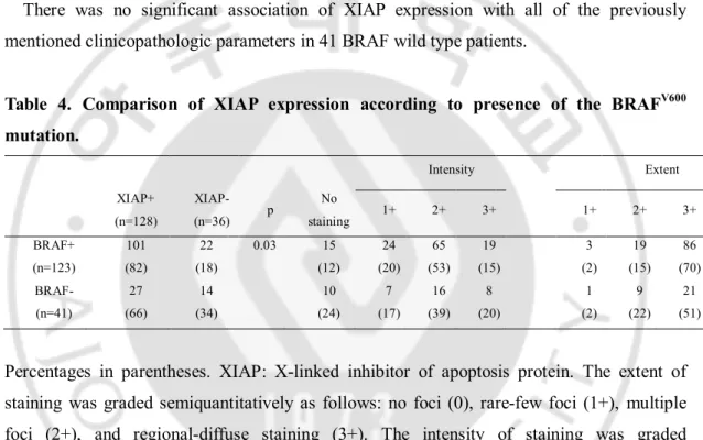
Table 4. Comparison of XIAP expression according to presence of the BRAFV600 mutation --- 9
1
I. INTRODUCTION
X-linked inhibitor of apoptosis protein (XIAP) is a member of the IAP protein family that selectively binds and inhibits caspases-3, -7 and -9. XIAP blocks the downstream portion of the apoptosis pathway and inhibits cell death in response to multiple stimuli (Holcik and Korneluk, 2001; Holcik et al, 2001; Hunter et al, 2007). All cells have a finite life-span, and cell death occurs mainly as a result of passive necrotic processes and/or active apoptosis (programmed cell death) (Bree et al, 2002; Fulda and Debatin, 2004). Apoptosis is the most common mechanism by which the body eliminates damaged or unnecessary cells without local inflammation from leakage of cell contents (Kasibhatla and Tseng, 2003; Fadeel and Orrenius, 2005). This process relies on a tightly balanced signaling pathway involving both pro- and anti-apoptotic proteins. Defective apoptosis represents a major causative factor in the development and progression of cancer (Lowe and Lin, 2000). The Inhibitor of Apoptosis (IAP) protein family plays critical roles in maintaining tight apoptogenic regularities.
XIAP expression is increased in a number of human malignancies, including colorectal, esophageal, ovarian, renal cell, hepatocellular, and prostate cancer (Holcik and Korneluk, 2001). Several reports have confirmed higher XIAP expression in thyroid cancer, compared to benign thyroid disorder (Xiao et al, 2007). Moreover, a recent study showed the utility of XIAP expression as a marker to predict the aggressiveness of papillary thyroid carcinoma (PTC) (Gu et al, 2010).
In the present study, we evaluated the expression patterns of XIAP in various neoplastic thyroid disorders and additionally aimed to delineate possible associations between XIAP expression and clinicopathologic parameters in conventional papillary thyroid carcinomas (PTCs), the most common type of thyroid cancer. We additionally evaluated the association of XIAP expression and BRAF mutation in conventional PTCs and analyzed the association of XIAP expression and tumor recurrence in BRAFV600E prevalent PTC population.
2
II. MATERIALS AND METHODS
A. Thyroid samples
We analyzed 217 formalin-fixed, paraffin-embedded thyroid tissue specimens obtained with thyroidectomy at Asan medical center between 1996 and 1998. The study specimens comprised 164 conventional PTCs,
patients of which
underwent total or near total thyroidectomy followed by immediate I-131 remnant ablation, and 40 other malignant thyroid tumors, including 10 follicular variant PTCs, 8 follicular thyroid carcinomas, 8 medullary thyroid carcinomas, 6 poorly differentiated thyroid carcinomas and 8 anaplastic thyroid carcinomas. The remaining 13 specimens were benign thyroid tumors, including 6 nodular hyperplasia and 7 follicular thyroid adenomas.All thyroid specimens were stained using hematoxylin and eosin, and reviewed by two pathologists. Diagnoses were confirmed according to the World Health Organization histologic classification of thyroid tumors.
B. Immunohistochemical analysis of XIAP
Sections of formalin-fixed, paraffin-embedded tumor tissue specimens were deparaffinized, treated with 3% hydrogen peroxide to block endogenous peroxidase activity, and microwaved in 0.01 mol/L citric acid (pH 6.0) for 6 min at 100℃, followed by slow cooling for antigen retrieval. Slides were incubated for 72 h at 4℃ with a monoclonal anti-XIAP antibody (BD Biosciences, Franklin Lakes, NJ, USA), stained using Envision-Plus reagents (DAKO, Carpinteria, CA, USA) and diaminobenzidine as chromogen, and counterstained with hematoxylin.
The extent of staining was graded semiquantitatively as follows: no foci (0), rare-few foci (1+), multiple foci (2+), and regional-diffuse staining (3+). Staining intensity was graded semiquantitatively as follows: negative (0), weak (1+), moderate (2+), and strong (3+). Specimens were classified as XIAP-positive when the sum of the extent score plus intensity score was equal to more than 4 points (Fig. 1).
3
D
Fig. 1. XIAP expression in PTC specimens. A, Weak-rare foci positive XIAP staining; B, Moderate multifocal positive XIAP staining; C, Strong diffuse positive XIAP staining; D, Expression of XIAP in 9 thyroid cancer cell lines
C. Clinicopathologic features of conventional papillary thyroid carcinoma
Patients diagnosed with conventional PTC and subjected to thyroid surgery at Asan medical center between 1996 and 1998 were retrospectively analyzed. Demographic data, including age and gender, and pathologic data, such as tumor size, multifocality, extrathyroidal invasion and AJCC TNM 2002 staging, were reviewed.
4
D. Statistics
Categorical variables are presented as numbers and percentages, and continuous variables as means ± standard deviations or median values with a range. Comparison of clinicopathologic parameters according to positivity of XIAP expression was performed using the Student's t test for continuous data and Fisher's exact test for categorical data. All P values were two-sided. Data were considered statistically significant at P <0.05. R version 2.11.1 and R libraries car and Cairo were used to analyze data (R Foundation for Statistical Computing, Vienna, Austria, http://www.R-project.org) (CoreTeam D, 2007)
E. Ethics statement
Formalin-fixed, paraffin-embedded tumor specimens were obtained with approval from the Institutional Review Board (2010-0477).
5
III. RESULTS
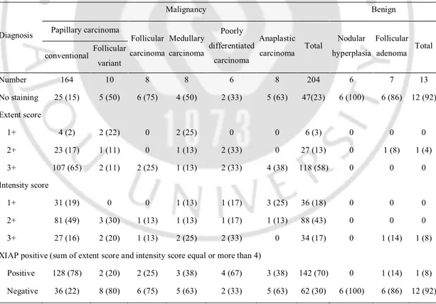
A. XIAP expression in malignant and benign thyroid tumors (Table 1)
Among the 164 conventional PTC specimens, 128 (78%) stained positive for XIAP. In terms of staining extent, 130 (82%) specimens scored 2+ or 3+. Intensity scores of 1+, 2+ and 3+ were conferred to 31 (19%), 81 (49%) and 27 (16%) specimens, respectively.
Positive XIAP staining was found in 2 (20%) of 10 follicular variant PTCs, 2 (25%) of 8 follicular thyroid carcinomas, 3 (38%) of 8 medullary thyroid carcinomas, 4 (67%) of 6 poorly differentiated thyroid carcinomas, and 3 (38%) of 8 anaplastic thyroid carcinomas.
Among the benign thyroid nodule specimens, none of the 6 nodular hyperplasias and only one of 7 (14%) follicular thyroid adenomas displayed XIAP-positive staining.
Table 1. Summary of XIAP immunostaining results for thyroid neoplasms and disorders.
Diagnosis Malignancy Benign Papillary carcinoma Follicular carcinoma Medullary carcinoma Poorly differentiated carcinoma Anaplastic carcinoma Total Nodular hyperplasia Follicular adenoma Total conventional Follicular variant Number 164 10 8 8 6 8 204 6 7 13 No staining 25 (15) 5 (50) 6 (75) 4 (50) 2 (33) 5 (63) 47(23) 6 (100) 6 (86) 12 (92) Extent score 1+ 4 (2) 2 (22) 0 2 (25) 0 0 6 (3) 0 0 0 2+ 23 (17) 1 (11) 0 1 (13) 2 (33) 0 27 (13) 0 1 (8) 1 (4) 3+ 107 (65) 2 (11) 2 (25) 1 (13) 2 (33) 4 (38) 118 (58) 0 0 0 Intensity score 1+ 31 (19) 0 0 1 (13) 1 (17) 3 (25) 36 (18) 0 0 0 2+ 81 (49) 3 (30) 1 (13) 1 (13) 1 (17) 1 (13) 88 (43) 0 0 0 3+ 27 (16) 2 (20) 1 (13) 2 (25) 2 (33) 0 34 (17) 0 1 (14) 1 (8) XIAP positive (sum of extent score and intensity score equal or more than 4)
Positive 128 (78) 2 (20) 2 (25) 3 (38) 4 (67) 3 (38) 142 (70) 0 1 (14) 1 (8) Negative 36 (22) 8 (80) 6 (75) 5 (63) 2 (33) 5 (63) 62 (30) 6 (100) 6 (86) 12 (92)
6
staining was graded semiquantitatively as follows: no foci (0), rare-few foci (1+), multiple foci (2+), and regional-diffuse staining (3+). The intensity of staining was graded semiquantitatively as follows: negative (0), weak (1+), moderate (2+), and strong (3+).
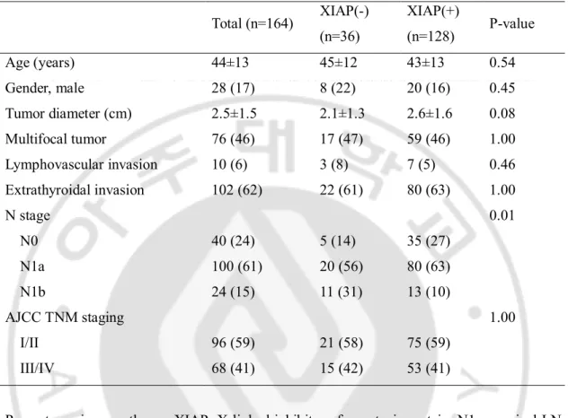
B. Clinicopathological factors of classic PTC according to positivity for XIAP expression (Table 2)
Among the 128 patients in the XIAP-positive group, 35 (27%) were in the N0 stage, 80 (63%) in the N1a stage and 13 (10%) in the N1b stage. Among the 34 patients in the XAIP-negative group, 5 (14%) patients were in the N0 stage, 20 (56%) in the N1a stage and 11 (33%) in the N1b stage. The incidence of metastasis to lateral lymph node was higher in patients with negative XIAP expression than those positive for XIAP (P=0.01).
No significant differences in age, sex, tumor diameter, multifocality, lymphovascular invasion, extrathyroidal extension, and AJCC TNM 2002 stage were observed between the two groups.
7
Table 2. Clinicopathological features of conventional-type papillary thyroid carcinoma according to XIAP expression.
Total (n=164) XIAP(-) (n=36) XIAP(+) (n=128) P-value Age (years) 44±13 45±12 43±13 0.54 Gender, male 28 (17) 8 (22) 20 (16) 0.45 Tumor diameter (cm) 2.5±1.5 2.1±1.3 2.6±1.6 0.08 Multifocal tumor 76 (46) 17 (47) 59 (46) 1.00 Lymphovascular invasion 10 (6) 3 (8) 7 (5) 0.46 Extrathyroidal invasion 102 (62) 22 (61) 80 (63) 1.00 N stage 0.01 N0 40 (24) 5 (14) 35 (27) N1a 100 (61) 20 (56) 80 (63) N1b 24 (15) 11 (31) 13 (10) AJCC TNM staging 1.00 I/II 96 (59) 21 (58) 75 (59) III/IV 68 (41) 15 (42) 53 (41)
Percentages in parentheses. XIAP: X-linked inhibitor of apoptosis protein. N1a, cervical LN metastasis at level VI (i.e., pretracheal, paratracheal, prelaryngeal/Delphian LN); N1b, cervical LN metastasis defined as unilateral, bilateral or contralateral cervical or superior mediastinal LNs, classified based on AJCC/UICC TNM staging; AJCC/UICC TNM, American Joint Committee on Cancer/International Union against Cancer Tumor-Node-Metastases classification
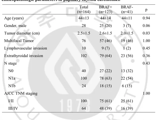
C. Association between clinicopathologic parameters and BRAFV600E mutation in PTCs
The BRAFV600E mutation was found in 123 of 164 conventional PTCs (75%). The study population was composed of 28 men (17%) and 136 women (83%), and their mean age was 44 ± 13 yrs. The Clinical and pathological characteristics of the study patients were shown in Table 3. The BRAFV600E mutation was significantly correlated with tumor size (p=0.03), and
8
there was a trend towards it being associated with gender (p=0.06). There was no significant association of BRAFV600E mutation with age, lymph node metastasis, and AJCC TNM 2002 stage.
Table 3. Correlations between presence of the BRAF
V600mutation and
clinicopathologic parameters in papillary thyroid carcinomas.
Total (n=164) BRAF+ (n=123) BRAF- (n=41) p Age (years) 44±13 44±14 44±11 0.94 Gender, male 28 25 (20) 3 (7) 0.06 Tumor diameter (cm) 2.5±1.5 2.6±1.5 2.0±1.5 0.03 Multifocal Tumor 76 57 (46) 19 (46) 1.00 Lymphovascular invasion 10 9 (7) 1 (2) 0.45 Extrathyroidal invasion 102 79 (64) 23 (56) 0.36 N stage 0.43 N0 40 27 (22) 13 (32) N1a 100 78 (63) 22 (54) N1b 24 18 (15) 6 (15) AJCC TNM staging 1.00 I/II 100 75 (61) 25 (61) III/IV 64 48 (39) 16 (39)Percentages in parentheses. XIAP: X-linked inhibitor of apoptosis protein. N1a, cervical LN metastasis at level VI (i.e., pretracheal, paratracheal, prelaryngeal/Delphian LN); N1b, cervical LN metastasis defined as unilateral, bilateral or contralateral cervical or superior mediastinal LNs, classified based on AJCC/UICC TNM staging; AJCC/UICC TNM, American Joint Committee on Cancer/International Union against Cancer Tumor-Node-Metastases classification
D. Association of XIAP expression and BRAF mutation in conventional PTCs
9
stained negative for XIAP (Table 4). In the XIAP-positive group, 23(23%) were in the N0 stage, 68 (67%) in the N1a stage and 10 (10%) in the N1b stage. In the XIAP-negative group, 4 (18%) were in the N0 stage, 10 (46%) in the N1a stage and 8 (36%) in the N1b stage. The incidence of metastasis to lateral neck lymph node was higher in patients with negative XIAP expression than those positive for XIAP (p=0.01). No significant differences in age, sex, tumor diameter, multifocality, lymphovascular invasion, extrathyroidal extension, lymph node metastasis, and AJCC TNM 2002 stage were observed between the two groups.
There was no significant association of XIAP expression with all of the previously mentioned clinicopathologic parameters in 41 BRAF wild type patients.
Table 4. Comparison of XIAP expression according to presence of the BRAFV600 mutation. Intensity Extent XIAP+ (n=128) XIAP- (n=36) p No staining 1+ 2+ 3+ 1+ 2+ 3+ BRAF+ (n=123) 101 (82) 22 (18) 0.03 15 (12) 24 (20) 65 (53) 19 (15) 3 (2) 19 (15) 86 (70) BRAF- (n=41) 27 (66) 14 (34) 10 (24) 7 (17) 16 (39) 8 (20) 1 (2) 9 (22) 21 (51)
Percentages in parentheses. XIAP: X-linked inhibitor of apoptosis protein. The extent of staining was graded semiquantitatively as follows: no foci (0), rare-few foci (1+), multiple foci (2+), and regional-diffuse staining (3+). The intensity of staining was graded semiquantitatively as follows: negative (0), weak (1+), moderate (2+), and strong (3+).
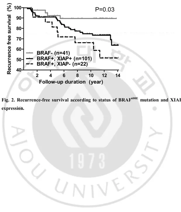
E. Recurrence and presence of BRAFV600E mutation and XIAP expression
Patients were followed-up for a median 11 years (1-14 years) after thyroidectomy. During this time, 38 (23%) patients had recurrences which involved 35 lateral lymph node recurrence, 7 central lymph node recurrence, 2 mediastinal lymph node recurrence, and 3 lung metastasis. The presence of BRAFV600E mutation and XIAP expression were significantly associated with lateral lymph node recurrence (log rang statistics = 7.05, df = 2, p = 0.03; Figure. 2).
10 40 50 60 70 80 90 100 BRAF- (n=41) BRAF+, XIAP- (n=22) BRAF+, XIAP+ (n=101) 2 4 6 8 10 12 14
P=0.03
Follow-up duration (year)
R ec u rr en ce f re e su rv iv al (% )
Fig. 2. Recurrence-free survival according to status of BRAF600E mutation and XIAP expression.
11
IV. DISCUSSION
The major historical thyroid cancer types are composed of differentiated thyroid carcinomas, such as PTC and follicular thyroid cancer, and undifferentiated thyroid carcinomas, such as medullary thyroid cancer or anaplastic thyroid cancer. PTC is the predominant thyroid cancer type in most parts of the world. Thyroid fine-needle aspiration cytology (FNAC) is a standard diagnostic tool for thyroid neoplasms (Baloch et al, 1998). However, diagnostic challenges of inadequate specimens and indeterminate cytology categories that fail to meet the criteria for definitive diagnosis of cancer present significant obstacles to clinicians (Elliott et al, 2006; Jemal et al, 2008). Data from the present study support the potential of XIAP as a molecular marker in thyroid cancer diagnosis. More recently, molecular diagnoses, such as galectin-3, HBME-1, cytokeratin-19 or B-type raf (BRAF) gene mutations have been introduced, but their clinical value is yet to be established (Kim et al, 2006; Jo et al, 2006; Kim et al, 2010). Some groups have shown that immunohistochemical testing for a combination of 2 or more markers improves the accuracy of diagnosis (Rossi et al, 2006; Scognamiglio et al, 2006; Nga et al, 2008; Wiseman et al, 2008). In our experiments, most specimens with benign pathology stained negative for XIAP. Thus, XIAP immunostaining from FNAC specimens, in combination with other potential markers, may be promising for the differential diagnosis of thyroid neoplastic disorders.
The incidence of positive XIAP expression was higher overall in thyroid cancer, particularly in cases of conventional PTC, compared with benign thyroid nodules. These findings are consistent with other studies suggesting that XIAP contributes to tumor cell survival as an apoptosis inhibitor (Holcik and Korneluk, 2001). However, XIAP immunostaining is inappropriate to differentiate between other histologic types of thyroid cancer and benign nodules. In particular, FTC and follicular adenoma, which are difficult to diagnose using cytology, showed similar patterns of XIAP expression in this study. However, the number of FTC and follicular adenoma samples analyzed was limited, and thus, a follow-up study on a larger scale is needed to evaluate the diagnostic value of XIAP expression in these diseases.
Interestingly, follicular variant PTCs, such as FTC, showed a lower incidence of positive XIAP expression than conventional PTC. Although PTC as a group constitutes tumors with
12
favorable biologic behavior that share characteristic microscopic features, increasing evidence suggests that they represent a heterogeneous group of tumors, and specific genetic alterations influence histologic variants and tumor behavior (Kondo et al, 2006). Adeniran et al. (Adeniran et al, 2006) reported that the molecular profile of the follicular variant PTC is close to that of FTC, with high prevalence of RAS and a very low BRAF mutation rate. Rivera and co-workers (Rivera et al, 2010) reported that the mutational patterns of encapsulated follicular variant PTC and FTC are similar. Infiltrative follicular variant PTC displays a BRAF and RAS genotype in between FTC and conventional PTC. Encapsulated follicular variant PTC has behavioral profile similar to that of FTC, while infiltrative follicular variant PTC has an invasive and behavioral profile similar to that of conventional PTC. XIAP appears to be associated with histologic variant and clinical behavior, similar to BRAF and RAS.
We further evaluated the potential association between XIAP expression and clinicopathological factors in patients with conventional PTC. XIAP expression was negatively associated with risk of lateral lymph node metastasis in our study. This result was inconsistent with a recent report from China (Gu et al, 2010). In the earlier study, XIAP expression was positively associated with the presence of lymph node metastasis in PTC. Several Korean groups have shown a high prevalence of the BRAF V600E mutation (around 70%-80%) in Korea (Kim et al, 2006; Jo et al, 2006; Kim et al, 2010), compared with other parts of the world. Additionally, Gu et al. (Gu et al, 2009) detected the BRAF V600E mutation in 34.1% of classical PTC samples. This regional difference in the prevalence of the BRAF mutation in PTC between Korea and China could explain the discrepancy regarding the association of XIAP with neck node metastasis. Further studies are required to explore the potential interactions between BRAF and XIAP.
Moreover, controversial results have been reported with regard to the association of XIAP expression with clinical outcomes in various cancer types other than thyroid cancer. A number of studies have shown that increased XIAP is correlated with decreased survival in acute myeloid leukemia, diffuse large B-cell lymphoma, and renal cell carcinoma (Xiang et al, 2009). In another study, high levels of XIAP were correlated with favorable outcomes in prostate cancer and early-stage non-small cell lung cancer (Ferreira et al, 2001). In the current investigation, XIAP expression was negatively associated with risk of lateral lymph
13
node metastasis. Lateral lymph node metastasis is one of the important prognostic factors for recurrence and disease-specific mortality in thyroid cancer patients (Ito and Miyauchi, 2009). Eventually, XIAP expression may be used as a surrogate marker to predict prognosis in thyroid cancer.
The possible mechanisms linking XIAP expression to clinical outcomes have been explored. The anti-apoptotic activity of XIAP is regulated by Smac/DIABLO. This protein is normally localized to mitochondria and released into the cytosol during the early stages of apoptosis, where it promotes caspase activity by inhibiting XIAP. Recent studies have reported that the relative proportion of XIAP, compared with Smac/DIABLO, rather than absolute value of XIAP expression, may determine tumor cell survival (Du et al, 2000; Verhagen et al, 2000; Ferreira et al, 2001). However, the present investigation focused solely on XIAP expression, and follow-up studies are required to establish the roles of XIAP and Smac/DIABLO in tumorigenesis and metastasis of thyroid cancer.
14
V. CONCLUSION
Immunohistochemical staining for XIAP as a potential novel molecular marker may aid in the differential diagnosis of thyroid cancer. Negative XIAP expression in conventional PTC is strongly associated with increased risk of lateral lymph node metastasis.
Furthermore, negative XIAP expression was associated with lateral lymph node metastases and an increased risk of recurrence in BRAF mutated PTC patients. XIAP immunohistochemical staining is useful for predicting patients’ prognosis in in BRAFV600E prevalent PTC population.
15
REFERENCES
1. Adeniran AJ, Zhu Z, Gandhi M, Steward DL, Fidler JP, Giordano TJ, Biddinger PW, Nikiforov YE: Correlation between genetic alterations and microscopic features, clinical manifestations, and prognostic characteristics of thyroid papillary carcinomas. Am J Surg
Pathol 30: 216-222, 2006
2. Baloch ZW, Sack MJ, Yu GH, Livolsi VA, Gupta PK: Fine-needle aspiration of thyroid: an institutional experience. Thyroid 8: 565-569, 1998
3. Bree RT, Stenson-Cox C, Grealy M, Byrnes L, Gorman AM, Samali A: Cellular longevity: role of apoptosis and replicative senescence. Biogerontology 3: 195-206, 2002
4. CoreTeam D: A language and environment for statistical computing. Available at http://www.R-progect.org. [accessed on December 5 2007].
5. Du C, Fang M, Li Y, Li L, Wang X: Smac, a mitochondrial protein that promotes cytochrome c-dependent caspase activation by eliminating IAP inhibition. Cell 102: 33-42, 2000
6. Elliott DD, Pitman MB, Bloom L, Faquin WC: Fine-needle aspiration biopsy of Hurthle cell lesions of the thyroid gland. Cancer Cytopathol 108: 102-109, 2006
7. Fadeel B, Orrenius S: Apoptosis: a basic biological phenomenon with wide-ranging implications in human disease. J Intern Med 258: 479-517, 2005
8. Ferreira CG, van der Valk P, Span SW, Ludwig I, Smit EF, Kruyt FA, Pinedo HM, van Tinteren H, Giaccone G: Expression of X-linked inhibitor of apoptosis as a novel prognostic marker in radically resected non-small cell lung cancer patients. Clin Cancer
Res 7: 2468-474, 2001
9. Fulda S, Debatin KM: Apoptosis signaling in tumor therapy. Ann N Y Acad Sci 1028: 150-156, 2004
10. Gu LQ, Li FY, Zhao L, Liu Y, Chu Q, Zang XX, Liu JM, Ning G, Zhao YJ: Association of XIAP and P2X7 receptor expression with lymph node metastasis in papillary thyroid carcinoma. Endocrine 38: 276-282, 2010
11. Gu LQ, Li FY, Zhao L, Liu Y, Zang XX, Wang TX, Chen HP, Ning G, Zhao YJ: BRAFV600E mutation and X-linked inhibitor of apoptosis expression in papillary thyroid carcinoma. Thyroid 19: 347-354, 2009
16
12. Holcik M, Korneluk RG: XIAP, the guardian angel. Nat Rev Mol Cell Biol 2: 550-556, 2001
13. Holcik M, Gibson H, Korneluk RG: XIAP: apoptotic brake and promising therapeutic target. Apoptosis 6: 253-261, 2001
14. Hunter AM, LaCasse EC, Korneluk RG: The inhibitors of apoptosis (IAPs) as cancer targets. Apoptosis 12: 1543-1568, 2007
15. Ito Y, Miyauchi A: Prognostic factors and therapeutic strategies for differentiated carcinomas of the thyroid. Endocr J 56: 177-192, 2009
16. Jemal A, Siegel R, Ward E, Hao Y, Xu J, Murray T, Thun MJ: Cancer statistics, 2008.
CA Cancer J Clin 58: 71-96, 2008
17. Jo YS, Li S, Song JH, Kwon KH, Lee JC, Rha SY, Lee HJ, Sul JY, Kweon GR, Ro H-k, Kim J-M, Shong M: Influence of the BRAF V600E Mutation on Expression of Vascular Endothelial Growth Factor in Papillary Thyroid Cancer. J Clin Endocrinol Metab 91: 3667-3670, 2006
18. Kasibhatla S, Tseng B: Why Target Apoptosis in Cancer Treatment? Mol Cancer Ther 2: 573-580, 2003
19. Kim SW, Lee JI, Kim J-W, Ki C-S, Oh YL, Choi Y-L, Shin JH, Kim HK, Jang HW, Chung JH: BRAFV600E Mutation Analysis in Fine-Needle Aspiration Cytology Specimens for Evaluation of Thyroid Nodule: A Large Series in a BRAFV600E-Prevalent Population. J Clin Endocrinol Metab 95: 3693-3700, 2010
20. Kim TY, Kim WB, Rhee YS, Song JY, Kim JM, Gong G, Lee S, Kim SY, Kim SC, Hong SJ, Shong YK: The BRAF mutation is useful for prediction of clinical recurrence in low-risk patients with conventional papillary thyroid carcinoma. Clin Endocrinol 65: 364-368, 2006
21. Kondo T, Ezzat S, Asa SL: Pathogenetic mechanisms in thyroid follicular-cell neoplasia.
Nat Rev Cancer 6: 292-306, 2006
22. Lowe SW, Lin AW: Apoptosis in cancer. Carcinogenesis 21: 485-495, 2000
23. Nga ME, Lim GS, Soh CH, Kumarasinghe MP: HBME-1 and CK19 are highly discriminatory in the cytological diagnosis of papillary thyroid carcinoma. Diagn
17
24. Rivera M, Ricarte-Filho J, Knauf J, Shaha A, Tuttle M, Fagin JA, Ghossein RA: Molecular genotyping of papillary thyroid carcinoma follicular variant according to its histological subtypes (encapsulated vs infiltrative) reveals distinct BRAF and RAS mutation patterns. Mod Pathol 23: 1191-1200, 2010
25. Rossi ED, Raffaelli M, Mule' A, Miraglia A, Lombardi CP, Vecchio FM, Fadda G: Simultaneous immunohistochemical expression of HBME-1 and galectin-3 differentiates papillary carcinomas from hyperfunctioning lesions of the thyroid.
Histopathology 48: 795-800, 2006
26. Scognamiglio T, Hyjek E, Kao J, Chen YT: Diagnostic usefulness of HBME1, galectin-3, CK19, and CITED1 and evaluation of their expression in encapsulated lesions with questionable features of papillary thyroid carcinoma. Am J Clin Pathol 126: 700-708, 2006
27. Verhagen AM, Ekert PG, Pakusch M, Silke J, Connolly LM, Reid GE, Moritz RL, Simpson RJ, Vaux DL: Identification of DIABLO, a mammalian protein that promotes apoptosis by binding to and antagonizing IAP proteins. Cell 102: 43-53, 2000
28. Wiseman SM, Melck A, Masoudi H, Ghaidi F, Goldstein L, Gown A, Jones SJ, Griffith OL: Molecular phenotyping of thyroid tumors identifies a marker panel for differentiated thyroid cancer diagnosis. Ann Surg Oncol 15: 2811-2826, 2008
29. Xiang G, Wen X, Wang H, Chen K, Liu H: Expression of X-linked inhibitor of apoptosis protein in human colorectal cancer and its correlation with prognosis. J Surg Oncol 100: 708-712, 2009
30. Xiao GQ, Unger PD, Burstein DE: Immunohistochemical detection of X-linked inhibitor of apoptosis (XIAP) in neoplastic and other thyroid disorders. Ann Diagn Pathol 11: 235-240, 2007
18
- 국문 요약 –
갑상선 종양에서
X-linked Inhibitor of Apoptosis Protein 의 발현 양상
아주대학교 대학원 의학과 윤 종 호
(지도교수: 소 의 영)
X-linked inhibitor of apoptosis protein (XIAP)는 8개의 IAP family의 하나로 caspase 3, 7, 9와 결합하여 세포 자멸사를 조절하고 억제하는 것으로 알려져 있는 단백질이 다. 본 연구는 표준형 유두 갑상선암과 그 밖의 갑상선암 그리고 양성 갑상선 결 절에서 XIAP 발현 양상을 알아보고, 표준형 유두 갑상선암 환자에서 임상적 특 징을 분석하여 XIAP 발현과 표준형 유두 갑상선암의 예후와의 관계에 대해 알아 보고자 하였다. 또한 XIAP 발현과 BRAFV600E 돌연변이 사이의 상관 관계를 알아 보고 BRAFV600E 돌연변이 환자에서 XIAP 발현과 암 재발의 상관 관계를 분석하 고자 하였다. 1996년에서 1998년까지 갑상선절제술을 통해 얻어진 217개의 갑상 선 종양 조직을 XIAP 항체로 면역조직화학염색을 하여 XIAP 발현 양상을 확인 하였다. 표준형 유두 갑상선암 164명에 대해서 XIAP 발현과 BRAFV600E 돌연변이 여부 및 임상병리학적 특징을 분석하였다. 표준형 유두 갑상선암의 78%에서 XIAP 양성 소견을 보인 반면에, 저분화 갑상선암은 67%, 미분화 갑상선암은 38% 만이 양성 소견을 보였다. 갑상선 여포성 변이 유두암 20%, 여포암 25%, 수질암 38%에서 XIAP 양성 소견을 보였으며, 양성 결절에서 XIAP 양성 소견은 결절성 과증식에서는 없었고, 여포성 선종에서 1예 (1/7, 8%) 만이 있었다. XIAP 발현 여 부와 림프절 전이를 제외한 임상적, 병리적 특징은 특별한 상관 관계를 보이지 않았다. 임파선 전이에 있어서 XIAP 음성 유두암은 N1b 33%, XIAP 양성인 유두 암은 N1b 10% 로 XIAP 양성 유두암에 비해 XIAP 음성인 유두암이 임파선 전이
19
병기가 더 높은 것으로 나타났다 (p=0.01). BRAFV600E 돌연변이는 표준형 유두 갑 상선암 환자의 75%에서 관찰되었고, BRAFV600E 돌연변이가 있는 유두 갑상선암에 서 XIAP 발현율이 82%로 BRAFV600E 돌연변이가 없는 경우에 비해 유의하게 높 았다 (p=0.03). 또한 BRAFV600E 돌연변이가 있는 환자 중 XIAP 음성인 경우 XIAP 양성인 경우에 비해 측경부 림프절 전이 및 재발의 빈도가 유의하게 높은 것으 로 조사되었다 (p = 0.03). XIAP는 갑상선암 감별진단을 위한 표지자로 사용될 수 있을 것으로 사료되며, 특히 BRAFV600E 돌연변이가 있는 표준형 유두 갑상선암 환자에서 측경부 림프절 전이 및 재발을 예측할 수 있는 유용한 예후 인자로서 역할을 할 수 있을 것으로 생각된다. 핵심어: X-linked inhibitor of apoptosis protein, BRAFV600E mutation, 유두 갑상선암, 갑