The ERK pathway involves positive and negative regulations
of HT-29 colorectal cancer cell growth by extracellular zinc
Ki-Sook Park,1 Nam-Gu Lee,1Ki-Hoo Lee,3Jeong Taeg Seo,4and Kang-Yell Choi1,2 1Department of Biotechnology, College of Engineering, and2Protein Network Research Center,
Yonsei University, Seoul 120-752;3Laboratory of Molecular Oncology, Korea Institute of
Radiological and Medical Sciences, Seoul 136-706; and4Department of Oral Biology,
College of Dentistry, Yonsei University, Seoul 120-752, South Korea
Submitted 28 January 2003; accepted in final form 17 June 2003 Park, Ki-Sook, Nam-Gu Lee, Ki-Hoo Lee, Jeong Taeg
Seo, and Kang-Yell Choi. The ERK pathway involves
pos-itive and negative regulations of HT-29 colorectal cancer cell growth by extracellular zinc. Am J Physiol Gastrointest Liver
Physiol 285: G1181–G1188, 2003. First published June 19,
2003; 10.1152/ajpgi.00047.2003.—Dietary zinc is an impor-tant trace element in the body and is related to both cell proliferation and growth arrest. A recent study found that extracellular zinc-sensing receptors trigger intracellular sig-nal transduction in HT-29 human colorectal cancer cells. However, the signaling mechanism causing this growth reg-ulation by extracellular zinc is not clearly understood. At 10-and 100-M levels of ZnCl2treatment, HT-29 cell growth and
proliferation increased and decreased, respectively, in a min-imally serum-starved medium (MSSM). A lack of significant increase in intracellular zinc levels after zinc treatment sug-gested that this differential growth regulation of HT-29 cells by extracellular zinc is acquired by receptor-mediated signal transduction. Moreover, this zinc-induced growth regulation was differentially affected by PD-98059, suggesting the in-volvement of the ERK pathway. Transient ERK activation and subsequent cyclin D1 induction were observed on adding 10M ZnCl2in MSSM in the presence of cell proliferation.
On the other hand, prolonged ERK activity was observed with a subsequent increase of cyclin D1 and p21Cip/WAF1on
adding 100M ZnCl2in MSSM, and this was associated with
nonproliferation. Moreover, this ERK activation and cyclin D1 and p21Cip/WAF1induction were abolished by PD-98059
pretreatment. The differential regulations of cell growth, ERK activities, and cyclin D1 and p21Cip/WAF1 inductions
were also observed in serum-enriched medium containing higher zinc concentrations. Therefore, differential cell cycle regulator induction occurs by a common ERK pathway in the differential growth regulation of HT-29 cells by extracellular zinc.
signal transduction; extracellular signal-regulated kinase; p21Cip/WAF1
ZINC IS INVOLVED IN THE STIMULATIONof cell growth, and a
zinc deficiency inhibits growth in animals, causes pro-liferation in cultured cells, and accelerates established tumor growth (23, 38). However, it has also been re-ported that zinc has an antitumor effect that is asso-ciated with growth inhibition (2, 9, 19, 39). The levels
of zinc in tumorous and normal tissues differ (8, 21, 37), and cell growth may be affected by zinc dosage (8, 29). The epithelium of the colonic epithelial mucosa undergoes continuous renewal, and the growth and tumorigenesis of colorectal cells are highly sensitive to zinc levels (5, 24, 25). However, the mechanism of the differential growth regulation of cells by zinc remains unknown.
Extracellular zinc is reported to activate signaling pathways related to cell growth regulation, such as MAPK and phosphoinositol 3-kinase pathways (17, 20, 28, 32), and the chelation of intracellular zinc does not change the activation status of MAPK by extracellular zinc (14). Moreover, cellular growth regulation by zinc is not likely to be mediated by direct interaction with intracellular metalloproteins, because symptoms of zinc deficiency precede changes in the intracellular zinc content (6, 20). Recently, Hershfinkel et al. (15) reported the presence of an extracellular zinc-sensing receptor (ZnR) in HT-29 colorectal cancer cells and suggested its involvement in extracellular receptor-mediated signaling pathway(s) for growth regulation.
In the present study, we investigated the mechanism of the differential regulation of HT-29 colorectal cancer cell growth at nontoxic micromolar concentrations of extracellular zinc. HT-29 colorectal cancer cell growth was found to be positively and negatively regulated by different extracellular zinc concentrations, which re-sulted from differential ERK activation. Moreover, cy-clin D1 and p21Cip/WAF1 cell cycle regulators were also
differentially induced, and this was also found to depend on ERK activation and to be related to the differential regulation of cell growth. No significant accumulation of intracellular zinc by the application of micromolar con-centrations of extracellular zinc suggests that the effects of zinc are mediated by receptor-mediated signal trans-duction rather than being due to complex effects associ-ated with intracellular zinc accumulation (18, 34).
METHODS
Cell culture. HT-29 cells were maintained in McCoy’s 5A
medium supplemented with 10% FBS, 100 U/ml penicillin,
Address for reprint requests and other correspondence: K.-Y. Choi, Dept. of Biotechnology, Yonsei Univ. College of Engineering, Seoul 120-752, South Korea (E-mail: [email protected]).
The costs of publication of this article were defrayed in part by the payment of page charges. The article must therefore be hereby marked ‘‘advertisement’’ in accordance with 18 U.S.C. Section 1734 solely to indicate this fact.
and 250 ng/ml streptomycin in 5% CO2 at 37°C. Unless
otherwise stated, the experiments were performed on cells at 70% confluence. To observe the effects of zinc, the cells were grown in McCoy’s 5A containing either 1 or 10% FBS for 16–20 h before zinc treatment. In required cases, 1, 5, or 10 M of the MEK inhibitor PD-98059 (Calbiochem, La Jolla, CA) was given 30 min before ZnCl2addition.
Cell counting and thymidine incorporation. HT-29
colorec-tal cells were grown in minimally serum-starved medium (MSSM) containing 1% FBS for 16 h. The cell culture me-dium was replaced with fresh 1 or 10% FBS meme-dium contain-ing different concentrations of ZnCl2 (0–500 M). The
at-tached cell numbers were counted by mounting 10l of the cell mixture (1:1 mixture of 0.4% trypan blue and HT-29 cell suspension) onto a Tiefe Depth Profondeur 0.0025-mm2
cell-counting plate (Superior) and observing plated cells under a microscope.
For thymidine incorporation, the HT-29 cells were plated in 12-well culture dishes at 1.3 ⫻ 105 cells/well and then
grown in MSSM for 16–20 h. The medium was then replaced with fresh MSSM containing different ZnCl2concentrations
(0, 10, or 100M), and the cells were further incubated for 40 h. When required, PD-98059 was also added 30 min before ZnCl2treatment. Finally, the incubations were continued in
the presence of [3H]thymidine (0.5Ci/well) for 8 h. After
cells were washed twice with 5% trichloroacetic acid, they were solubilized by the addition of 0.2 N NaOH, and DNA-associated3H activity was counted by using a liquid
scintil-lation counter.
Western blot analysis. Cell extracts were prepared from
harvested cells as previously described (28). Samples of 10– 50g protein from the whole cell extracts were separated by SDS-PAGE, and the blots were prepared on a Protran mem-brane. The following antibodies were used as probes: for ERK, phosphospecific anti-ERK (New England Biolabs, Bev-erly, MA) and anti-ERK (Stratagene, La Jolla, CA) antibod-ies; for cyclin D1 and p21Cip/WAF1, anti-cyclin D1 and -p21Cip/ WAF1 antibodies (Santa Cruz Biotechnology, Santa Cruz,
CA). The blots were probed with horseradish peroxidase-conjugated secondary antibodies (Transduction Laboratories, San Diego, CA), and the proteins were visualized by en-hanced chemiluminescence.
Measurement of intracellular zinc. The intracellular zinc
concentration ([Zn2⫹]i) in HT-29 cells was measured by using
a modification of a method described previously (34). HT-29 cells were plated onto coverslips at a density of 3 ⫻ 105
cells/coverslip into six-well plates, and the cells were grown overnight in McCoy’s 5A medium containing 1 or 10% FBS and treated with ZnCl2at 1, 100, or 500M. Cells were then
loaded with the fluorescent indicator mag-fura-2 at 37°C in a 5% CO2incubator by including 3 M of the acetoxymethyl
ester of mag-fura-2 (mag-fura-2 AM; Molecular Probes, Eu-gene, OR) for 20 min in a HCO3⫺-buffered solution containing
(in mM): 110 NaCl, 4.5 KCl, 1 NaH2PO4, 1 MgSO4, 1.5 CaCl2,
5 HEPES-Na, 5 HEPES-free acid, 25 NaHCO3, and 10 D -glucose (pH 7.4). Cells were then rinsed twice and incubated in the HCO3⫺-buffered solution for at least 20 min before use.
[Zn2⫹]iwas measured on the stage of an inverted
micro-scope (Nikon, Tokyo, Japan) by spectrofluorometry (Photon Technology International, New Brunswick, NJ) while cells were superfused at a constant perfusion rate of 2 ml/min with HCO3⫺-buffered solution equilibrated with 95% O2-5% CO2to
maintain a pH of 7.4. All experiments were performed at 37°C. The excitation wavelength alternated between 340 and 380 nm, and emission fluorescence was recorded at 510 nm. The values of [Zn2⫹]iwere calculated by using a modification
of the equation described by Grynkiewicz et al. (12): [Zn2⫹]i⫽
Kd䡠Sf2/Sb2[(R ⫺ Rmin)/(Rmax ⫺ R)], in which Kd of
mag-fura-2 for Zn2⫹ was assumed to be 20 nM (36), R is the observed 340/380 fluorescence ratio, Rmin is the 340/380
fluorescence ratio value determined in mag-fura-2-loaded HT-29 cells exposed to 100M TPEN, Rmaxis the 340/380
fluorescence ratio value [obtained by adding 20 M 1-hy-droxypyridine-2-thione (pyrithione; Sigma, St. Louis, MO), a Zn2⫹ionophore, in the presence of 1 mM Zn2⫹], Sf2 is the fluorescence intensity at 380 nm at Rmin, and Sb2 is the
fluorescence intensity at 380 nm at Rmax.
Statistical analysis. The statistical significances of
differ-ences between the positive- and negative-regulated groups were determined by using a one-way ANOVA and Bonferro-ni’s test. A P value⬍0.05 was considered statistically signif-icant. The results are expressed as means⫾ SD.
RESULTS
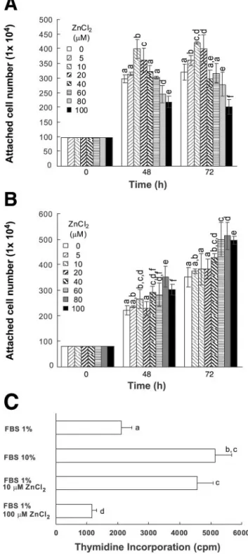
HT-29 cell growth and proliferation were positively or negatively regulated by 5–100 M of ZnCl2 in MSSM. The number of cells was measured after the
extracellular zinc treatments to determine the role of zinc in regulating the growth of HT-29 cells. The num-bers of HT-29 cells increased up to 72 h after a ZnCl2
treatment at ⬍10 M in MSSM containing 1% FBS (Fig. 1A). On the other hand, the number of cells decreased after increasing the ZnCl2 concentration to
80–100M (Fig. 1A). Unlike the cells grown in MSSM, the number of cells increased as a result of the treat-ments in the range of 5–100 M ZnCl2 in a medium
containing 10% FBS (Fig. 1B). The lack of growth inhibition by 100M ZnCl2in the medium containing
10% FBS suggests that ZnCl2 at this concentration is
not toxic to HT-29 cells. Because 10 and 100M ZnCl2
resulted in the most significant effects in terms of the positive and negative regulation of HT-29 cell growth in MSSM, we used these two zinc concentrations in further studies involving MSSM.
The level of DNA synthesis was measured by deter-mining the extent of [3H]thymidine incorporation and
was found to increase in cells by more than twofold on adding 10M ZnCl2to MSSM (Fig. 1C). The amount of
thymidine incorporated by 10 M ZnCl2 was only
slightly lower than that level acquired by growing cells in medium containing 10% FBS without zinc treatment (Fig. 1C). However, the thymidine incorporation level was reduced by adding 100M ZnCl2to cells grown in
MSSM.
Positive and negative regulation of zinc-treated HT-29 cell growth by PD-98059 in MSSM. The ERK
pathway is involved in cell cycle arrest (7, 35, 40) and is well known to be a function of cell cycle progression (4, 7). The ERKs were previously found to be strongly activated in a prolonged manner by 100 M ZnCl2
treatments in MSSM, and this activation was related to the antiproliferation of colorectal cancer cells, in-cluding HT-29 cells (28). However, ERK activities were also found to be transiently and weakly activated by extracellular zinc treatment (14, 33). This growth reg-ulation was not further investigated with respect to differential ERK regulation. In this study, we investi-gated the growth and proliferation induced by the
different zinc concentrations and their relationship with the ERK activation profiles.
Cell numbers, which had been previously increased by 10 M ZnCl2 in MSSM, were dose-dependently
decreased by treatment with the MEK inhibitor PD-98059 (Fig. 2A), suggesting that ERK activation affects the proliferation of HT-29 cells by 10 M ZnCl2 in
MSSM. Surprisingly, cell numbers reduced by 100M ZnCl2 treatment in MSSM were increased by
subse-quently treating the cells with 1 M PD-98059 (Fig. 2B). These results suggest that the ERK activation by 100M ZnCl2in MSSM, which caused growth
inhibi-tion (28), was blocked by 1 M PD-98059. A further increase in PD-98059 to 5 and 10 M in the cells treated with 100M ZnCl2resulted in a further
reduc-tion in the number of cells (Fig. 2B). These results suggest that the ERK pathway may be involved in both the positive and negative regulation of cell growth and further suggest that the zinc-induced growth regula-tion of HT-29 cells may be highly sensitive to the level of ERK activation. The upregulation of cell numbers by PD-98059 in 100 M ZnCl2-treated cells in MSSM
confirms that 100M ZnCl2is not a toxic concentration
to HT-29 cells. The thymidine incorporation level, which was increased by adding 10M ZnCl2, was also
dose-dependently inhibited by pretreating the cells with PD-98059 in MSSM (Fig. 2C). However, the thy-midine incorporation level was also increased by the 1 M PD-98059 cotreatment in the cells treated with 100 M ZnCl2and was reduced by cotreatment with 10M
PD-98059 (Fig. 2C) compared with 1 M PD-98059 cotreatment. Therefore, cell numbers in this system were found to be highly correlated with proliferation status. The differential modulations of cell growth and proliferation by PD-98059 suggest that the ERK path-way participates in both the proliferation and the an-tiproliferation of cellular growth.
The cyclin D1 and p21Cip/WAF1are differentially in-duced by 10 and 100 M ZnCl2 in MSSM and are dependent on ERK activations. The ERK pathway is
involved in the progression and inhibition of the G1
-to-S phase progression by the differential activations of Raf-13 MEK 3 ERK cascade kinases, which is fol-lowed by the differential induction of cell cycle regula-tors such as cyclin D1 and p21Cip/WAF1(35, 40).
In this study, we monitored the inductions of cyclin D1 and p21Cip/WAF1 at 10 min and 9 h after treating
HT-29 cells with 10 and 100M ZnCl2in MSSM, which
induced transient and prolonged ERK activations, re-spectively (28). Cyclin D1 was induced 9 h after treat-ment with 10 or 100M ZnCl2, and this was higher in
cells treated with the higher concentration of ZnCl2
(Fig. 3). On the other hand, the p21Cip/WAF1was
signif-icantly induced only in cells treated with 100M ZnCl2
(Fig. 3). The cyclin D1 and p21Cip/WAF1 inductions by
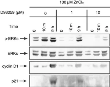
100M ZnCl2 were reduced by subsequently treating
cells with PD-98059 (Fig. 4), which suggests that both cyclin D1 and p21Cip/WAF1inductions are dependent on
ERK activations.
The effects of zinc on ERK activation, as well as cyclin D1 and p21Cip/WAF1 induction, resulted in different
Fig. 1. Effects of different concentrations of extracellular zinc on HT-29 cell growth. Positive and negative regulation of cell growth and proliferation were affected by extracellular zinc treatment. The HT-29 colorectal cells were minimal serum starved (the cells were grown in McCoy’s 5A media containing 1% FBS, called MSSM) for 16 h, and the medium was replaced with MSSM (A) or 10% FBS medium (B) with different concentrations of ZnCl2(0, 5, 10, 20, 40,
60, 80, and 100M). The numbers of attached cells were counted at 0, 48, and 72 h after zinc treatments. C: HT-29 cells were cultured as shown in A in MSSM, and [3H]thymidine levels were measured 48 h
after replacing medium with media containing different concentra-tions of zinc as described inMETHODS. Values are means⫾ SD. Means with different lowercase letters are significantly different at P ⬍ 0.05.
sensitivities to the zinc concentrations in an extracellu-lar serum concentration-dependent manner. In contrast
to the cells grown in MSSM, we did not observe inhi-bition of cell growth by 100 M ZnCl2 in 10% FBS
medium (Fig. 1B). To further characterize the role of ERK activation in zinc-induced growth regulation, we measured the effects of 100M ZnCl2on ERK
activa-tion and the subsequent cyclin D1 and p21Cip/WAF1
inductions in media containing 10% FBS. We did not observe prolonged ERK activation by 100M ZnCl2in
this medium; only transient ERK activation was ob-served 10 min after the zinc treatment (Fig. 5). Fur-thermore, we observed the weak induction of cyclin D1 9 h after treatment with 100 M ZnCl2 in medium
containing 10% FBS, without significant p21Cip/WAF1
induction. These observations of the effects of zinc, including prolonged ERK activation and p21Cip/WAF1
induction in MSSM, may have been caused by an inhibitory factor(s) present in the serum or because the effect of zinc may have been attenuated by signaling homeostasis of the cells grown in 10% FBS medium. Both the transient and prolonged ERK activations, detected within 10 min and 9 h of zinc treatment, respectively, were redetected in cells grown in 10% FBS medium containing 500 M ZnCl2 (Fig. 5). In
addition, cyclin D1 and p21Cip/WAF1 inductions were
also observed in cells grown in 10% FBS medium
con-Fig. 2. Effects of PD-98059 on the zinc-induced pos-itive and negative regulations of cell growth and proliferation. Essentially, these experiments are the same as those in Fig. 1A. In the required cases, cells were incubated with different PD-98059 (PD) con-centrations (0, 1, 5, or 10 M) for 30 min before treatment with 10M ZnCl2(A) or 100M ZnCl2
(B). C: [3H]thymidine was measured 48 h after
treatment with zinc. In the required cases, the cells were incubated with 1 or 10M PD-98059 for 30 min before zinc treatment. Values are means⫾ SD. Means with different lowercase letters are signifi-cantly different at P⬍ 0.05.
Fig. 3. ERK activations and inductions of cyclin D1 and p21Cip/WAF1
by 10 and 100M ZnCl2. A: serum-starved HT-29 cells grown in
MSSM were treated with 10 or 100M ZnCl2. The cells were then
harvested at different time points (0 min, 10 min, and 9 h), and Western blot analysis was performed using phospho-ERK (p-ERK), ERK, cyclin D1, or p21Cip/WAF1(p21) antibodies. B: same experiment
as in A except that samples were harvested over a greater period of time to detect cyclin D1 or p21Cip/WAF1.
taining 500M ZnCl2. The patterns of ERK activations
and subsequent cyclin D1 and p21Cip/WAF1 inductions
by 100 and 500M ZnCl2 in 10% FBS medium were
similar to the patterns observed in cells grown in MSSM containing 10 and 100 M ZnCl2, respectively
(Fig. 3A).
Differential growth regulations by zinc in 10% FBS medium were associated with prolonged ERK activa-tion and subsequent p21Cip/WAF1induction. To measure
the differential effects of zinc in more detail, HT-29 cells were treated with a range of ZnCl2concentrations
(0, 100, 200, 300, and 500 M) in 10% FBS medium, and the effects of different zinc concentrations on cell growth were measured. As observed in MSSM, the positive and negative regulations of cell growth were also observed by the higher zinc concentrations in the
10% FBS medium. The number of HT-29 cells was increased by 100M ZnCl2(Fig. 6A). However, the cell
numbers were unaffected by 200 and 300 M ZnCl2
and were reduced as a result of the 300 and 500 M ZnCl2treatments (Fig. 6A). p21Cip/WAF1protein levels
were found to be increased in 10% FBS medium by only 200–500 ZnCl2, and this occurred concomitantly with
prolonged ERK activation (Fig. 6B). On the other hand, cyclin D1 protein induction was weak at 100M ZnCl2
and at higher zinc concentrations (200–500M ZnCl2)
in 10% FBS medium. Both cyclin D1 and p21Cip/WAF1
inductions by zinc in 10% FBS medium were inhibited by pretreatment with 10M PD-98059 (Fig. 6B).
Extracellular zinc was not significantly taken up into HT-29 cells. To identify uptake level of extracellular
zinc in HT-29 colorectal cancer cells, we measured the
Fig. 6. Positive and negative regulations of cell growth and the differential activation of ERK and induction of p21Cip/WAF1in 10%
FBS medium by ZnCl2at concentrations⬎ 100 M ZnCl2. A:
exper-iments are as in Fig. 1B except that higher ZnCl2concentrations (0,
100, 200, 300, and 500M) were used. Values are means ⫾ SD of 3 independent experiments. Means with different lowercase letters are significantly different at P⬍ 0.05. B: experiments are essentially the same as in Fig. 5, except that a broader range of ZnCl2
concentra-tions (0, 100, 200, 300, or 500M) was used. When required, 10 M PD-98059 was added 1 h before zinc treatment. Cells were harvested at 9 h after treatment with zinc for Western blot analysis.
Fig. 4. Effects of PD-98059 on cyclin D1 and p21Cip/WAF1induction.
Serum-starved HT-29 cells grown in MSSM were treated with 100 M ZnCl2. Cells were incubated with 1 or 10M PD-98059 for 1 h
before zinc treatment. Cells were then harvested, and Western blot analysis was performed as described in Fig. 3A.
Fig. 5. Prolonged ERK activations and cyclin D1 and p21Cip/WAF1
induction were not observed in 10% FBS medium but were observed again at a higher extracellular zinc level. Experiments shown are essentially the same as in Fig. 3A except for the higher ZnCl2
intracellular zinc levels after adding 10 or 100 M ZnCl2to cells grown in MSSM and after adding 500M
ZnCl2to cells grown in 10% FBS medium. Intracellular
zinc levels were not found to be significantly increased by treating cells grown in MSSM with 10 and 100M ZnCl2or in cells grown in 10% FBS containing 500M
ZnCl2 (Fig. 7). However, the intracellular zinc levels
started to increase soon after adding the Zn2⫹
iono-phore pyrithione to cells previously treated with 100 or 500 M ZnCl2. Therefore, the high levels of zinc
ap-plied to the culture medium were not significantly taken up by HT-29 colorectal cancer cells regardless of the serum levels, and ERK activation may have been effected via an extracellular zinc sensor such as ZnR (15) rather than via the activation of intracellular zinc-sensing protein(s).
DISCUSSION
Zinc is a trace element in the human body and is known to participate like a growth factor in cellular proliferation (13, 27, 30). However, zinc has also been reported to have a growth-inhibitory role (3, 9, 19). The physiological roles and the mechanisms of zinc partic-ipation in the regulation of cell growth are not yet fully understood. The intestinal mucosa undergoes continu-ous renewal and needs zinc constantly (39). In addi-tion, zinc was suggested to be a primary determinant of risk in human colorectal cancer (5, 24, 25, 37), and low zinc intake may increase the incidence of colorectal cancer (22). Therefore, it is possible that zinc levels in the extracellular plasma environment may affect the growth of colorectal tumors (1, 18).
We recently identified growth arrest in colorectal cancer cells grown in MSSM containing 100M ZnCl2
and found that this may have been mediated by the activation of p21Cip/WAF1 via ERK pathway activation
(28). In the current study, we also observed the growth stimulation of HT-29 colorectal cancer cells in MSSM
containing 10M ZnCl2and in MSSM with 10% FBS
containing 100M ZnCl2. Therefore, extracellular zinc
is involved in both the positive and negative regulation of colorectal cancer cell growth, and the direction of this regulation is dependent on the levels of extracel-lular zinc. The pH was slightly changed during the culture period and was not significantly affected by zinc or FBS concentrations. Therefore, the differential cell growth induced by zinc may not have been caused by nonspecific effects such as pH change.
To identify an intracellular signaling mechanism for the differential regulation of cell growth by extracellu-lar zinc, we used HT-29 colorectal cancer cells, which retain putative ZnR (15). These observations of the positive and negative regulations of HT-29 cell growth and proliferation by 10 and 100 M ZnCl2 in MSSM
are surprising and suggest that the extracellular zinc-mediated signal transduction in colorectal cancer cells is highly dependent on the extracellular zinc level. Moreover, in MSSM, this cell growth switching point lies in the range of 10–20M ZnCl2. The positive and
negative regulations of HT-29 cell growth by 10 and 100M ZnCl2 were further modulated by subsequent
treatment with PD-98059, suggesting the involvement of the ERK pathway. The growth inhibition of HT-29 cells by 100 M ZnCl2 in MSSM was abrogated by 1
M PD-98059, which suggests that at 100 M ZnCl2is
not toxic to HT-29 cells. Moreover, the ERK pathway is known to be involved in both cellular proliferation and growth arrest (35, 40). In those studies, only cyclin D1 was weakly induced by the moderate activation of the ERK pathway, and this induced G1-to-S phase cell
cycle progression. However, both cyclin D1 and p21Cip/ WAF1were substantially induced by strong ERK
path-way activation, and this blocked G1-to-S phase
progres-sion (40).
The patterns of cyclin D1 and p21Cip/WAF1inductions
by 10 and 100 M ZnCl2 in MSSM were similar to
those reported in a study of the transient and sus-tained activation of the ERK pathway (40). The inhi-bition of cyclin D1 and p21Cip/WAF1 induction by
PD-98059 suggest that these inductions occur via ERK activation. Furthermore, the regulation of cell growth and proliferation by different zinc treatments was highly correlated with the G1-to-S phase progression
observed for different levels of ERK pathway activation (16, 40). Therefore, we suggest that the ERK pathway is involved in the positive and negative regulation of cell growth and proliferation by extracellular zinc and that this regulation is achieved via the differential inductions of cell cycle regulators, such as cyclin D1 and p21Cip/WAF1. It is not known why cyclin D1 was
induced with p21Cip/WAF1in growth- and
proliferation-inhibited cells. The effects of zinc on ERK activation and cyclin D1 and p21Cip/WAF1induction were found to
be highly sensitive to serum levels and were not ob-served in cells grown in 10% FBS medium containing 10 and 100M ZnCl2. However, the positive and
neg-ative regulation of cell growth were reobserved in 10% FBS medium containing 100 and 500M ZnCl2. These
results suggest that the lack of the differential zinc
Fig. 7. Profile of intracellular zinc accumulations caused by extra-cellular zinc treatment in HT-29 cells and the effect of 1-hydroxy-pyridine-2-thione (pyrithione). Cells were grown on coverslips MSSM or 10% FBS medium and were treated with ZnCl2at 1, 100,
or 500M. Intracellular zinc concentration ([Zn2⫹]i) was monitored
vs. time. Changes in [Zn2⫹]iwere measured by using a ratiometric
fluorescence-recording technique (33). After 30 min, 10 M pyri-thione was added and zinc uptake was monitored.
effects in cells treated with 10 and 100 M ZnCl2 in
10% FBS medium may be due to an intracellular ho-meostasis, which reduces zinc sensitivity.
Correlation between p21Cip/WAF1 induction and
pro-longed ERK activation and the inhibition of p21Cip/ WAF1 induction by PD-98059 confirmed that p21Cip/ WAF1induction occurs after prolonged ERK activation.
The serum-modulated effects of zinc on cyclin D1 and p21Cip/WAF1induction were found to correlate well with
cellular growth. For example, cellular growth was not inhibited but slightly enhanced by 100 M ZnCl2 in
10% FBS medium, and under this condition cyclin D1 was induced and p21Cip/WAF1 was not. On the other
hand, the level of cell growth was reduced by treatment with 300–500M ZnCl2, where the p21Cip/WAF1levels
were most increased.
The zinc concentrations (100–500M of ZnCl2) used
in the present study to activate ERKs and differential cell growth in media containing 10% FBS were higher than the zinc levels in plasma and in the luminal surface of enterocytes, where relatively high zinc con-centration was observed (11). However, we found that intracellular zinc levels were not increased by treat-ment with 100–500 M ZnCl2. These results suggest
that the zinc-mediated differential growth regulation of HT-29 colorectal cancer cells may be achieved via extracellular receptor-mediated ERK pathway activa-tion and the subsequent inducactiva-tion of cell cycle regula-tors such as p21Cip/WAF1.
Our identification of positive and negative growth regulation of colorectal cancer cells by extracellular zinc and other epidemiological studies related to zinc (9, 10, 26, 37) suggest that an adequate intake and the right dosage of zinc is important to maintain good health and prevent disease.
DISCLOSURES
This work was supported by the following grants to K.-Y. Choi: The Korean Science and Engineering Foundation through the Pro-tein Network Research Center at Yonsei University; The Basic Research Program of the Korean Science and Engineering Founda-tion (1999-1-212-001-5); The Hallym Academy of Science; and The Ministry of Commerce, Industry and Energy (IMT 2000 Grant and Clean Technology Grant).
REFERENCES
1. Baer MT and King JC. Tissue zinc levels and zinc excretion during experimental zinc depletion in young men. Am J Clin
Nutr 39: 556–570, 1984.
2. Barch DH and Iannaccone PM. Role of zinc deficiency in carcinogenesis. Adv Exp Med Biol 206: 517–527, 1986. 3. Beljanski M and Crochet S. Differential effects of ferritin,
calcium, zinc, and gallic acid on in vitro proliferation of human glioblastoma cells and normal astrocytes. J Lab Clin Med 123: 547–555, 1994.
4. Blenis J. Signal transduction via the MAP kinases: proceed at your own RSK. Proc Natl Acad Sci USA 90: 5889–5892, 1993. 5. Carter JW, Lancaster H, Hardman WE, and Cameron IL.
Zinc deprivation promotes progression of 1,2-dimethylhydrazine-induced colon tumors but reduces malignant invasion in mice.
Nutr Cancer 27: 217–221, 1997.
6. Chesters JK. Trace element-gene interactions with particular reference to zinc. Proc Nutr Soc 50: 123–129, 1991.
7. Cook SJ, Azix N, and McMahon M. The repertoire of Fos and Jun proteins expressed during the G1 phase of the cell cycle is
determined by the duration of mitogen-activated protein kinase activation. Mol Cell Biol 19: 330–341, 1999.
8. Ellwood KC, Roebuck BD, and Hathcock JN. Marginal zinc status does not exacerbate pancreatic carcinogenesis associated with dietary soybean trypsin inhibitor concentrate in rats. J
Nutr 124: 894–900, 1994.
9. Fong LY, Farber JL, and Magee PN. Zinc replenishment reduces esophageal cell proliferation and N-nitrosomethylben-zylamine (NMBA)-induced esophageal tumor incidence in zinc-deficient rats. Carcinogenesis 19: 1591–1596, 1998.
10. Fong LY, Li JX, Farber JL, and Magee PN. Cell proliferation and esophageal carcinogenesis in the zinc-deficient rat.
Carcino-genesis 17: 1841–1848, 1996.
11. Fraker PJ and Telford WG. A reappraisal of the role of zinc in life and death decisions of cells. Proc Soc Exp Biol Med 215: 229–236, 1997.
12. Grynkiewicz G, Poenie M, and Tsien RY. A new generation of Ca2⫹indictors with greatly improved fluorescence properties. J Biol Chem 260: 3440–3450, 1985.
13. Huang JS, She QB, Crilly KS, and Kiss Z. Ethanol, Zn⫹2and
insulin interact as progression factors to enhance DNA synthesis synergistically in the presence of Ca⫹2 and other cell cycle initiators in fibroblasts. Biochem J 346: 241–247, 2000. 14. Hansson A. Extracellular zinc ions induce mitogen-activated
protein kinase activity and protein tyrosine phosphorylation in bombesin-sensitive swiss 3T3 fibroblasts. Arch Biochem Biophys 328: 233–238, 1996.
15. Hershfinkel M, Moran A, Grossman N, and Sekler I. A zinc-sensing receptor triggers the release of intracellular Ca2⫹
and regulates ion transport. Proc Natl Acad Sci USA 98: 11749– 11754, 2001.
16. Kerkhoff E and Rapp UR. Cell cycle targets of Ras/Raf sig-naling. Oncogene 17: 1457–1462, 1998.
17. Kim J, Jung Y, Kim D, Koh H, and Jung J. Extracellular zinc activates p70 S6 kinase through the phosphatidylinositol 3-kinase signaling pathway. J Biol Chem 275: 25979 – 25984, 2000.
18. Lee HH, Prasad AS, Brewer GJ, and Owyang C. Zinc ab-sorption in human small intestine. Am J Physiol Gastrointest
Liver Physiol 256: G87–G91, 1989.
19. Liang JY, Liu YY, Zou J, Franklin RB, Costello LC, and Feng P. Inhibitory effect of zinc on human prostatic carcinoma cell growth. Prostate 40: 200–207, 1999.
20. MacDonald RS. The role of zinc in growth and cell prolifera-tion. J Nutr 130: 1500S-1508S, 2000.
21. Margalioth EJ, Schenker JG, and Chevion M. Copper and zinc levels in normal and malignant tissues. Cancer 52: 868–872, 1983. 22. Martin Mateo MC and Martin G. Influence of metallic carci-nogenesis in lung and colorectal neoplasia. Metals in neoplastic processes. Clin Physiol Biochem 6: 321–326, 1988.
23. Mills B, Broghamer WL, Higgins PJ, and Lindeman RD. Inhibition of tumor growth by zinc depletion of rats. J Nutr 114: 746–752, 1984.
24. Mulder TP, Verspaget HW, Janssens AR, Bruin PA, Grif-fioen G, and Lamers CB. Neoplasia-related changes of two copper (Cu)/zinc (Zn) proteins in the human colon. Free Radic
Biol Med 9: 501–516, 1990.
25. Nelson RL. Dietary minerals and colon carcinogenesis.
Antican-cer Res 7: 259–269, 1987.
26. Newberne PM and Schrager T. Promotion of gastrointestinal tract tumors in animals: dietary factors. Environ Health Perspect 50: 71–83, 1983.
27. Nishi Y. Zinc and growth. J Am Coll Nutr 15: 340–344, 1996. 28. Park KS, Ahn Y, Kim JA, Yun MS, Seong BL, and Choi KY.
Extracellular zinc stimulates ERK-dependent activation of p21Cip/WAF-1and inhibits proliferation of colorectal cancer cells. Br J Pharmacol 137: 597–607, 2002.
29. Prasad AS. Clinical, biochemical, and pharmacological role of zinc. Annu Rev Pharmacol Toxicol 19: 393–426, 1979.
30. Prasad AS. Zinc: an overview. Nutrition 11: 93–99, 1995. 31. Pumiglia KM and Decker SJ. Cell cycle arrest mediated by
the MEK/mitogen-activated protein kinase pathway. Proc Natl
32. Oh SY, Park KS, Kim JA, and Choi KY. Differential modu-lation of zinc-stimulated p21Cip/WAF1 and cyclin D1 induction by inhibition of PI3 kinase in HT29 colorectal cancer cells. Exp
Mol Med 34: 27–31, 2002.
33. Samet JM, Graves LM, Quay J, Dailey LA, Devlin RB, Ghio AJ, Wu W, Bromberg PA, and Reed W. Activation of MAPKs in human bronchial epithelial cells exposed to metals. Am J Physiol Lung Cell Mol Physiol 275: L551 – L558, 1998.
34. Seo SR, Chong SA, Lee SI, Sung JY, Ahn YS, Chung KC, and Seo JT. Zn⫹-induced ERK activation mediated by reactive
oxygen species causes cell death in differentiated PC12 cells.
J Neurochem 78: 600–610, 2001.
35. Sewing A, Wiseman B, Lloyd AC, and Land H. High-inten-sity Raf signal causes cell cycle arrest mediated by p21Cip1. Mol
Cell Biol 17: 5588–5597, 1997.
36. Simons TJ. Measurement of free Zn2⫹ion concentration with
the fluorescent probe mag-fura-2 (furaptra). J Biochem Biophys
Methods 27: 25–37, 1993.
37. Song MK, Heng MC, Rolandelli R, Ament ME, and Heng MK. Possible link between zinc intake and colon cancer. J Natl
Cancer Inst 85: 667–669, 1993.
38. Thornton WH Jr, MacDonald RS, Wollard-Biddle LC, Brown-ing JD, and O’Dell BL. Chelation of extracellular zinc inhibits proliferation in 3T3 cells independent of insulin-like growth factor-I receptor expression. Proc Soc Exp Biol Med 219: 64–68, 1998. 39. Vallee BL and Falchuk KH. The biochemical basis of zinc
physiology. Physiol Rev 73: 79–118, 1993.
40. Woods D, Parry D, Cherwinski H, Bosch E, Lees E, and McMahon M. Raf-induced proliferation or cell cycle arrest is determined by the level of Raf activity with arrest mediated by p21Cip. Mol Cell Biol 17: 5598–5611, 1997.