저작자표시-비영리-변경금지 2.0 대한민국 이용자는 아래의 조건을 따르는 경우에 한하여 자유롭게 l 이 저작물을 복제, 배포, 전송, 전시, 공연 및 방송할 수 있습니다. 다음과 같은 조건을 따라야 합니다: l 귀하는, 이 저작물의 재이용이나 배포의 경우, 이 저작물에 적용된 이용허락조건 을 명확하게 나타내어야 합니다. l 저작권자로부터 별도의 허가를 받으면 이러한 조건들은 적용되지 않습니다. 저작권법에 따른 이용자의 권리는 위의 내용에 의하여 영향을 받지 않습니다. 이것은 이용허락규약(Legal Code)을 이해하기 쉽게 요약한 것입니다. Disclaimer 저작자표시. 귀하는 원저작자를 표시하여야 합니다. 비영리. 귀하는 이 저작물을 영리 목적으로 이용할 수 없습니다. 변경금지. 귀하는 이 저작물을 개작, 변형 또는 가공할 수 없습니다.
Safety evaluation of Chlorella vulgaris
cultivated under heterotrophic conditions
by single and repeated oral dose toxicity tests
in animal models
Byung Gon Kim
The Graduate School
Yonsei University
Safety evaluation of Chlorella vulgaris
cultivated under heterotrophic conditions
by single and repeated oral dose toxicity tests
in animal models
A Dissertation
Submitted to the Department of Science for Aging
and the Graduate School of Yonsei University
in partial fulfillment of the
requirements for the degree of
Doctor of Philosophy
Byung Gon Kim
감사의 글
박사 학위논문이 나오기까지 물심양면으로 도와주신 모든 분들께 감사드 립니다. 앞으로 제가 받은 이 감사한 마음을 잊지 않고, 제가 가진 능력을 나 눌 수 있는 사람이 되도록 노력하겠습니다. 박사과정 동안 회사일에 쫒겨 찾아뵙지 못해 죄송한 마음뿐인데 제 사정 을 이해해주시고 격려해주신 이종호 지도교수님께 특히 감사드립니다. 또한 귀중한 시간 내셔서 심사하시고 조언해주신 이승민 교수님, 김민식 교수님, 박유경 교수님께도 감사드립니다. 그리고 이 논문이 나오는 마지막까지 꼼꼼 하게 챙겨주신 김민주 교수님께 마음 깊숙히 감사드립니다. 더불어 배움의 즐 거움을 알게 해주신 정용섭 교수님께도 감사의 말씀을 드립니다. 2014 년 12 월 면접보는 날부터 지금까지 만학도인 저를 입학동기라고 챙 겨준 민호쌤과 화진쌤, 그리고 2020 년에 같이 졸업하자며 응원해준 졸업동 기 지숙쌤에게도 감사한 마음을 전합니다. 본인의 경험을 후배에게 아낌없이 풀어주신 장혜윤 박사님, 행정업무에 도움을 주신 구정임 조교선생님과 정지 수 조교선생님께도 감사인사를 드립니다. 입학할 때 그리고 졸업을 하는 지금도 많은 배려해주신 전진영 팀장님, 부족한 영어에 도움 주신 강성준 박사님, 든든한 응원군이신 한재갑 부장님을 비롯한 회사 동료분들께도 글로나마 감사의 말씀을 드립니다. 또한 이 연구에 참여하신 ㈜켐온 관계자들의 노고와 실험동물들의 희생을 감사히 생각하겠습 니다.언제나 우리딸 믿는다고 말씀하시며 제가 세상에 당당히 맞설 용기를 한 없이 불어넣어 주시는 부모님, 시도 때도 없이 논문 작업을 도와준 홍근이, 그리고 지난해 하늘로 가신 아버님과 홀로되신 어머님께도 그동안 못 전한 감 사의 마음을 전달합니다. 저 보다 더 이 논문의 완성을 기다리며 집안일과 육 아로 힘들었을 서영덕님과 공부하는 엄마 때문에 섭섭했을 우리집 보물 1 호 재인이에게도 고마움과 사랑을 듬뿍 드립니다. 마지막으로 이 논문을 끝이 아닌 또 다른 시작으로 여기며, 몸과 마음이 건강한 김병곤이 되라고 저에게 응원을 보냅니다. 2020 년 6 월 김 병 곤
i
CONTENTS
LIST OF TABLES
··· vLIST OF FIGURES
··· viiiABBREVIATIONS
··· ixABSTRACT ···
xiI. INTRODUCTION
··· 1II. BACKGROUD
··· 3 1. Characteristics of chlorella ··· 3 2. Nutrients of chlorella ··· 63. Health benefits of chlorella ··· 10
III. MATERIALS AND METHODS
··· 121. Single oral dose toxicity test in Sprague-Dawley (SD) rats ··· 13
1.1. Material and excipient ··· 13
1.2. Animal husbandry and maintenance ··· 14
1.3. Administration ··· 15
1.4. Observation of general symptoms and animal deaths ··· 17
ii
1.6. Autopsy observation ··· 17
1.7. Statistical analysis ··· 17
2. Single oral dose toxicity test in beagle dogs ··· 18
2.1. Material and excipient ··· 18
2.2. Animal husbandry and maintenance ··· 18
2.3. Administration ··· 19
2.4. Observation of general symptoms and animal deaths ··· 20
2.5. Weight measurement ··· 21
2.6. Autopsy observation ··· 21
2.7. Statistical analysis ··· 21
3. Thirteen-week repeated oral doses toxicity test in SD rats ··· 22
3.1. Material and excipient ··· 22
3.2. Animal husbandry and maintenance ··· 22
3.3. Administration ··· 23
3.4. Observation of general symptoms ··· 25
3.5. Weight measurement ··· 25
3.6. Measurement of food intake ··· 25
3.7. Measurement of water intake ··· 25
3.8. Eye test ··· 26
3.9. Urinalysis ··· 26
iii
3.11. Blood coagulation time test ··· 29
3.12. Blood biochemical test ··· 29
3.13. Autopsy ··· 31
3.14. Organ weight measurement ··· 31
3.15. Histopathological test ··· 31
3.16. Statistical analysis ··· 32
IV. RESULTS
··· 331. Single oral dose toxicity test in SD rats ··· 33
1.1. Mortality ··· 33
1.2. General symptoms ··· 34
1.3. Body weight ··· 37
1.4. Autopsy observation ··· 39
2. Single oral dose toxicity test in beagle dogs ··· 40
2.1. Mortality ··· 40
2.2. General symptoms ··· 41
2.3. Body weight ··· 44
2.4. Autopsy observation ··· 47
3. Thirteen-week repeated oral dose toxicity test in SD rats ··· 48
3.1. Mortality ··· 48
iv
3.3. Body weight ··· 48
3.4. Food and water consumption ··· 51
3.5. Eye test ··· 55
3.6. Urinalysis ··· 55
3.7. Hematological test ··· 58
3.8. Blood coagulation time test ··· 58
3.9. Blood biochemical parameters ··· 62
3.10. Autopsy results ··· 65
3.11. Organ weight ··· 66
3.12. Histopathological observation ··· 71
V. DISCUSSION
··· 761. Single oral dose toxicity test in SD rats ··· 76
2. Single oral dose toxicity test in beagle dogs ··· 77
3. Thirteen-week repeated oral doses toxicity test in SD rats ··· 78
VI. CONCLUSION
··· 82REFERENCES
··· 85v
LIST OF TABLES
Table 1. Taxonomic classification of C. vulgaris ··· 4
Table 2. Comparison of components between indoor (heterotrophic) and outdoor (autotrophic) chlorella ··· 5
Table 3. Comparison of biochemical properties and composition of chlorella, spinach, milk, and chicken egg (per 100g) ··· 7
Table 4. Comparison of the amino acid composition of chlorella and beef (per 100g) ·· 8
Table 5. Potential pigments content in C. vulgaris under different growth conditions ·· 9
Table 6. Composition of C. vulgaris used in this study ··· 13
Table 7. Test group organization of single oral doses toxicity test to SD rats ··· 16
Table 8. Test group organization of single oral dose toxicity test to beagle dogs ··· 20
Table 9. Test group organization of repeated oral doses toxicity test to SD rats ··· 24
Table 10. Hematological test item and method ··· 28
Table 11. Blood coagulation time test item and method ··· 29
Table 12. Biochemical blood test item and method ··· 30
Table 13. Mortality after a single oral dose in rats ··· 33
Table 14. General symptoms after a single oral dose in male rats ··· 35
Table 15. General symptoms after a single oral dose in female rats ··· 36
Table 16. Gross findings after a single oral dose in male rats ··· 39
vi
Table 18. Mortality after a single oral dose in beagle dogs ··· 40
Table 19. General symptoms after a single oral dose in male dogs ··· 42
Table 20. General symptoms after a single oral dose in female dogs ··· 43
Table 21. Gross findings after a single oral dose in male dogs ··· 47
Table 22. Gross findings after a single oral dose in female dogs ··· 47
Table 23. General symptoms after repeated oral dose in male rats ··· 49
Table 24. General symptoms after repeated oral dose in female rats ··· 49
Table 25. Urinalysis of male and female rats after repeated oral dose ··· 56
Table 26. Hematological values of male rats after repeated oral dose ··· 59
Table 27. Hematological values of female rats after repeated oral dose ··· 60
Table 28. Reticulocyte counts values of male and female rats after repeated oral dose ··· 61
Table 29. Plasma coagulation values of male and female rats after repeated oral dose ··· 61
Table 30. Serum biochemical values of male rats after repeated oral dose ··· 63
Table 31. Serum biochemical values of female rats after repeated oral dose ··· 64
Table 32. Gross findings after repeated oral dose in male rats ··· 65
Table 33. Gross findings after repeated oral dose in female rats ··· 65
Table 34. Absolute and relative organ weights of male rats after repeated oral dose ·· 67
Table 35. Absolute and relative organ weights of female rats after repeated oral dose ··· 69
vii
Table 36. Histopathological findings after repeated oral dose in male rats ··· 72 Table 37. Histopathological findings after repeated oral dose in female rats ··· 74
viii
LIST OF FIGURES
Figure 1. Optical micrograph and electron micrograph of C. vulgaris cultivated under heterotrophic conditions ··· 3 Figure 2. Body weight changes of rats given a single dose of C. vulgaris powder ···· 38 Figure 3. Body weight changes of male dogs given a single dose of C. vulgaris
Powder ··· 45 Figure 4. Body weight changes of female dogs given a single dose of C. vulgaris
powder ··· 46 Figure 5. Body weight changes of rats administered C. vulgaris containing diet for 13- week ··· 50 Figure 6. Food intake of rats administered C. vulgaris containing diet for 13-week ·· 52 Figure 7. Water intake of male rats administered C. vulgaris containing diet for 13-
week ··· 53 Figure 8. Water intake of female rats administered C. vulgaris containing diet for 13- week ··· 54
ix
ABBREVIATIONS
ADI Acceptable daily intake A/G ratio Albumin/globulin ratio ALB Albumin
ALP Alkaline phosphatase ALT Alanine aminotransferase
APTT Active partial thromboplastin time AST Aspartate aminotransferase BUN Blood urea nitrogen Ca Calcium
CHO Total cholesterol Cl Chloride CPK Creatine phosphokinase CRE Creatinine GLU Glucose HCT Hematocrit HGB Hemoglobin concentration IP Inorganic phosphorus K Potassium
x
MCH Mean corpuscular hemoglobin
MCHC Mean corpuscular hemoglobin concentration MCV Mean corpuscular volume
MLD Minimum lethal dose Na Sodium
NOAEL No observable adverse effect level PLT Platelet
PRO Total protein PT Prothrombin time RBC Red blood cell
SPF Specific pathogen free T-BIL Total bilirubin
TG Triglyceride WBC White blood cell
xi
ABSTRACT
Safety evaluation of Chlorella vulgaris
cultivated under heterotrophic conditions
by single and repeated oral dose toxicity tests
in animal models
Byung Gon Kim Dept. of Science for Aging The Graduate School Yonsei University
Aims: Chlorella is a unicellular green algae that is mainly used as a dietary supplement or food. There are several species in the Chlorella genus, including Chlorella vulgaris. The study aimed to evaluate the safety of C. vulgaris cultivated under heterotrophic conditions as a food supplement.
Methods: The chlorella sample (C. vulgaris) used in this study was obtained from Daesang Corp. (Seoul, Korea). It was cultured under heterotrophic conditions with
xii
glucose as a carbon source. A single oral dose toxicity test was conducted to evaluate the acute toxicity of C. vulgaris in rodents and non-rodents. The subacute toxicity was examined by repeated oral dose toxicity test in rodents for 13 weeks. In a single oral dose toxicity test in Sprague-Dawley (SD) male (n=15) and female (n=15) rats, C. vulgaris was administered orally at 0, 5,000, and 10,000 mg/kg and then mortality rate, general symptoms, changes in body weight, and autopsy observation were observed for 2 weeks after treatment. For the single oral dose toxicity test in male (n=6) and female (n=6) beagle dogs, C. vulgaris was administered orally at 0, 2,000, and 5,000 mg/kg. In the repeated oral doses toxicity test, SD male (n=40) and female (n=40) rats were treated with C. vulgaris at doses of 0, 300, 1,000, and 2,000 mg/kg/day; moreover, mortality, general symptoms, body weight, food and water intake, and organ weight were measured. Eye test, urinalysis, hematological test, blood coagulation time test, blood biochemical test, autopsy observation, and histopathological test were conducted.
Results: In a single oral dose toxicity test in SD rats, there were no animal deaths in all test groups. Although Polyuria was observed in all test groups and chlorella-colored feces was observed in the groups treated with 5,000 and 10,000 mg/kg of test substance. There were no significant changes in body weight and autopsy results. Thus, the minimum lethal dose (MLD) of C. vulgaris in rats was determined at more than 10,000 mg/kg. In beagle dogs, 5,000 mg/kg dose administration caused chlorella-colored feces and diarrhea. But there were no animal deaths and no abnormal observations in all test groups.
xiii
Therefore, the MLD of C. vulgaris in dogs was more than 5,000 mg/kg. In the repeated oral doses toxicity test, there were no animal deaths and no significant changes caused by
C. vulgaris. Therefore, the no observed adverse effect level of C. vulgaris in rats was
found to be more than 2,000 mg/kg/day, based on the highest dose.
Conclusion: The safety tests results showed that C. vulgaris led to no animal deaths and no significant toxic effects in our tested conditions. Therefore, C. vulgaris might be considered safe as a food and dietary supplement under the present dosage conditions. In addition, to estimate the acceptable daily intake, further studies are needed to subacute toxicity test for excess amount of chlorella and chronic toxicity test.
Keywords: Chlorella, Chlorella vulgaris, Acute toxicity, Subacute toxicity, Toxicity, Safety
1
I. INTRODUCTION
Microalgae or microscopic algae are usually found in freshwater and marine environments and have the ability to convert solar energy into chemical energy. Microalgae are widely consumed as health foods, carotenoid supplements, fatty acid supplements, and animal feed. Representative commercial products of microalgae as health foods or dietary supplements are chlorella, spirulina, and dunaliella. Commercial large-scale culture of chlorella was started in the early 1960’s in Japan by “Nihon Chlorella” [1, 2].
Chlorella have been widely consumed as food [1, 2]. It contains essential amino acids, proteins, vitamins, minerals, and bioactive substances, such as chlorophyll, lutein, and β-carotene [2-5]. Chlorella has been shown to have health benefits including skin health-improving [6, 7], antioxidant [8-10], blood cholesterol-lowering [11-14], and immune-enhancing [15-18] effects.
Several species, including Chlorella vulgaris, C. protothecoides, C. sorokiniana, and
C. pyrenoidosa, have been identified [19-21]. Among them, C. protothecoides strain S106 was generally recognized as safe (GRAS) by the US FDA 22]. There has been no report of chlorella’s serious negative effects in humans [1, 10, 14, 15, 23, 24]. However, the components of chlorella may be different based on the culture conditions and strains [25, 26]. Safety evaluation studies have been performed on C. sorokiniana strain CK-22 [27], protein from C. protothecoides [28], high-lipid biomass from C. protothecoides [29],
2
carotenogenic C. vulgaris of orange color (which results from the carotenogenesis process) [30], They were reported that was safe under the tested conditions. But these studies were safety evaluation for chlorella cultured in different species or in different conditions from C. vulgaris conducted in this study. Neumann et al. [31] assessed the safety about histological parameter of C. vulgaris for 14-day repeated oral dose. There have been no studies on single and 13-week repeated oral dose toxicity test on biomass of green C. vulgaris, which is the most consumed chlorella supplement in Korea [32].
Therefore, this study investigated the safety of green C. vulgaris cultivated under heterotrophic conditions as a food and dietary supplement through single oral dose toxicity test in rodents and non-rodents as well as through repeated oral dose toxicity test in rodents.
3
II. BACKGROUND
1. Characteristics of chlorella
Chlorella is a eukaryotic microalgae that grows mainly in freshwaters, such as ponds and lakes. It has existed on the earth since the Precambrian period. It was discovered by a Dutch microbiologist, Martinus W. Beijerinck in 1890 [33]. It is spherical and approximately 2 to 10 μm in diameter. The reproductive rate of chlorella is faster than that of other industrial microalgae, capable of renewing into four new cells within 24 h [5]. The name chlorella was taken from “Chloros”, meaning green in Greek, and “'ella”, meaning small in Latin [34]. The characteristic green color and peculiar odor of chlorella are due to its high chlorophyll content. However, depending on the culture conditions, it may have a different colors, such as orange, rather than green [26, 30].
Figure 1. Optical micrograph of C. vulgariscultivated under heterotrophic conditions
4
Table 1. Taxonomic classification of C. vulgaris Domain Eukaryota Phylum Chlorophyta Class Trebouxiophyceae Order Chlorellales Family Chlorellaceae Genus Chlorella
Species Chlorella vulgaris
Adapted from NCBI database [19] and Champenois et al. [20]
Chlorella belongs to the phylum Chlorophyta (Table 1), and several species, including C. vulgaris, C. protothecoides, C. sorokiniana, and C. pyrenoidosa, have been identified [19-21]. C. vulgaris can grow under autotrophic, heterotrophic, or mixotrophic
condition [5, 31]. Autotrophic cultivation is a method of cultivation using light without a carbon source. It is generally cheaper than heterotrophic cultivation, which involves artificial supply of a carbon source, but has low productivity. Heterotrophic cultivation uses an organic carbon source, such as glucose. Thus, it is more productive, but costs more than autotrophic cultivation [25]. However, the economic efficiency of heterotrophic cultivation can be increased compared with that of autotrophic cultivation by developing strains and improving the culture conditions. Mixotrophic cultivation is a mixture of autotrophic and heterotrophic cultivations, with high productivity and reduced amount of the organic substrate used for biomass growth. However, it has not been used industrially owing to facility difficulties. There are differences in component between heterotrophic (indoor) and autotrophic (outdoor) cultivations (Table 2) [25, 35].
5
According to Yang et al. [36] energetics and carbon metabolism during the growth of microalgae vary depending on the culture conditions. Therefore, components such as pigment and lipid composition of chlorella are different depending on the culture conditions, even within the same species [26].
Table 2. Comparison of components between indoor (heterotrophic) and outdoor (autotrophic) chlorella
Component Indoor Chlorella (mg/100 g) Outdoor Chlorella (mg/100 g) Chlorophyll 2,800 - 3,600 1,000 - 3,000 Carotene 70 - 120 5 - 50 Vitamin C 30 - 60 8 - 91 Calcium 80 - 1,200 80 - 170 Iron 40 - 50 70 - 200 Extract 17,000 - 21,000 14,000 - 26,000 Digestibility 82% 82%
6
2. Nutrients of chlorella
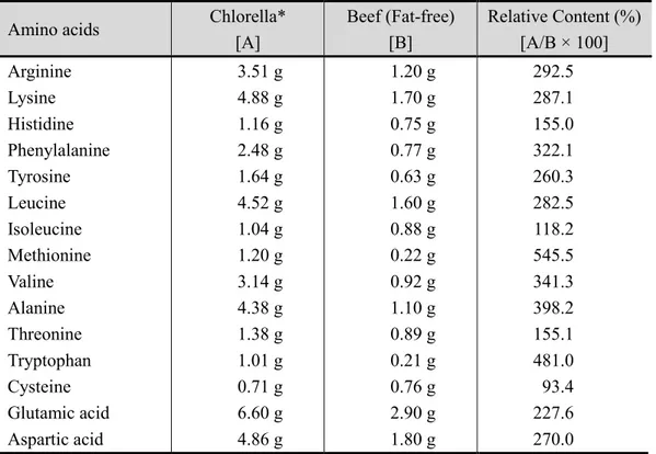
Chlorella contains various nutrients, including proteins, essential amino acids, vitamins, and minerals, as well as many phytochemicals, such as chlorophyll [1, 2, 5]. Chlorella has a solid cell wall, which is mainly composed of cellulose, hemicellulose, proteins, lipids, and minerals. The sugar composition of the cell wall is a mixture of rhamnose, galactose, glucose, xylose, arabinose, and mannose [5]. The protein content of chlorella is 42%–61% of the dry cell weight [37-39]. It is a high-protein food, with higher protein content than that of chicken eggs (Table 3) [39]. Chlorella contains more amino acids than beef, except for cysteine (Table 4) [39]. It also contains lipids (14%–22% of the dry mass), carbohydrates (12%–17% dry weight) [40], and bioactive pigment substances, such as chlorophyll, lutein, astaxanthin, and β-carotene [5, 41, 42] (Table 5). Chlorella has high content of magnesium, calcium, and iron, which are needed for maintaining normal heart functions, blood formation, and circulation [5]. Chlorella is an excellent food that can improve the unbalanced diet of modern people, as it contains various nutrients.
7
Table 3. Comparison of biochemical properties and composition of chlorella, spinach, milk, and chicken egg (per 100g)
Component Chlorella* Spinach Milk Chicken egg Protein Carbohydrate Fat Ash Fiber Vitamin A Vitamin B1 Vitamin B2 Niacin Vitamin C Vitamin E Energy 60.6 g 3.7 g 12.8 g 4.5 g 13.0 g 58,900 IU 1.29 mg 4.55 mg 32.1 mg 74 mg 22.8 mg 372 kcal 3.3 g 3.6 g 0.2 g 1.7 g 3.5 g 2,900 IU 0.13 mg 0.23 mg 0.6 mg 65 mg 2.1 mg 25 kcal 2.9 g 4.5 g 3.2 g 0.7 g - 1,100 IU 0.03 mg 0.15 mg 0.1 mg 0 mg 0.1 mg 59 kcal 12.3 g 0.9 g 11.2 g 0.9 g - 640 IU 0.08 mg 0.48 mg 0.1 mg 0 mg 1.1 mg 162 kcal
8
Table 4. Comparison of the amino acid composition of chlorella and beef (per 100g) Amino acids Chlorella*
[A] Beef (Fat-free) [B] Relative Content (%) [A/B × 100] Arginine Lysine Histidine Phenylalanine Tyrosine Leucine Isoleucine Methionine Valine Alanine Threonine Tryptophan Cysteine Glutamic acid Aspartic acid 3.51 g 4.88 g 1.16 g 2.48 g 1.64 g 4.52 g 1.04 g 1.20 g 3.14 g 4.38 g 1.38 g 1.01 g 0.71 g 6.60 g 4.86 g 1.20 g 1.70 g 0.75 g 0.77 g 0.63 g 1.60 g 0.88 g 0.22 g 0.92 g 1.10 g 0.89 g 0.21 g 0.76 g 2.90 g 1.80 g 292.5 287.1 155.0 322.1 260.3 282.5 118.2 545.5 341.3 398.2 155.1 481.0 93.4 227.6 270.0
9
Table 5. Potential pigments content in C. vulgaris under different growth conditions Pigments ㎍/g (dw) β-Carotene 7 - 12,000 Astaxanthin 555,000 Cantaxanthin 362,000 Lutein 52 - 3,830 Chlorophyll-a 250 - 9,630 Chlorophyll-b 72 - 5,770 Pheophytin-a 2,310 - 5,640 Violoxanthin 10 - 37
10
3. Health benefits of chlorella
Because chlorella contains various nutrients, chlorella has been investigated as a future food to solve the food shortage problem caused by population growth after World War II [43]. Since various health benefits of chlorella have been studied, it is now consumed as a dietary supplement, and not as food [44-46].
In Korea, four health claims of chlorella have been approved by the Ministry of Food and Drug Safety (MFDS): skin health-improving [6, 7], antioxidant [8-10], blood cholesterol-lowering [11-14], and immune-enhancing [15-18] effects. Chlorella has a skin-protective effect against UVB via production of MMP-1 and degradation of procollagen genes in human skin fibroblasts by [6], and protects against UVC-induced cytotoxicity [7]. Chlorella improves antioxidative capacity in rats with oxidative stress by reducing the production of radicals and promoting the capture and elimination of radicals [8, 9]. Ryu et al. [14] revealed that chlorella has beneficial health effects on the serum lipid profiles of mildly hypercholesterolemic subjects by improving serum carotenoid profiles in humans. Chlorella enhances the activity of NK cells and produces interferon-γ and interleukin-12 in humans [15]. Chlorella also increases the production of Th1-type cytokines, such as IFN-γ and IL-12, while not affecting Th2-type cytokines, such as IL-4,
in vivo [18].
According to studies in rats, dietary chlorella has a protective effect on the toxicity of heavy metals, such as cadmium-induced liver damage and lead-induced oxidative
11
stress [47-49]. Moreover, it excretes dioxin-like PCB-138 and PCB-153 via urine [50]. In addition, it protects rats from heterocyclic amine-induced aberrant gene expression by increasing fecal excretion of unmetabolized PhIP [51].
Jung et al. [52] proved that chlorella could be a source of lutein, which helps maintain eye health; similar to lutein extracted from marigold flower, lutein from chlorella is absorbed from plasma.
12
III. MATERIALS AND METHODS
To evaluate the safety of substances for the human body, it is dangerous to test it directly on the human body; thus, a toxicity test is conducted on animals. General toxicity test (single- and repeated-dose) and genetic toxicity study are basics for safety assessment. Reproductive toxicity study, immunotoxicity study, and carcinogenicity study are conducted according to the characteristics of the raw materials [53]. In this study, acute toxicity was tested by a single dose toxicity study in rodents, the most widely used test animal in general toxicity studies. However, as the bioreaction to the test substance may be different depending on the type of animal, a single toxicity test was additionally performed in beagle dogs. Among non-rodents, beagle dog is easy to test, and many basic test data on beagle dog have accumulated; thus, these data can be used for interpreting and evaluating our test result. In addition, subacute toxicity test was performed through repeated 13-week administration in rodents to evaluate the toxicity that may occur during continuous medium-term intake.
The research was pre-approved by the animal care and use committee at Preclinical Research Center of ChemOn Corporation (06-RA-097, 06-DA-098, 06-RR-100) and was carried out in accordance with the “Toxicity Test Standards of Drug Medicine and the Like” of Korea’s MFDS Notification No. 2017-71 (August 30, 2017) and “Good Laboratory Practice” of Korea’s MFDS Notification No. 2018-93 (November 21, 2018).
13
1. Single oral dose toxicity test in Sprague-Dawley (SD) rats
1.1. Material and excipient
The chlorella sample (C. vulgaris) used in this study was obtained from Daesang Corp. (Seoul, Korea). C. vulgaris was isolated from freshwater samples collected in South Korea, and was cultured under heterotrophic conditions with glucose as a carbon source. After the culture was completed, the biomass was separated from the culture medium by centrifugation, and then prepared into a powder using a spray-dryer. It was green-colored due to chlorophyll. The nutritional components of chlorella used in this study are shown in Table 6. The content of the ingredient was measured according to the methods recorded in the Food Code [54] and the Health Functional Food Code [55], which were published by the MFDS of Korea.
Table 6. Composition of C. vulgaris used in this study Ingredients Content (per 100 g powder) Protein Ash Fat β-carotene Vitamin B2 Vitamin C Chlorophyll Letein 56.4 g 3.5 g 15.4 g 69.6 mg 4.6 mg 84.0 mg 2,550 mg 336.0 mg
14
Sterile water for injection (Daihan Pharm. Co., Ltd., Seoul, Korea) was used as a vehicle and kept refrigerated after opening. The chlorella sample used in this study was suspended in sterile water for injection according to the doses of each group assigned in the single dose toxicity studies before administration.
1.2. Animal husbandry and maintenance
In this study, specific pathogen-free (SPF) SD rats at 7 weeks of age, which were used for the single oral dose toxicity study, were provided by Koatech Co., Ltd., (Pyeongtaek, Korea). After 1 week of acclimation, the test substance was administered when the rats were 8 weeks of age.
Throughout the study period, the animals were housed in a room controlled at a temperature of 23 ± 3°C, relative humidity of 55% ± 15%, number of air changes of 10 to 20 times/h, lighting hour of 12 h (lights on: 8 a.m.; lights out: 8 p.m.), and illumination intensity of 150 to 300 Lux. During the test, the temperature and humidity in the animal room were measured every hour by using an auto-temperature and humidity measurer utilizing a computer system. The environmental conditions, such as the number of air changes and illumination intensity, were regularly measured.
The animals were acclimated in a stainless-steel net feeding cage (215 W × 355 L × 200 H mm) with a suitable amount of bedding materials. The animals were raised under a condition of five animals/feeding cage for the quarantine and adaptation periods, and under a condition of less than or equal to five animals/feeding cage for the administration
15
period. The feeding cages were distinguished by an individual identity card that had the test number and the animal number written.
The test animals were fed solid feeds sterilized by irradiation (Teklad global 18% protein rodent diet, 2918C; Harlan Co., Ltd., USA), supplied by Folas International. The animals were provided free access to the food.
The animals were also provided underground water sterilized by a UV sterilizer and a microfiltration system. Water was freely provided via a water bottle.
1.3. Administration
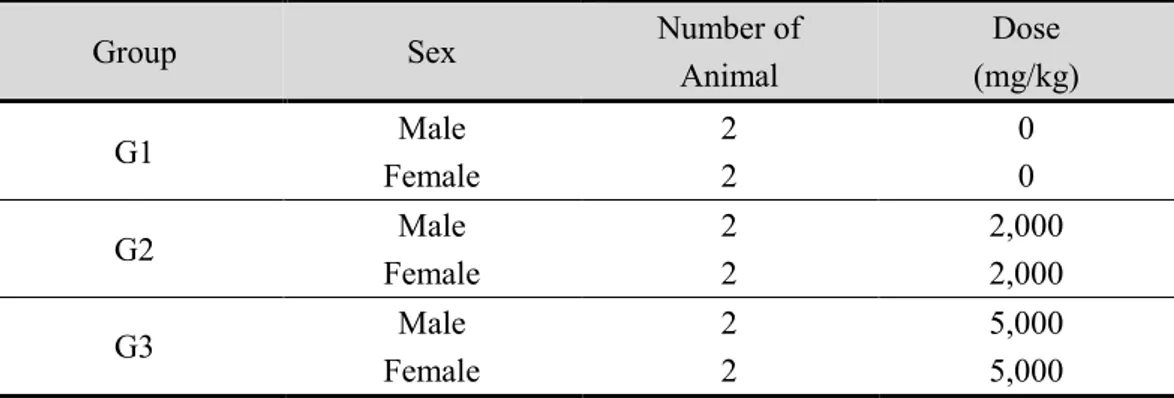
In a preliminary test, one animal each from the male and female groups was allocated as a high-dose group and administered 10,000 mg/kg; three other groups were administered the test substance at lower doses. No animal deaths nor specific symptoms were observed until 4 days after administration; thus, the group administered the maximum dose of 10,000 mg/kg was set as group 3, and one group receiving a lower dose was established as group 2. To observe the effect of liquid (sterile water for injection) overdose, an excipient-administered group was set as group 1 (Table 7).
16
Table 7. Test group organization of single oral doses toxicity test to SD rats Group Sex Number of
Animal Liquid Dose (mL/kg) Dose (mg/kg) G1 Male Female 5 5 40 40 0 0 G2 Male Female 5 5 40 40 5,000 5,000 G3 Male Female 5 5 40 40 10,000 10,000
G1: Control material-administered group (Sterilized injection water-administered group), G2, G3: Test sample-administered group
As the test sample was to be used for oral administration in humans, the test sample was orally administered to rats. During test sample administration, the animals were in a state of an empty stomach owing to one-night fasting. To administer the test sample, the abdominal skin of the animal was grasped with the hand, and then the test sample was orally administered into the stomach using a metal sonde. Half of the total administered volume was administered twice/day at hourly intervals on the day of treatment. The liquid dose was calculated based on the animal’s weight, which was weighed just before sample administration. The liquid dose was calculated to be 40 mL/kg.
17
1.4. Observation of general symptoms and animal deaths
General symptoms were observed immediately after sample administration once a day over the whole administration period. They were observed every hour for 6 h later on the day of administration.
1.5. Weight measurement
The weight of all animals was measured before administration, and on the 1st, 3rd, 7th, and 14th days after administration.
1.6. Autopsy observation
Animals were anesthetized with ether on the 14th day after administration, and the postcavas and abdominal aorta were removed to exsanguinate the animals. Organs in the abdominal and thoracic cavities were then observed with the naked eye.
1.7. Statistical analysis
In the case of body weight increment, the mean and standard deviation were calculated. Next, the test sample-treated group was compared with the control group. T-test was used as a statistical method. The computer program SPSS v25 (IBM Corp, Armonk, New York, USA) was used for statistical analysis.
18
2. Single oral dose toxicity test in beagle dogs
2.1. Material and excipient
The chlorella sample (C. vulgaris) used in this study was obtained from Daesang Corp. Gelatin capsule (Torpac Inc., Fairfield, NJ, USA) was used as vehicle for oral administration.
2.2. Animal husbandry and maintenance
Beagle dogs are suitable and widely used experimental non-rodents for general toxicity testing. In this study, the beagle dogs, aged 5.5 months, for the single oral dose toxicity study were provided by Jung Ang Lab. Animal Inc. (Seoul, Korea). After 15 days of acclimation, the animals were administered the test substance at 6 months of age.
Throughout the study period, the animals were housed in a room with controlled conditions: temperature, 23 ± 3°C; relative humidity, 55% ± 15%; number of air changes, 10 to 20 times/h; lighting hour, 12 h (lights on, 8 a.m.; lights out, 8 p.m.); and illumination intensity, 150 to 300 Lux. During the test, the temperature and humidity in the animal room were measured every hour by using an auto-temperature and humidity measurer utilizing a computer system. The environmental conditions, such as the number of air changes and the illumination intensity, were regularly measured.
The animals were acclimated in a stainless-steel net feeding cage (895 W × 795 L × 765 H mm) during the adaptation and quarantine, administration, and observation periods. One animal each was raised for the quarantine and observation period after administration.
19
The feeding cages were distinguished by an individual identity card that had the test and animal numbers written.
The dogs were fed solid feeds for a pet dog (PuppyMac) supplied from BioMac Corp. (Suwon, Korea) at 300 g per day. Underground water sterilized by a UV sterilizer and a microfiltration system was provided freely to the dogs via an automatic waterer.
2.3. Administration
A preliminary test revealed that administration of the test sample at 1,000, 2,000, 5,000, and 10,000 mg/kg did not result in abnormal changes in male and female animals (one animal each). Accordingly, 2,000 and 5,000 mg/kg, which were generally used as a limit dose for a one-time oral administration [56], were set as the administration doses for the subsequent test. In addition, an excipient control group, which was administered an empty capsule, was established (Table 8).
20
Table 8. Test group organization of single oral dose toxicity test to beagle dogs Group Sex Number of
Animal Dose (mg/kg) G1 Male Female 2 2 0 0 G2 Male Female 2 2 2,000 2,000 G3 Male Female 2 2 5,000 5,000
G1: Control material-administered group (Empty gelatin capsule-administered group), G2, G3: Test sample-administered group
As the test sample is to be used for oral administration in humans, the test sample was orally administered in this study. The animals were fasted overnight, and then administered the test sample. First, an animal was naturally positioned in the feeding cage, its mouth was opened, the capsule was put inside its inner tongue, and then its mouth was closed. After that, the pharyngolarynx of the animal was gently touched to ensure swallowing of the capsule. The test sample was administered once on the day of administration.
2.4. Observation of general symptoms and animal deaths
General symptoms were observed once a day in all animals over the whole administration period. However, on the day of administration, it was observed every hour from immediately after to 6 h after administration.
21
2.5. Weight measurement
The weight of all animals was measured before administration, and on the 1st, 3rd, 7th, and 14th days after administration.
2.6. Autopsy observation
All surviving animals were anesthetized by oral administration of pentobarbital (45 mg/kg; Entobar; Hanlim Pharm. Co., Ltd., Seoul, Korea) on the 14th day after administration, and then the animals were exsanguinated by cutting the armpit artery. All organs were then observed with the naked eye.
2.7. Statistical analysis
For body weight, the mean and standard deviation per group was calculated, and then the test sample-treated group was compared with the control group. T-test was used as a statistical method. The program SPSS v25 (IBM Corp.) was used for statistical analysis.
22
3. Thirteen-week repeated oral doses toxicity test in SD rats
3.1. Material and excipient
The chlorella sample (C. vulgaris) used in this study was obtained from Daesang Corp. Sterile water for injection (Daihan Pharm. Co., Ltd.) was used as the vehicle and was kept refrigerated after opening. The chlorella sample used in this study was suspended in sterile water for injection according to the doses of each group assigned in the repeated-dose toxicity study.
3.2. Animal husbandry and maintenance
In this study, SPF SD rats at 5 weeks of age were provided by Koatech Co., Ltd. After 1 week of acclimation, the animals were administered the test substance at 6 weeks of age.
Throughout the study period, the animals were housed in a room with controlled conditions: temperature, 23 ± 3°C; relative humidity, 55% ± 15%; number of air changes, 10 to 20 times/h; lighting hour, 12 h (lights on, 8 a.m.; lights out, 8 p.m.); and illumination intensity, 150 to 300 Lux. During the test, the temperature and humidity in the animal room were measured every hour by using an auto-temperature and humidity measurer utilizing a computer system. The environmental conditions, such as the number of air changes and the illumination intensity, were regularly measured.
The animals were acclimated in a polycarbonate feeding cage (235 W × 380 L × 175 H mm) with a suitable amount of bedding materials. The animals were raised under the
23
condition of less than or equal to five animals/feeding cage for the quarantine period and of two animals/feeding cage for the administration and observation periods. The feeding cages were distinguished by an individual identity card with the test and animal numbers written.
The animals were provided free access to solid feeds sterilized by irradiation (Teklad global 18% protein rodent diet, 2918C; Harlan Co., Ltd.), supplied by Folas International. The animals were also provided underground water sterilized by a UV sterilizer and a microfiltration system. Water was provided ad libitum via a water bottle.
3.3. Administration
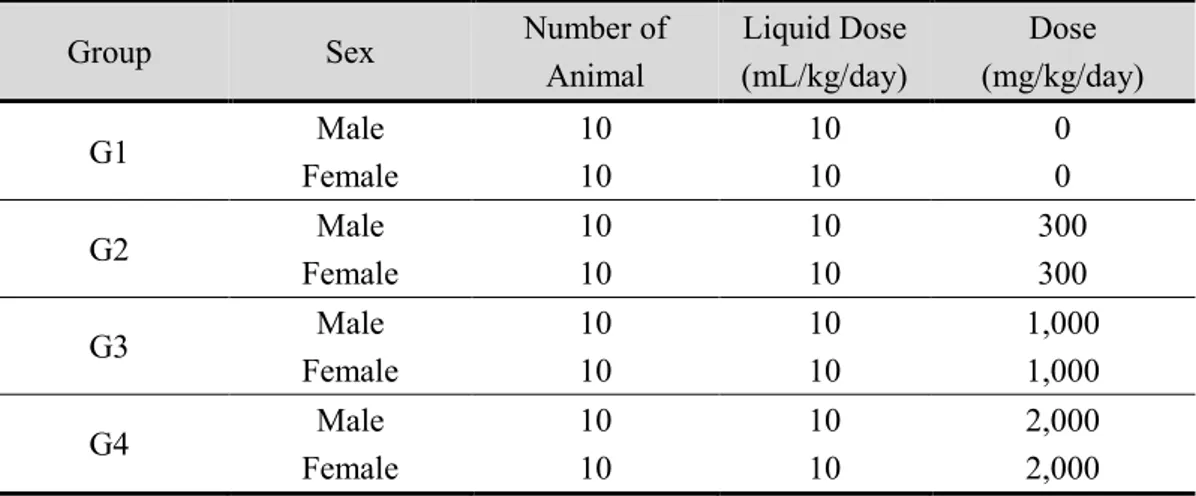
The preliminary test showed no remarkable toxicological changes, except that BUN was significantly increased in a male group administered with the test substance at 2,000 mg/kg/day. Accordingly, for this test, the high-dose was set to be 2,000 mg/kg/day, which was two-times higher than 1,000 mg/kg/day, the limit dose in the OECD Guidelines for toxicity testing of repeated oral administration [57], and the low-dose was set to be 300 mg/kg/day, which was two-times higher than the planned clinical dose (150 mg/kg/day). A medium-dose was set to be 1,000 mg/kg/day, i.e., half the high-dose. The control group was an excipient control group administered only sterile water (Table 9).
24
Table 9. Test group organization of repeated oral doses toxicity test to SD rats Group Sex Number of
Animal Liquid Dose (mL/kg/day) Dose (mg/kg/day) G1 Male Female 10 10 10 10 0 0 G2 Male Female 10 10 10 10 300 300 G3 Male Female 10 10 10 10 1,000 1,000 G4 Male Female 10 10 10 10 2,000 2,000
G1: Control material-administered group (Sterilized injection water-administered group), G2, G3, G4: Test sample-administered group
As the test sample is to be used for oral administration in humans, the test sample was orally administered in this study. For administration of the test sample, the abdominal skin of the animal was grasped with one hand, and then the test sample was orally administered into the stomach using a metal sonde. The test sample was generally administered in the morning on the administration day. The liquid dose was calculated based on the weight of the animal on the administration day, and was determined to be 10 mL/kg/day.
The number and period of administration was once per day, 7 days per week, for 13 weeks.
25
3.4. Observation of general symptoms
General symptoms were observed immediately after the once-a-day administration over the whole administration period. The type, expression, and degree of the symptoms were recorded per individual animal as general symptoms.
3.5. Weight measurement
The weight of all animals was measured at the start of administration, once per week during the test period, and on the autopsy day.
3.6. Measurement of food intake
Food intake was measured at the administration day and once per week over the whole test period. The amounts of feed supplied and remaining were measured each day after weight measurement, and then calculated as an average intake amount per one animal (g/rat/day).
3.7. Measurement of water intake
Water intake was measured on the administration day. The amounts of water supplied and remaining were measured each day and then calculated as an average intake amount per one animal (g/rat/day).
26
3.8. Eye test
The eyes of all animals were observed upon their allocation into groups. The pupils were dilated using a mydriatic (Ocuhomapine, Lot 013118; Samil Pharm. Co., Ltd., Seoul, Korea), and then the eyeground parts of the eyes were observed and photographed using an eyeground camera (Genesis; Gowa Co., Ltd., Japan) at the final week of observation, and compared between the excipient control group and the high-dose group.
3.9. Urinalysis
At the final week of administration, each group of five animals were put in a metabolic cage, and 1.0 mL of new urine was collected for 3 to 4 h. Next, 0.3 mL urine sample was smeared on a urinalysis test paper (Multistix 10SG; Bayer, Elkhart, IN, USA) and a urine auto-analyzer (CliniTek 100; Bayer, Elkhart, IN, USA) to measure glucose, bilirubin, ketone body, specific gravity, occult blood, pH, protein, urobilinogen, nitrite, and leukocyte content in the urine. The color of 0.7 mL urine sample was observed both with the naked eye and by a urine auto-analyzer (CliniTek 100; Bayer). The same 0.7 mL sample was centrifuged for 5 min at 1,500 rpm in a centrifuge (MF300; Hanil Scientific Inc., Gimpo, Korea), and the sediment was then microscopically examined for the presence of red blood cells, leukocytes, epithelial cells, and cylinder epithelium. Urine amount was measured after collection for 24 h [59].
27
3.10. Blood test
Blood test was performed on the animals to be sacrificed by using a blood corpuscle auto-measuring instrument (Cell-Dyn 3700; Abbot, Illinois , USA) (Table 10). The animals were fasted overnight and anesthetized, and then blood was collected via the postcava for the blood test. EDTA-2K was used as an anticoagulant [58].
28
Table 10. Hematological test item and method
Item Unit Method
WBC
(White blood cell count)* ´ 10 3
/mm3 Electrical resistance method
RBC
(Red blood cell count)* ´ 10
6
/mm3 Electrical resistance method
HGB
(Hemoglobin conc.)* g/dL
Cyanmethemoglobin conversion method
HCT
(Hematocrit)* % Calculation from RBC and MCV
MCV
(Mean corpuscular volume)* fL Electrical resistance method MCH
(Mean corpuscular hemoglobin)* pg Calculation from RBC and HGB MCHC
(Mean corpuscular hemoglobin conc.)* g/dL Calculation from HGB and HCT
Platelet* ´ 103/mm3 Electrical resistance method
Red blood cell percentage* % Light Scattering Detection Method
Reticulocyte** ea/1,000 Smear method
* They were tested by using a blood corpuscle auto-measuring instrument (Cell dyn 3700; Abbot, Illinois , USA).
29
3.11. Blood coagulation time test
Blood coagulation time test was performed on the same animals subjected to the blood test. Prothrombin time (PT) and active partial thromboplastin time (APTT) were measured by using a blood coagulation time analyzer (CA-50; Sysmex, Japan) on the autopsy day (Table 11). As an anticoagulant for a blood coagulation time test, 3.2% sodium citrate was used [58].
Table 11. Blood coagulation time test item and method
Item Unit Method
PT
(Prothrombin time)* second Photometric scattered light detection method & Percentage test endpoint detection method
APTT
(Active partial thromboplastin time)* second
*They were tested by using a blood coagulation time analyzer (CA-50; Sysmex, Japan).
3.12. Blood biochemical test
Blood biochemical test was performed on the same animals subjected to the blood test. Blood collected from the postcava was centrifuged at 3,000 rpm for 10 min to obtain serum for the blood biochemical test. They were measured by using an auto-analyzer (AU400; Olympus, Japan) and an electrolyte auto-analyzer (644 Na, K, Cl Analyzer; Ciba-Corning, USA) (Table 12) [58].
30
Table 12. Biochemical blood test item and method
Item Unit Method
AST (Aspartate aminotransferase)* lU/L IFCC method
ALT (Alanine aminotransferase)* lU/L IFCC method
ALP (Alkaline phosphatase)* lU/L P-NPP method
BUN (Blood urea nitrogen)* mg/dL Urease-UV method
CRE (Creatinine)* mg/dL Jaffe method
GLU (Glucose)* mg/dL UV method
CHO (Total cholesterol)* mg/dL Enzyme method
PRO (Total protein)* g/dL Biuret method
CPK (Creatine phosphokinase)* lU/L UV-Rate method
ALB (Albumin)* g/dL BCG method
BIL (Total bilirubin)* mg/dL Evelyn-Malloy method
A/G ratio* Calculation from PRO and ALB
TG (Triglyceride)* mg/dL Enzyme method
IP (Inorganic phosphorus)* mg/dL Enzyme method
Ca (Calcium)* mg/dL O-CPC method
Cl (Chloride)** mmol/L Electrode method
Na (Sodium)** mmol/L Electrode method
K (Potassium)** mmol/L Electrode method
*They were measured by using an auto-analyzer (AU400; Olympus, Japan).
31
3.13. Autopsy
The postcavas of the same animals subjected to the blood test were cut to exsanguinate the animals. Next, all organs in the abdominal and thoracic cavities as well as the head were observed.
3.14. Organ weight measurement
After the autopsy, the following organs were removed and then weighed with an electronic scale. In the case of organs that are present on both sides of the body, both organs were measured. The organs included the brain, heart, lungs, liver, spleen, kidney, thymus, adrenal gland, thyroid gland, pituitary gland, prostate, testis, epididymis, uterus, and ovary.
3.15. Histopathological test
Microscopic examinations were performed on the organs and tissues collected from
the excipient control group (0 mg/kg/day) and high-dose group (2,000 mg/kg/day). The organs of all the treated animals were fixed in 10% neutral formalin solution; the tissues were embedded in paraffin and sectioned at 5 μm thickness. Representative sections of each specified organ were stained with hematoxylin-eosin for microscopic examination.
32
3.16. Statistical analysis
The data obtained from the group administered with the test sample was compared with those from the excipient control group by using parametric and non-parametric multiple comparison tests. Regression analysis was used to confirm whether or not a dose correlation was significant concerning the test item of interest. The generation rate was expressed as a percentage. All statistical analyses were performed using SPSS v25 (IBM Corp.).
33
IV. RESULTS
1. Single oral dose toxicity test to SD rats
1.1. Mortality
There were no animal deaths during the whole test period in all test groups (Table 13).
Table 13. Mortality after a single oral dose in rats
Dose (mg/kg)
Days after dosing No. death
/No. dosed 0 1 2 3 4 5 6 7 8~14 Male CV 0 0* 0 0 0 0 0 0 0 0 0/5 CV 5,000 0 0 0 0 0 0 0 0 0 0/5 CV 10,000 0 0 0 0 0 0 0 0 0 0/5 Female CV 0 0 0 0 0 0 0 0 0 0 0/5 CV 5,000 0 0 0 0 0 0 0 0 0 0/5 CV 10,000 0 0 0 0 0 0 0 0 0 0/5
*Number of dead animals. Day 0: The day of dosing CV: C. vulgaris powder
34
1.2. General symptoms
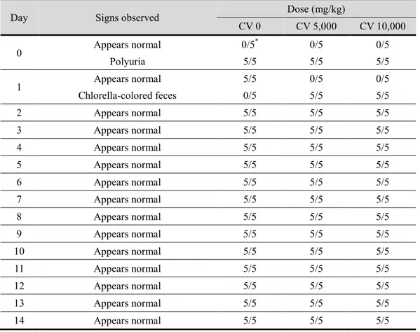
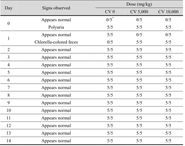
Polyuria was observed between 1 and 3 h after administration in all test groups at the day of administration, and chlorella-colored feces was observed in both male and female rats in the groups treated with 5,000 and 10,000 mg/kg of test substance on the 1st day after administration (Table 14, 15).
35
Table 14. General symptoms after a single oral dose in male rats
Day Signs observed Dose (mg/kg)
CV 0 CV 5,000 CV 10,000 0 Appears normal Polyuria 0/5* 5/5 0/5 5/5 0/5 5/5 1 Appears normal Chlorella-colored feces 5/5 0/5 0/5 5/5 0/5 5/5 2 Appears normal 5/5 5/5 5/5 3 Appears normal 5/5 5/5 5/5 4 Appears normal 5/5 5/5 5/5 5 Appears normal 5/5 5/5 5/5 6 Appears normal 5/5 5/5 5/5 7 Appears normal 5/5 5/5 5/5 8 Appears normal 5/5 5/5 5/5 9 Appears normal 5/5 5/5 5/5 10 Appears normal 5/5 5/5 5/5 11 Appears normal 5/5 5/5 5/5 12 Appears normal 5/5 5/5 5/5 13 Appears normal 5/5 5/5 5/5 14 Appears normal 5/5 5/5 5/5
*Number of animals with the sign/Number of animals examined. Day 0: The day of dosing
36
Table 15. General symptoms after a single oral dose in female rats
Day Signs observed Dose (mg/kg)
CV 0 CV 5,000 CV 10,000 0 Appears normal Polyuria 0/5* 5/5 0/5 5/5 0/5 5/5 1 Appears normal Chlorella-colored feces 5/5 0/5 0/5 5/5 0/5 5/5 2 Appears normal 5/5 5/5 5/5 3 Appears normal 5/5 5/5 5/5 4 Appears normal 5/5 5/5 5/5 5 Appears normal 5/5 5/5 5/5 6 Appears normal 5/5 5/5 5/5 7 Appears normal 5/5 5/5 5/5 8 Appears normal 5/5 5/5 5/5 9 Appears normal 5/5 5/5 5/5 10 Appears normal 5/5 5/5 5/5 11 Appears normal 5/5 5/5 5/5 12 Appears normal 5/5 5/5 5/5 13 Appears normal 5/5 5/5 5/5 14 Appears normal 5/5 5/5 5/5
*Number of animals with the sign/Number of animals examined. Day 0: The day of dosing
37
1.3. Body weight
There were no significant changes in weight in female rats treated with the test sample (Figure 2). In contrast, the weight of male rats administered 10,000 mg/kg of the test sample significantly increased (p < 0.05) on the 1st day after administration, compared with that of the excipient control group counterpart.
38 Time (days) 0 2 4 6 8 10 12 14 B o d y W ei g h t (g ) 150 200 250 300 350 0 mg/kg 5,000 mg/kg 10,000 mg/kg Male Female
*
Figure 2. Body weight changes of rats given a single dose of C. vulgaris powder. Male and female rats were administered 0 (●), 5,000 (▼), 10,000 (■) mg/kg body weight of C.
vulgaris powder. The values represent the means ± standard deviation (n=5).
39
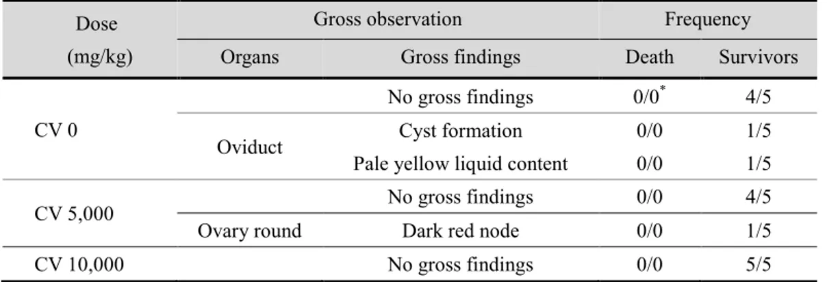
1.4. Autopsy observation
There were no specific changes in autopsy results between all male rats treated with the test sample (Table 16). On the contrary, female rats in the excipient control group showed pale yellow liquid and cyst formations in the oviducts. There was a dark red node around the ovaries in the group administered 5,000 mg/kg of the test substance (Table 17).
Table 16. Gross findings after a single oral dose in male rats
Dose (mg/kg)
Gross observation Frequency
Organs Gross findings Death Survivors
CV 0 No gross findings 0/0* 5/5
CV 5,000 No gross findings 0/0 5/5
CV 10,000 No gross findings 0/0 5/5
*Number of animals with the sign/Number of animals examined. CV: C. vulgaris powder
Table 17. Gross findings after a single oral dose in female rats
Dose (mg/kg)
Gross observation Frequency
Organs Gross findings Death Survivors
CV 0
No gross findings 0/0* 4/5 Oviduct Cyst formation
Pale yellow liquid content
0/0 0/0
1/5 1/5
CV 5,000 No gross findings 0/0 4/5
Ovary round Dark red node 0/0 1/5
CV 10,000 No gross findings 0/0 5/5
*Number of animals with the sign/Number of animals examined. CV: C. vulgaris powder
40
2. Single oral dose toxicity test in beagle dogs
2.1. Mortality
There were no animal deaths during the whole test period in all the test groups (Table 18).
Table 18. Mortality after a single oral dose in beagle dogs
Dose (mg/kg)
Days after dosing No. death
/No. dosed 0 1 2 3 4 5 6 7 8~14 Male CV 0 0* 0 0 0 0 0 0 0 0 0/2 CV 2,000 0 0 0 0 0 0 0 0 0 0/2 CV 5,000 0 0 0 0 0 0 0 0 0 0/2 Female CV 0 0 0 0 0 0 0 0 0 0 0/2 CV 2,000 0 0 0 0 0 0 0 0 0 0/2 CV 5,000 0 0 0 0 0 0 0 0 0 0/2
*Number of dead animals Day 0: The day of dosing CV: C. vulgaris powder
41
2.2. General symptoms
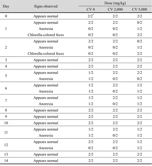
Chlorella-colored feces was observed on the 1st and 2nd day after administration in both male and female dogs administered 5,000 mg/kg of the test sample, and diarrhea was observed in male dogs of the 5,000 mg/kg group (Table 19, 20). In addition, sporadic symptoms of anorexia were observed in several groups, including the control group.
42
Table 19. General symptoms after a single oral dose in male dogs
Day Signs observed Dose (mg/kg)
CV 0 CV 2,000 CV 5,000 0 Appears normal 2/2* 2/2 2/2 1 Appears normal Diarrhea Chlorella-colored feces 2/2 0/2 0/2 2/2 0/2 0/2 0/2 1/2 2/2 2 Appears normal Diarrhea Chlorella-colored feces 2/2 0/2 0/2 2/2 0/2 0/2 0/2 1/2 2/2 3 Appears normal 2/2 2/2 2/2 4 Appears normal 2/2 2/2 2/2 5 Appears normal 2/2 2/2 2/2 6 Appears normal 2/2 2/2 2/2 7 Appears normal 2/2 2/2 2/2 8 Appears normal 2/2 2/2 2/2 9 Appears normal 2/2 2/2 2/2 10 Appears normal 2/2 2/2 2/2 11 Appears normal Anorexia 1/2 1/2 1/2 1/2 2/2 0/2 12 Appears normal Anorexia 1/2 1/2 1/2 1/2 2/2 0/2 13 Appears normal 2/2 2/2 2/2 14 Appears normal 2/2 2/2 2/2
*Number of animals with the sign/Number of animals examined Day 0: The day of dosing
43
Table 20. General symptoms after a single oral dose in female dogs
Day Signs observed Dose (mg/kg)
CV 0 CV 2,000 CV 5,000 0 Appears normal 2/2* 2/2 2/2 1 Appears normal Anorexia Chlorella-colored feces 2/2 0/2 0/2 2/2 0/2 0/2 0/2 1/2 2/2 2 Appears normal Anorexia Chlorella-colored feces 2/2 0/2 0/2 2/2 0/2 0/2 0/2 1/2 2/2 3 Appears normal 2/2 2/2 2/2 4 Appears normal 2/2 2/2 2/2 5 Appears normal Anorexia 1/2 1/2 2/2 0/2 2/2 0/2 6 Appears normal Anorexia 1/2 1/2 2/2 0/2 1/2 1/2 7 Appears normal Anorexia 1/2 1/2 2/2 0/2 1/2 1/2 8 Appears normal 2/2 2/2 2/2 9 Appears normal 2/2 2/2 2/2 10 Appears normal 2/2 2/2 2/2 11 Appears normal Anorexia 1/2 1/2 2/2 0/2 1/2 1/2 12 Appears normal Anorexia 2/2 0/2 2/2 0/2 1/2 1/2 13 Appears normal 2/2 2/2 2/2 14 Appears normal 2/2 2/2 2/2
*Number of animals with the sign/Number of animals examined Day 0: The day of dosing, CV: C. vulgaris powder
44
2.3. Body weight
There were no significant changes in weight following administration of the test sample in both male and female dogs over the whole test period, compared with that in the control group (Figure 3, 4).
However, in female dogs of the control group, there was a slight decrease in weight on the autopsy day, compared with that on the day of administration; however, this decrease was not significant.
45 Time (days) 0 2 4 6 8 10 12 14 B o d y W ei g h t (k g ) 0 2 4 6 8 10 12 0 mg/kg 2,000 mg/kg 5,000 mg/kg
Figure 3. Body weight changes of male dogs given a single dose of C. vulgaris powder. Male dogs were administered 0 (●), 2,000 (▼), 5,000 (■) mg/kg body weight of C.
46 Time (days) 0 2 4 6 8 10 12 14 B o d y w ei g h t (k g ) 0 2 4 6 8 10 12 0 mg/kg 2,000 mg/kg 5,000 mg/kg
Figure 4. Body weight changes of female dogs given a single dose of C. vulgaris powder. Female dogs were administered 0 (●), 2,000 (▼), 5,000 (■) mg/kg body weight of C.
47
2.4. Autopsy observation
There were no abnormal autopsy results in all animals (Table 21, 22).
Table 21. Gross findings after a single oral dose in male dogs
Dose (mg/kg)
Gross observation Frequency
Organs Gross findings Death Survivors
CV 0 No gross findings 0/0* 2/2
CV 2,000 No gross findings 0/0 2/2
CV 5,000 No gross findings 0/0 2/2
*Number of animals with the sign/Number of animals examined CV: C. vulgaris powder
Table 22. Gross findings after a single oral dose in female dogs
Dose (mg/kg)
Gross observation Frequency
Organs Gross findings Death Survivors
CV 0 No gross findings 0/0* 2/2
CV 2,000 No gross findings 0/0 2/2
CV 5,000 No gross findings 0/0 2/2
*Number of animals with the sign/Number of animals examined CV: C. vulgaris powder
48
3. Thirteen-week repeated oral dose toxicity test in SD rats
3.1. Mortality
There were no animal deaths during the whole test period in all test groups (data not shown).
3.2. General symptoms
There was a temporary, partial loss of teeth in two animals from the 77th to the 81st day after administration in the male excipient control group (Table 23, 24).
3.3. Body weight
There were no significant changes in body weight caused by the test sample in both the male and female groups (Figure 5).
49
Table 23. General symptoms after repeated oral dose in male rats
Day Signs observed
Dose (mg/kg) CV 0 CV 300 CV 1,000 CV 2,000 0 Appears normal 10/10* 10/10 10/10 10/10 1~76 Appears normal 10/10 10/10 10/10 10/10 77 Appears normal Loss of teeth 8/10 2/10 10/10 0/10 10/10 0/10 10/10 0/10 78 Appears normal Loss of teeth 8/10 2/10 10/10 0/10 10/10 0/10 10/10 0/10 79 Appears normal Loss of teeth 8/10 2/10 10/10 0/10 10/10 0/10 10/10 0/10 80 Appears normal Loss of teeth 8/10 2/10 10/10 0/10 10/10 0/10 10/10 0/10 81 Appears normal Loss of teeth 8/10 2/10 10/10 0/10 10/10 0/10 10/10 0/10 82~91 Appears normal 10/10 10/10 10/10 10/10
*Number of animals with the sign/Number of animals examined CV: C. vulgaris powder
Table 24. General symptoms after repeated oral dose in female rats
Day Signs observed
Dose (mg/kg) CV 0 CV 300 CV 1,000 CV 2,000 0 Appears normal 10/10* 10/10 10/10 10/10 1~91 Appears normal 10/10 10/10 10/10 10/10
*Number of animals with the sign/Number of animals examined CV: C. vulgaris powder
50 Time (Weeks) 0 2 4 6 8 10 12 14 B o d y W ei g h t (g ) 100 200 300 400 500 0 mg/kg/day 300 mg/kg/day 1,000 mg/kg/day 2,000 mg/kg/day Male Female
Figure 5. Body weight changes of rats administered C. vulgaris containing diet for 13-week. Male and female rats were administered 0 (●), 300 (▼), 1,000 (■), 2,000 (◆) mg/kg body weight/day of C. vulgaris powder. The values represent the means ± standard deviation (n=10).
51
3.4. Food and water consumption
There were no significant changes in food intake related to the test sample in both the male and female groups (Figure 6).
There was a significant increase (p < 0.05) in water intake during the 1st week after administration in male rats of the 2,000 mg/kg/day group (Figure 7). There were no significant changes in water intake in the female group (Figure 8).
52 Time (Weeks) 0 2 4 6 8 10 12 14 F ee d i n ta k e (g /d ay ) 5 10 15 20 25 0 mg/kg/day 300 mg/kg/day 1,000 mg/kg/day 2,000 mg/kg/day Male Female
Figure 6. Food intake of rats administered C. vulgaris containing diet for 13-week. Male and female rats were administered 0 (●), 300 (▼), 1,000 (■), 2,000 (◆) mg/kg body weight/day of C. vulgaris powder. The values represent the means ± standard deviation (n=10).
53 Time (Weeks) 0 2 4 6 8 10 12 14 W at er i n ta k e (g /d ay ) 0 10 20 30 40 50 0 mg/kg/day 300 mg/kg/day 1,000 mg/kg/day 2,000 mg/kg/day
*
Figure 7. Water intake of male rats administered C. vulgaris containing diet for 13-week. Male rats were administered 0 (●), 300 (▼), 1,000 (■), 2,000 (◆) mg/kg body weight/day of C. vulgaris powder. The values represent the means ± standard deviation (n=10).
54 Time (Weeks) 0 2 4 6 8 10 12 14 W at er i n ta k e (g /d ay ) 0 10 20 30 40 50 0 mg/kg/day 300 mg/kg/day 1,000 mg/kg/day 2,000 mg/kg/day
Figure 8. Water intake of female rats administered C. vulgaris containing diet for 13-week. Female rats were administered 0 (●), 300 (▼), 1,000 (■), 2,000 (◆) mg/kg body weight/day of C. vulgaris powder. The values represent the means ± standard deviation (n=10).
55
3.5. Eye test
There were no remarkable changes in eye test results related to the test sample (data not shown).
3.6. Urinalysis
There was a significant decrease in urine pH (p<0.05) in the male rats of the 2,000 mg/kg/day group (Table 25). There were no significant changes cause by the test sample in the female group.
Table 25. Urinalysis of male and female rats after repeated oral dose Parameter Result Dose (mg/kg/day) Male Female CV 0 CV 300 CV 1,000 CV 2,000 CV 0 CV 300 CV 1,000 CV 2,000
No. of animals examined 5 5 5 5 5 5 5 5
Specific gravity ≤1.005 0 0 0 0 0 1 0 0 1.010 0 0 1 0 1 0 0 1 1.015 3 1 1 0 0 1 0 1 1.020 1 3 2 1 2 2 4 1 1.025 0 1 0 3 2 0 1 2 ≥ 1.030 1 0 1 1 0 1 0 0 pH 6.5 0 0 1 0 0 0 0 0 7.0 0 0 0 0 0 0 0 0 7.5 0 0 0 0 0 0 1 0 8.0 0 0 0 3* 0 0 0 0 8.5 1 2 3 2 4 4 3 2 ≥ 9.0 4 3 1 0 1 1 1 3 Glucose - 5 5 5 5 5 5 5 5 +/- 0 0 0 0 0 0 0 0 Bilirubin - 5 5 5 5 5 5 5 5 0 0 0 0 0 0 0 0
*Significantly different from control value (p<0.05) CV: C. vulgaris powder
5