The role of MMP and E-cadherin in the
peritoneal metastasis of Gastric cancer
Thesis by
Chung Sub Kim
Department of Medical Science
The Graduate School, Yonsei University
The role of MMP and E-cadherin in the
peritoneal metastasis of Gastric cancer
Directed by Professor Sung Hoon Noh
The Master's Thesis
submitted to the Department of Medicine Science
the Graduate School of Yonsei University
in partial fulfillment of the requirements for the
degree of Master of Medical Science
Chung Sub Kim
June 2003
This certifies that the Master's
Thesis of Chung Sub Kim is
approved
---
[Thesis Supervisor : Sung Hoon Noh]
---
[Thesis Committee Member]
---
[Thesis Committee Member]
The Graduate School
Yonsei University
Acknowledgments
항상 지도와 관심을 보아주시는 노성훈 교수님께 먼저 감사를 전합니다. 아울러 논문 심사위원으로 수고해주신 최승호 교수님과 이용찬 교수님 그리고 많은 가르침을 주셨던 백자현 교수님께 감사 의 말씀 드립니다. 듬직한 동료이자 선배였던 김병진 선생님, 조상 래 선생님께 감사를 전합니다. 그리고 같은 연구센터에 있으면서 같이 많은 것을 생각하고 느꼈던 안연희, 김량여, 길미화, 김성재, 이일선, 김용년, 배미현, 최승훈, 박정범, 고혜경, 박기찬, 윤성환, 김 명윤, 홍성이, 김정민 선생님께도 감사를 전합니다. 또한 많은 도움 을 주신 이면희 선생님과 박사 후 과정에 있는 이송철 선생님께 고 마운 마음을 전합니다. 한 식구처럼 대해주신 5층 실험실 사람들인 김용군 선생님, 이은진 선생님, 황난주 선생님, 박민정 선생님, 그리 고 박기청 선생님께도 감사의 말씀 전합니다. 항상 걱정과 격려를 해주던 여자친구 효숙이에게 무엇보다 깊 은 감사의 말을 전합니다. 여러모로 신경 써주고 사랑으로 지켜봐 주시는 부모님, 누나, 매형, 그리고 효숙이 어머님, 정환, 현주, 희정 이에게 감사를 전하면서 이 논문을 마칩니다.2003년 6월
김 청 섭
Contents
Abstract……….……...………….……….9
Ⅰ
. Introduction……….……….……..………….……….12
Ⅱ
. Materials and Methods……….………17
1. Materials………..………17
2. Cell culture and western blot ……….….………17
3. RNA Preparation and RT-PCR .………19
4. Zymograph …………..……….………19
5. Stable cell line……….……….………21
6. In vivo tumor growth assay………..………21
Ⅲ
. Results………23
1. Western blot analysis of E-cadherin in human gastric cancer
c e l l l i n e s … … … . … … . … … … . . … … … … . 2 3
2. RT-PCR analysis of E-cadherin, α-catenin, β-catenin, and
γ-catenin in human gastric cancer cell line ………..…………24
3. Expression of invasion genes MMP-2, MMP-7, MMP-9 and
Mt1-MMP in human gastric cancer cell lines………..……27
4. Establishment of human E-Cadherin transfectants ………29
5. Expression of invasion candidate genes MMP-2, MMP-7 and
MMP-9 in
stable transfectants YCC-3-Ecad andKATOIII-Ecad………..……….
….…31
6. Suppression of peritoneal metastasis by E-cadherin
YCC-3Ecad transfectant………33
Ⅳ
. Discussion………..……….35
Ⅴ
. Conclusion………..…39
References………..……..……40
LIST OF FIGURES
Figure 1.
Western blot analysis of E-cadherin expression in human
gastric cancer cell lines……....………24
Figure 2. RT-PCR analysis of E-cadherin, α-catenin, β-catenin, and
γ-catenin in human gastric cancer cell line ………….26
Figure 3. A. Gelatin zymography and casein zymography of
conditioned medium from human gastric cancer cell
lines
B. RT-PCR analysis of MMP-2, MMP-7, MMP-9 and
Mt1-MMP in human gastric cancer cell lines…....…28
Figure 4. Restoration of E-cadherin expression in human gastric
cancer cell line YCC-3 and KATOIII cell lines.30
Figure 5.
Gelatin zymography and casein zymography of Invasioncandidate genes in protein level and RT-PCR analysis of Invasion candidate genes in mRNA level in stable transfectants YCC-3-Ecad and KATOIII-Ecad…………....32
Figure 6
Human gastric cancer cell line YCC-3 and YCC-3Ecadtransfectant was injected into the peritoneal cavity of
LIST OF TABLE
ABSTRACT
The role of MMP and E-cadherin in the
peritoneal metastasis of Gastric cancer
Chung Sub Kim
Department of Medical Science
The Graduate School, Yonsei University
(Directed by Professor Sung Hoon Noh)
E-cadherin (E-CD) germ-line mutations have recently been described as a molecular basis of early-onset familial gastric cancer. The loss of cadherin expression or mutation of
E-cadherin has been associated with accelerated tumorigenesis and progression. However, whether E-cadherin is involved the peritoneal metastasis in gastric cancer is not certain. To assess the role of E-cadherin (Ecad), the cell-cell adhesion protein, in peritoneal dissemination of gastric cancer, it was used human gastric cancer cell lines YCC-3 (Ecad-) and KATOIII (Ecad-) which are not expressed cadherin(Ecad). The full length E-cadherin was cloned into pCEP4 and transfected into two human gastric cancer cell line YCC-3 (Ecad-) and KATOIII (Ecad-) and it was isolated stable transfectant clones. Expression of E-cadherin in transfected cell lines was confirmed by RT-PCR and western blot analysis. To explore the peritoneal dissemination of gastric cancer, it was injected wild type cell line and transfectants expressing exogenous E-cadherin into the abdominal cavity of nude mice. These results demonstrated that over-expression of E-cadherin in human gastric cancer cell line YCC-3 (Ecad-) significantly decreased the peritoneal dissemination of gastric cancer. We are now studying the mechanism by which the E-cadherin is involved the peritoneal metastasis of gastric cancer.
Key words: gastric cancer, E-cadherin, MMP, peritoneal metastasis
The role of MMP and E-cadherin in the
peritoneal metastasis of Gastric cancer
(Directed by Professor Sung Hoon Noh)
Department of Medical Science
The Graduate School, Yonsei University
Chung Sub Kim
Ⅰ
. Introduction
Cell–cell adhesion is an essential component of epithelial morphology and function. Epithelial cells adhere tightly to each other, and several specialized adhesive structures ensure the appropriate integrity and tensile strength of epithelial sheets. These adhesive structures are connected either to intermediate filaments (desmosomes)
or to microfilaments (adherens junctions and tight junctions). This association with the cytoskeletal network is necessary for stable cell–cell adhesion and for the integration of cell–cell contacts1. Among the many types of cell-cell adhesion molecules, cadherins play a critical role in establishing adherens-type junctions by mediating Ca2+-dependent cell-cell adhesion2,3,4. Cadherin-based cell-cell adhesion is critically involved in early embryonic morphogenesis, as exemplified by the early embryonic lethality of mice lacking E-cadherin, a prototype cadherin5,6. Cadherin-mediated cell-cell adhesion is accomplished by homophilic protein-protein interactions of extracellular cadherin domains in a zipper-like fashion. The intracellular domain of cadherins interacts with various proteins, collectively termed catenins, which assemble the cytoplasmic cell adhesion complex (CCC) that is critical for the formation of extracellular cell-cell adhesion. -catenin and -catenin bind directly to
-catenin, which links the CCC to the actin cytoskeleton. p120ctn has been implicated both in cell-cell adhesion and in cell migration12, and recent studies suggest that p120ctn promotes cell migration by recruiting and activating Rho-family GTPases13.
It has long been known that cell-cell adhesion is dramatically changed during the development of malignant cancer. In particularly, in
most if not all cancers of epithelial origin, E-cadherin-mediated cell-cell adhesion is lost concomitantly with progression towards malignancy, and it has been proposed that the loss of E-cadherin-mediated cell-cell adhesion is a prerequisite for tumour cell invasion and metastasis formation14. Multiple mechanisms are found to underlie the loss of E-cadherin function during tumourigenesis: mutations or deletions of the E-cadherin gene itself, mutations in the -catenin gene, transcriptional repression of the E-cadherin gene. Recent reports have highlighted that the DNA binding protein Snail acts as a strong repressor of E-cadherin gene expression in tumour cells, thus inducing tumour malignancy15,16,17,18. The observation that E-cadherin function is frequently lost in malignant cancers prompted an examination of the functional role of E-cadherin in tumour progression,19,20,21. Perhaps the strongest evidence in support of a causal role for cadherin alterations in cancer pathogenesis is the observation that germline mutations in the gene encoding E-cadherin strongly predispose affected individuals to diffuse-type gastric cancer22,23,24,25. In some kindreds segregating a germline CDH1 mutation, individuals have been identified with colorectal23,26, breast24,25, and prostatecancers22.
other organs, it must locally degrade the extracellular matrix (ECM) components that are the physical barriers for cell migration. The ECM holds cells together and maintains the three-dimensional structure of the body27. It also plays critical roles in cell growth, differentiation, survival and motility28. The key enzymes responsible for ECM breakdown are matrix metalloproteinases (MMPs)29. ECM degradation by MMPs not only enhances tumour invasion, but also affects tumour cell behaviour and leads to cancer progression28,29,30,31. Recent studies on the gastric cancer have demonstrated that the expression of MMP-2 and MMP-9 which can degrade type IV collagen of basement membrane - the first barrier for cancer invasion are elevated in peritoneal disseminated metastasis of gastric cancer in association with low expression of E-cadherin32, 33,34,35,36. Furthermore, it has been suggested that the expression of MMP-7 is associated with the formation of peritoneal dissemination in gastric cancer37.
To elucidate the role of E-cadherin in the formation of peritoneal dissemination in gastric cancer, stable human gastric cancer cell lines expressing human E-cadherin was used. We are now studying the mechanism by which the E-cadherin is involved the peritoneal metastasis of gastric cancer.
Ⅱ
. Materials and Methods
1. Materials
Human gastric cancer cell lines MKN-28(80102), MKN-45(80103), NCI-N87 (CRL-5822), AGS (21739), SNU-216(00216), SNU-638(00638), SNU-668(00668), SNU-719(00719) was purchased from KCLB(Korean Cell Line Bank) and Human gastric cancer cell line KATO-III (30103) was purchased from ATCC(USA), and Human gastric cancer cell line YCC-3 was from Cancer Metastasis Research Center in University of Yonsei (Korea). Human E-cadherin cDNA was kindly provided by Dr. David L. Rimm (University of Yale, New Heaven, USA). LipofectamineTM 2000 was purchased from Invitrogen (USA). Hygromycine B was purchased Invitrogen (USA). Pro-PrepTM Protein Extraction Solution was purchased iNtRON Biotechnology (Korea). Taq polymerase was purchased TaKaRa (Japan). Zymogram gel was purchased Invitrogen (USA). Monoclonal Anti-Human E-cadherin was purchased TaKaRa (Japan).
2. Cell culture and Western Blot.
Human gastric cancer cell line MKN-28, MKN-45, AGS, KATOIII, SNU-216, SNU-638, SNU-668, SNU-719 cell lines were maintained in
RPMI 1640 medium supplemented with 10% FBS, 100 mg/ml streptomycin sulfate, 100 units/ml penicillin G and 250 mg/ml amphotericin B under 5% CO2 in air at 37° and NCI-N87 cell line was maintained in DMEM medium supplemented with 10% FBS, 100 mg/ml streptomycin sulfate, 100 units/ml penicillin G and 250 mg/ml amphotericin B under 5% CO2 in air at 37° and YCC-3 cell line was maintained in MEM medium supplemented with 10% FBS, 100 mg/ml streptomycin sulfate, 100 units/ml penicillin G and 250 mg/ml amphotericin B under 5% CO2 in air at 37°. 70% ~ 80% confluent cells in 100-mm culture plates were incubated at 37°C and were lysed in lysis buffer Pro-PrepTM Protein Extraction Solution. Samples were sonicated 4 times for 5 second each, centrifuged at 10,000g at 4°C for 10 min and the supernatant was collected. The proteins were separated on 10% SDS– polyacrylamide gel and transferred to Immobilon-P membranes (Millipore). The blots were incubated with 5% dried milk powder in TBST (10 mM Tris, pH 8.0, 150 mM NaCl, 0.05% Tween 20; also used for all incubations and washing steps) for 30 min. Next, the blots were incubated for 1h with Monoclonal Anti-Human E-cadherin. The blots were subsequently incubated with anti mouse-IgG antibody(Amersham Life Science NA931). After washing, signals were visualized using the
Enhanced ChemiLuminescence detection system (ECL, Amersham).
3. RNA preparation and RT-PCR
The total cellular RNA from the Human gastric cancer cell lines was isolated by using LiCl RNA extraction buffer. For reverse transcription, 2 μg aliquot of the total RNA was primed with a hexadeoxyribonucleotide of the random primers (Gibco/BRL) and the first strand was synthesized using ImProm-IITM Reverse Transcription System(Promega) according to the manufacturer’s protocol. The cDNA/mRNA hybrids were amplified with the sense and antisense primers by PCR. PCR amplification was performed for 25 cycles under the following conditions: (a) denaturing at 95°C for 1 min; and (b) polymerization at 72°C for 1 min. Each annealing condition for amplification of these cDNAs is included.
4. Zymograph
가
. Gelatin Zymography
NOVEX 4–16% zymogram gelatin gels (Invitrogen, Carlsbad,CA) were used to detect MMP-2 and MMP-9 enzymatic levels. Conditioned Serum Free media was loaded on a gel. The gel wasrun in Tris/glycine
SDS running buffer under nondenaturing conditions. The gels were washed twice in 2.5% (v/v) Triton X-100 (TX-100)for 30 min at room temperature to remove SDS. Zymograms weresubsequently developed by incubation 72 h at 37°C in zymogramdeveloping buffer [0.2 M NaCl, 5 mM CaCl2, 1% Triton X-100 and0.02% NaN3 in 50 mM Tris-HCl (pH 7.4). Zymograms were stained with Coomassie R-250 for 30 minutes. And then gels were destained with destaining solution. Enzymatic activity was visualized as a clear band against a dark background of stainedgelatin.
나
. Casein Zymography
NOVEX 4–16% zymogram blue casein gels (Invitrogen, Carlsbad,CA) were used to detect MMP-7 enzymatic levels. Conditioned Serum Free media was loaded on a gel. The gel wasrun in Tris/glycine SDS running buffer under nondenaturing conditions.The gels were washed twice in 2.5% (v/v) Triton X-100 (TX-100)for 30 min at room temperature to remove SDS. Zymograms weresubsequently developed by incubation 72 h at 37°C in zymogramdeveloping buffer [0.2 M NaCl, 5 mM CaCl2, 1% Triton X-100 and0.02% NaN3 in 50 mM Tris-HCl (pH 7.4)]. Enzymatic activity wasvisualized as a clear band against a dark background of stainedgelatin.
5. Stable cell lines
For establishing stable cell lines, Human gastric cancer cell line YCC-3 and KATOIII (2 × 10⁶) were transfected with 4 μg of plasmid pCEP4-hEcad(Hygromycin B) using LipofectAmineTM reagent (Invitrogen, USA) according to the manufacturer’s instructions. These transfectants were selected by supplementing the medium with 200 μg/ml of Hygromycin B(Invitrogen. USA). Single cell clones were isolated from the bulk transfectants by limited dilution or picking up cloning in 96-well plates, and characterized by Western blot analysis and RT-PCR as described above.
6. In vivo tumor growth assay
Male BALB/c-nu/nu mice weighing 20-30g (6-8 weeks old) were maintained in cages with controlled temperature (21℃-23℃). After adapting to the environment for a week, they were used for the following experiments. A total of 1 X 10⁷ cells of the YCC-3 cells and stable transfectant cells(YCC-3Ecad) inoculated into the peritoneal cavity of the nude mice. At 4 weeks after inoculation, the mice were sacrificed and the presence of disseminated nodules was evaluated.
Table1. PCR Primers
Gene
Primer
Sequence
bp
5’ 5’ -TCCATTTCTTGGTCTACGCC-3’ 361 Human E-cadherin 3’ 5’ -CACCTTCAGCCAACCTGTTT-3’ α-catenin 5’ 5’ -GTCATTCAGTAGTCACCTCA-3’ 301 3’ 5’ -TTCTGACATCAAAATCTTCTGTC-3’ β-catenin 5’ 5’ -AAGGTCTGAGGAGCAGCTTC-3’ 668 3’ 5’ -TGGACCATAACTGCAGCCTT-3’ Plakoglobin 5’ 5’ -ATGGAGGTGATGAACCTGATGG-3’ 284 (γ-catenin) 3’ 5’ -CCTGACACACCAGGGCACAT-3’ MMP-2 5’ 5’ -CACCTACACCAAGAACTTCC-3’ 515 3’ 5’ -AACACAGCCTTCTCCTCCTG-3’ MMP-7 5’ 5’ -CGACTCACCGTGCTGTGTGCT-3’ 680 3’ 5’ -TCAGAGGAATGTCCCATACCC-3’ MMP-9 5’ 5’ -CCCTTCACTTTCCTGGGTAAG-3’ 850 3’ 5’ -CATCTTCCCCCTGCCACTCC-3’ Mt1-MMP 5’ 5 - CCCTATGCCTACATCCGTGA -3 572 3’ 5 - TCCATCCATCACTTGGTTAAT-3 GAPDH 5’ 5’ -GAAGGTGAAGGTCGGAGTC-3’ 226 3’ 5’ -GAAGATGGTGATGGGATTTC- 3’
Ⅲ
. Results
1. Western blot analysis of E-cadherin in Human gastric cancer cell
lines.
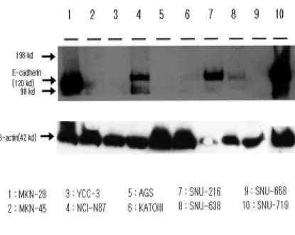
To identify the expression of the E-cadherin in Human gastric cancer cell lines, 10 human gastric cancer cell lines, MKN-28, MKN-45, NCI-N87, AGS, KATOIII, SNU-216, SNU-638, SNU-668, SNU-719, YCC-3 cells were used. To distinguish between the expression of E-cadherin and no expression of E-cadherin in 10 human gastric cancer cell lines, it was assayed by western blot analysis. It was shown that MKN-28 and NCI-N87 expressed E-cadherin at the expected molecular weight of 120 kDa38. In particular, it has been reported that the expression of E-cadherin in MKN-28 cell line was in normal form and should be used as positive control in this study38. As shown in Figure 1, MKN-28, NCI-N87, SNU-216 and SNU-719 cells highly expressed E-cadherin at 120 kDa. On the other hand, MKN-45, KATOIII, AGS, and SNU-638 cell lines slightly expressed E-cadherin while YCC-3 and SNU-668 cell lines did not expresse E-cadherin. In MKN-45, it was shown as one band at the expected molecular weight of 120 kDa and the second at 80 kDa, which is likely to represent the extracellular tryptic product of E-cadherin41. The lymph node metastatic model by gastric cancer cell line MKN-45 was
established by Matsuoka T et al.42. It can be associated with the peritoneal metastasis. In KATOIII, it has been reported that two protein bands reactive for E-cadherin were detected41. However, In this study, it was hard to find the expression of E-cadherin. In YCC-3, it was not expressed at the expected molecular weight of 120 kDa.
Fig. 1 Western blot analysis of E-cadherin in Human gastric cancer cell lines. 40 ug nuclear extracts from the cell lines were used in the assay.
2. RT-PCR analysis of E-cadherin, α-catenin, β-catenin, and
γ-catenin in human gastric cancer cell line.
γ-catenin) which consist of cadherin-catenin complex7,8,9, the RT-PCR analysis of E-cadherin, α-catenin, β-catenin, and γ-catenin in 10 human gastric cancer cell lines were performed. E-cadherin mRNA was not expressed in YCC-3 cell line. However, other human gastric cancer cell lines expressed E-cadherin mRNA in various level. Among them, even though two gastric cancer cell lines MKN-45 and KATOIII showed the expression of E-cadherin mRNA, MKN-45 has 18-bp deletion in exon 6-intron 6 boundary in E-cadherin gene39 and KATOIII cell line also has G to A base substitution of the last 3’ nucleotide of exon 7 in E-cadherin gene39. Therefore, it might have an influence on the aberrant expression of E-cadherin in protein level38. All human gastric cancer cell lines expressed α-catenin while SNU-719 cell line showed small expression . In β-catenin, only NCI-N87 cell line showed weak expression, but others showed normal expression level. In γ-catenin, NCI-N87, SNU-216, and SNU-638 cell lines showed high expression level, but others expressed it normally. Through RT-PCR analysis of E-cadherin and catenines, MKN-45, YCC-3 and KATOIII cell lines which showed absence or aberrant expression E-cadherin(figure 1) and normal expression of α-catenin, β-catenin and γ-β-catenin normally were chosen for the identification of the role of E-cadherin.
Fig 2. RT-PCR analysis of E-cadherin, α-catenin, β-catenin, and γ-catenin in Human gastric cancer cell lines. E-cadherin, α-catenin, β-catenin, and γ-catenin mRNA expressions in the 10 human gastric cancer cell lines. Total RNA samples were prepared by LiCl extraction buffer. Equal amounts of total RNA from individual samples (2 ug) were used for RT-PCR. After reverse transcription, specific primers for Human E-cadherin, α-catenin, β-catenin,
and γ-catenin were applied to the reaction system. Amplified genes were stained by ethidium bromide. M, molecular marker (100-bp ladder).
3. Expression of invasion candidate genes MMP-2, MMP-7, MMP-
9and Mt1-MMP in human gastric cancer cell lines.
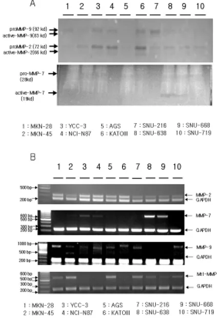
It has been reported that invasion candidate genes MMP-2, MMP-7, MMP-9 and Mt1-MMP are associated with the E-cadherin expression in several papers32,33,34,35,36,40. To identify the expression of invasion candidate genes in 10 human gastric cancer cell lines, the direct protein activity was assayed by zymogram. According to its substrate, MMP-2 and MMP-9 are measured by gelatin zymograph and MMP-7 is measured by casein zymograph. All human gastric cancer cell lines expressed MMP-2 and MMP-9 in various levels while 638 and SNU-668 showed the expression of MMP-7. However, it was difficult to identify MMP-7 expression through casein zymograph in human gastric cancer cell lines. In order to identify MMP-2, MMP-7, MMP-9 and Mt1-MMP mRNA levels in each cell lines, it was examined by RT-PCR using specific primers for them (Table 1). As shown in Figure 3 (B), mRNA expressions of MMP-2 and MMP-9 through RT-PCR by using specific primers for them have correlation with protein expression by zymogram. In MMP-7 mRNA level, YCC-3 cell line showed expression but MKN-45 and
KATOIII showed no expression. In Mt1-MMP mRNA level, MKN-45 showed high expression while YCC-3 and KATOIII showed no expression.
cancer cell lines. Conditioned media samples (20 µl/lane) were examined directly. Arrows: gelatinolytic activities at Mr 92, 83, 72, and 66 kDa, caseinolytic activities at Mr 19 kDa. (B) RT-PCR analysis of invasion candidate genes MMP-2, MMP-7, MMP-9 and Mt1-MMP in human gastric cancer cell lines. Total RNA samples were prepared by LiCl extraction buffer. Equal amounts of total RNA from individual samples (2 ug) were used for RT-PCR
4. Establishment of human E-Cadherin transfetant.
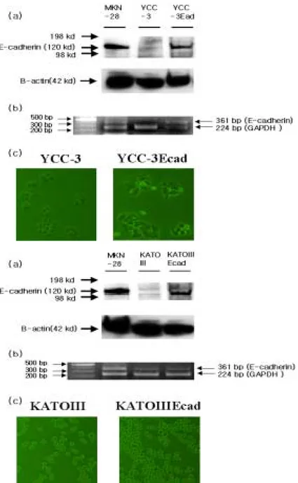
To identify the role of E-cadherin in the peritoneal metastasis in gastric cancer, it was constructed the stable cell lines YCC-3Ecad and KATOIIIEcad respectively. As shown in figure 4, the stable transfectants (YCC-3Ecad and KATOIIIEcad) were confirmed by western blot and RT-PCR. Lane 1 used positive control (MKN-28). Lane 2 is E-cadherin-negative parent cells. Lane 3 is stable transfectants. It has been reported that the effects of E-cadherin expression induce cell adhesion and decrease the cell proliferation in cancer cell line40. KATOIII cell line which is a suspension cell line and displays aberrant E-cadherin expression inclines to turn into a adhesion cell line (Figure 4B (c)) .
Fig 4. Restoration of E-cadherin expression in Human gastric cancer cell line YCC-3 and KATOIII cell lines. A.(a,b) Western blot analysis and RT-PCR analysis of E-cadherin in YCC-3 cell line (c) Morphology of YCC-3and YCC-3Ecad cell lines. B.(a,b) Western blot analysis and RT-PCR analysis of
E-cadherin in Kato3 cell line. (c) Morphology of KATOIII and KATOIIIEcad cell lines.
5. Expression of invasion candidate genes MMP-2, MMP-7 and
MMP-9 in
stable transfectants YCC-3-Ecad and KATOIIIEcad.To investigate whether E-cadherin is associated with invasion
candidate genes MMP-2, MMP-7, MMP-9, and Mt1-MMP, it was
studied by Zymogram and RT-PCR analysis. As shown in figure 5
(A), direct protein activity was measured by gelatin zymograph
which can assay MMP-2 and MMP-9 and casein zymograph which
can measure MMP-7. In MMP-2 and MMP-9 protein activity,
parent cell lines (YCC-3 and KATOIII) and stable transfectants
(YCC-3Ecad and KATOIIIEcad) showed no different expressions
them. On the other hand, in MMP-7 protein activity, between
parent cell line YCC-3 and stable transfectant YCC-3Ecad have
different expression of it. However, other parent cell line KATOIII
and stable transfectant KATOIIIEcad have no E-cadherin
expression. In order to check the expression of invasion candidate
genes in mRNA level, it was examined by RT-PCR analysis. As
shown in figure 5 (B), in MMP-7 mRNA level, 3 and
YCC-3Ecad showed different expression of MMP7 while other invasion
candidate genes MMP-2, MMP-9, and Mt1-MMP showed no
difference in expression levels between parent cell lines (YCC-3 and
KATOIII) and stable transfectants (YCC-3Ecad and
KATOIIIEcad).
Fig 5. Inhibition of MMP-7 expression but MMP-2, MMP-9 by
E-cadherin in stable transfectants YCC-3-Ecad. A.
Gelatinzymography and casein zymography of invasion candidate genes in protein level and B. RT-PCR analysis of Invasion candidate genes in mRNA level in stable transfectants YCC-3-Ecad and KATOIIIEcad.
6. Suppression of peritoneal metastasis by E-cadherin YCC3-Ecad
transfectant
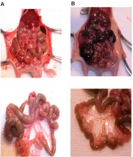
Stable transfectant (YCC-3Ecad) showed a significantly lower level of peritoneal dissemination than parent cell line(YCC-3) . The frequency and the degree of the dissemination was decreased in Human E-cadherin cDNA transfectants in comparison with E-cadherin-deficiency cell line after inoculation of the peritoneal cavity of the nude mice (n=4)(Figure 6)
Fig 6. Human gastric cancer cell line YCC-3 and YCC-3Ecad transfectant was injected into the peritoneal cavity of BALB/c-nu/nu mice. Mice were sacrificed 4 weeks later. One of YCC-3Ecad transfectants injected group(A) showed no peritoneal metastases. In contrast, one of parent cell line YCC-3 injected group (B) showed massive peritoneal metastases.
Ⅳ
. Discussion
E-cadherin is a cell adhesion molecule normally expressed at adherent junctions between epithelial cells. Recently, it has been reported that E-cadherin(Ecad) germ-line mutations were associated with the early-onset familial gastric cancer22,23,24,25. Many studies have demonstrated that the loss of cadherin expression or mutation of E-cadherin has been related with accelerated tumorigenesis and progression22,23,24,25,26. Although an exact mechanism on the peritoneal metastasis in gastric cancer regulated by E-cadherin is still unclear, some reports suggested that the decreased expression of E-cadherin attenuates the expression of protease and thus cancer cells can invade their environment44,45,46,47.
10 human gastric cancer cell lines MKN-28, MKN-45, YCC-3, NCI-N87, AGS, KATOIII, SNU-216, SNU-638, SNU-668, SNU-719 were used in order to assess the role of E-cadherin (E-CD), the cell-cell adhesion protein, in peritoneal dissemination of gastric cancer, and was screened which human gastric cancer cell lines among them did not express E-cadherin but had intact catenins(α-catenin, β-catenin, and γ-catenin) using western blot analysis (figure 1) and RT-PCR analysis(figure 2). Many studies demonstrated that MKN-45 and KATOIII gastric cancer
cell lines had aberrant mRNA expression due to E-cadherin(Ecad) mutations while other intracellular molecules such as α-catenin, β-catenin, and γ-catenin connected with E-cadherin are intact in MKN-45 and KATOIII38.
The overexpression of E-cadherin in human gastric cancer cell line YCC-3 and KATOIII altered the shape of the cell-cell interaction. Compared with parent cell lines, both YCC-3Ecad cell line and KATOIIIEcad cell line showed slightly tight cell-cell contacts and strong cell adhesion (Figure 4(A)(B)).
It was recently demonstrated that the down-regulation of E-cadherin enhances a migratory and invasive phenotype linked to invasion candidate genes such as MMP-2, MMP-7, MMP-9 and Mt1-MMP. 32,33,34,35,36,40. Many studies demonstrated that the loss of E-cadherin showed increased levels of invasion candidate genes (MMPs), motility in
vitro, and metastatic potential in vivo32,33,34,35,36,40. However, it remains to
be clarified whether the E-cadherin (CDH1) and invasion candidate genes are associated with the peritoneal metastasis in gastric cancer. Miyaki et al. mentioned that a colon cancer cell line which was transfected with mouse E-cadherin cDNA demonstrated decreased invasiveness with concomitant reduction of 62 kDa gelatinase
expression45. Luo et al. reported that the rat prostate cancer cell lines transfected with mouse E-cadherin cDNA were decreased in MMP-2 activity, thereby being less invasive than their parental cells47.
The association of E-cadherin with invasion candidate genes in parent cell lines3 and KATOIII) and stable transfectants (YCC-3Ecad and KATOIIIEcad) were examined by zymograph analysis and RT-PCR analysis(Figure 3 (A),(B)). The amount of MMP-2, MMP-9 and Mt1-MMP expression and their activities were almost identical among parent cell lines 3 and KATOIII) and stable transfectants (YCC-3Ecad and KATOIIIEcad) in RT-PCR and Zymogram. These results demonstrated that MMPs, at least MMP-2, MMP-9 and Mt1-MMP, did not affect the invasive phenotype of these cell lines. However, in MMP-7 level, there is a difference between parent cell line (YCC-3) and stable transfectant (YCC-3Ecad). The expression of MMP-7 might be inhibited by E-cadherin in stable transfectant (YCC-3Ecad). Furthermore, it was recently reported that MMP-7 may have a large role in the formation of peritoneal dissemination in gastric cancer37. These data strongly suggested that the loss of E-cadherin may be involved in the formation of the peritoneal dissemination in gastric cancer.
overexpression in YCC-3 gastric cancer cell line demonstrate the reduced potential of peritoneal metastasis. Some mechanisms by which E-cadherin is a metastasis-suppressor gene have been suggested, but further study should be continued.
Ⅴ
. Conclusion
In the present study, it is investigated whether invasion candidate genes are associated with E-cadherin expression for the peritoneal metastasis in gastric cancer.
1. The effect of E-cadherin expression in KATOIIIEcad cell line displays the induction of early-time cell adhesion.
2. Human gastric cancer cell line KATOIII injected into the peritoneal cavity of BALB/c-nu/nu mice didn’t induce any cancers in nude mice.
3.
Invasion candidate gene MMP-7 expression was decreased by
E-cadherin using Zymograph and RT-PCR in stable
transfectant YCC-3Ecad cell line.
4. E-cadherin overexpression in YCC-3 gastric cancer cell line demonstrates the reduced potential of peritoneal metastasis.
5.
Some mechanisms by which E-cadherin is one of the metastasis-suppressor gene, have been suggested, but further study should be continued.References
1. Takeichi M. Cadherin cell adhesion receptors as a morphogenetic regulator.
Science. 1991 Mar 22;251(5000):1451-5.
2. M. Takeichi, Morphogenetic roles of classic cadherins. Curr. Opin. Cell
Biol. 7 (1995), pp. 619–627.
3. O. Huber, R. Korn, J. McLaughlin, M. Ohsugi, B.G. Herrmann and R. Kemler, Nuclear localization of beta-catenin by interaction with transcription factor LEF-1. Mech. Dev. 59 (1996), pp. 3–10.
4. T. Yagi and M. Takeichi, Cadherin superfamily genes: functions, genomic organization, and neurologic diversity. Genes Dev. 14 (2000), pp. 1169–1180.
5. D. Riethmacher, V. Brinkmann and C. Birchmeier, A targeted mutation in the mouse E-cadherin gene results in defective preimplantation development.
Proc. Natl. Acad. Sci. USA 92 (1995), pp. 855–859.
6. L. Larue, M. Ohsugi, J. Hirchenhain and R. Kemler, E-cadherin null mutant embryos fail to form a trophectoderm epithelium. Proc. Natl. Acad. Sci. USA 91 (1994), pp. 8263–8267.
7. I.S. Nathke, L. Hinck, J.R. Swedlow, J. Papkoff and W.J. Nelson, Defining interactions and distributions of cadherin and catenin complexes in polarized epithelial cells. J. Cell Biol. 125 (1994), pp. 1341–1352.
8. M. Ozawa, H. Baribault and R. Kemler, The cytoplasmic domain of the cell adhesion molecule uvomorulin associates with three independent proteins structurally related in different species. EMBO J. 8 (1989), pp. 1711–1717.
9. L.L. Witcher, R. Collins, S. Puttagunta, S.E. Mechanic, M. Munson, B. Gumbiner and P. Cowin, Desmosomal cadherin binding domains of plakoglobin. J. Biol. Chem. 271 (1996), pp. 10904–10909.
10. A.S. Yap, C.M. Niessen and B.M. Gumbiner, The juxtamembrane region of the cadherin cytoplasmic tail supports lateral clustering, adhesive strengthening, and interaction with p120ctn. J. Cell Biol. 141 (1998), pp. 779– 789.
11. M. Ozawa, Identification of the region of alpha-catenin that plays an essential role in cadherin-mediated cell adhesion. J. Biol. Chem. 273 (1998), pp. 29524–29529.
12. P.Z. Anastasiadis and A.B. Reynolds, The p120 catenin family: complex roles in adhesion, signaling and cancer. J. Cell Sci. 113 (2000), pp. 1319– 1334
13. N.K. Noren, B.P. Liu, K. Burridge and B. Kreft, p120 catenin regulates the actin cytoskeleton via Rho family GTPases. J. Cell Biol. 150 (2000), pp. 567–580.
14. W. Birchmeier and J. Behrens, Cadherin expression in carcinomas: role in the formation of cell junctions and the prevention of invasiveness. Biochim.
15. E. Batlle, E. Sancho, C. Franci, D. Dominguez, M. Monfar, J. Baulida, A. Garcia and De Herreros, The transcription factor snail is a repressor of E-cadherin gene expression in epithelial tumour cells. Nat. Cell Biol. 2 (2000), pp. 84–89.
16. A. Cano, M.A. Perez-Moreno, I. Rodrigo, A. Locascio, M.J. Blanco, M.G. del Barrio, F. Portillo and M.A. Nieto, The transcription factor snail controls epithelial-mesenchymal transitions by repressing E-cadherin expression. Nat.
Cell Biol. 2 (2000), pp. 76–83.
17. I. Poser, D. Dominguez, A.G. de Herreros, A. Varnai, R. Buettner and A.K. Bosserhoff, Loss of E-cadherin expression in melanoma cells involves up-regulation of the transcriptional repressor Snail. J. Biol. Chem. 276 (2001), pp. 24661–24666.
18. K. Yokoyama, N. Kamata, E. Hayashi, T. Hoteiya, N. Ueda, R. Fujimoto and M. Nagayama, Reverse correlation of E-cadherin and snail expression in oral squamous cell carcinoma cells in vitro. Oral. Oncol. 37 (2001), pp. 65–71.
19. Schwartz GK, Wang H, Lampen N, Altorki N, Kelsen D, Albino AP. Defining the invasive phenotype of proximal gastric cancer cells. Cancer. 1994 Jan 1;73(1):22-7.
20. Cheng L, Nagabhushan M, Pretlow TP, Amini SB, Pretlow TG. Expression of E-cadherin in primary and metastatic prostate cancer. Am J
Pathol. 1996 May;148(5):1375-80.
21. Bringuier PP, Umbas R, Schaafsma HE, Karthaus HFM, Debruyne FMJ, Schalken JA. Decreased E-cadherin immunoreactivity correlates with poor
survival in patients with bladder tumors. Cancer Res. 1993 Jul 15;53(14):3241-5.
22. Gayther SA, Gorringe KL, Ramus SJ, Huntsman D, Roviello F, Grehan N, Machado JC, Pinto E, Seruca R, Halling K, MacLeod P, Powell SM, Jackson CE, Ponder BAJ, Caldas C. 1998. Identification of germ-line E-cadherin mutations in gastric cancer families of European origin. Cancer Res 58: 4086-4089.
23. Guilford P, Hopkins J, Harraway J, McLeod M, McLeod N, Harawira P, Taite H, Scoular R, Miller A, Reeve AE. 1998. E-cadherin germline mutations in familial gastric cancer. Nature 392: 402-405.
24. Guilford PJ, Hopkins JB, Grady WM, Markowitz SD, Willis J, Lynch H, Rajput A, Wiesner GL, Lindor NM, Burgart LJ, Toro TT, Lee D, Limacher JM, Shaw DW, Findlay MP, Reeve AE. 1999. E-cadherin germline mutations define an inherited cancer syndrome dominated by diffuse gastric cancer.
Hum Mutat 14: 249-255.
25. Keller G, Vogelsang H, Becker I, Hutter J, Ott K, Candidus S, Grundei T, Becker K-F, Mueller J, Siewert JR, Hofler H. 1999. Diffuse type gastric and lobular breast carcinoma in a familial gastric patient with an E-cadherin germline mutation. Am J Pathol 155: 337-342.
26. Richards FM, McKee SA, Rajpar MH, Cole TR, Evans DG, Jankowski JA, McKeown C, Sanders DS, Maher ER. 1999. Germline E-cadherin gene (CDH1) mutations predispose to familial gastric cancer and colorectal cancer.
27. Liotta , L. A., S. ABE, P. G. Robey, and G. R. Martin. Preferential digestion of basement membrane collagen by an enzyme derived from a metastatic murine tumor. Proc Natl Acad Sci U S A. 1979 May;76(5):2268-72 28. Albelda S, Buck C. Integrins and other cell adhesion molecules. FASEB J. 1990 Aug;4(11):2868-80. Review.
29. Woessner, J. F. Jr. Matrix metalloproteinases and their inhibitors in connective tissue remodeling. 1991 FASEB J. 5:2145-2154.
30. Liotta LA, Kohn E. Cancer invasion and metastases. JAMA. 1990 Feb 23;263(8):1123-6. Review
31. Lyons JG, Birkedal-Hansen B, Moore WG, O'Grady RL, Birkedal-Hansen H. Characteristics of a 95-kDa matrix metalloproteinase produced by mammary carcinoma cells. Biochemistry. 1991 Feb 12;30(6):1449-56.
32. Wroblewski LE, Pritchard DM, Carter S, Varro A. Gastrin-stimulated gastric epithelial cell invasion: the role and mechanism of increased matrix metalloproteinase 9 expression. Biochem J. 2002 Aug 1;365(Pt 3):873-9.
33. Torii A, Kodera Y, Ito M, Shimizu Y, Hirai T, Yasui K, Morimoto T, Yamamura Y, Kato T, Hayakawa T, Fujimoto N, Kito T. Matrix metalloproteinase 9 in mucosally invasive gastric cancer. Gastric Cancer. 1998 Mar;1(2):142-145.
34. Monig SP, Baldus SE, Hennecken JK, Spiecker DB, Grass G, Schneider PM, Thiele J, Dienes HP, Holscher AH. Expression of MMP-2 is associated with progression and lymph node metastasis of gastric carcinoma.
35. Mizutani K, Kofuji K, Shirouzu K. The significance of MMP-1 and MMP-2 in peritoneal disseminated metastasis of gastric cancer. Surg Today. 2000;30(7):614-21.
36. Parsons SL, Watson SA, Collins HM, Griffin NR, Clarke PA, Steele RJ. Gelatinase (MMP-2 and -9) expression in gastrointestinal malignancy. Br J
Cancer. 1998 Dec;78(11):1495-502.
37. Yonemura Y, Endou Y, Fujita H, Fushida S, Bandou E, Taniguchi K, Miwa K, Sugiyama K, Sasaki T. Role of MMP-7 in the formation of peritoneal dissemination in gastric cancer. Gastric Cancer. 2000 Sep 29;3(2):63-70.
38. AU Jawhari,M Noda, MJG Farthing and M Pignatelli. Abnormal expression and function of the E-cadherin-catenin complex in gastric carcinoma cell lines. British Journal of Cancer. 1999. 80(3/4),322-330
39. Yokozaki H. Molecular characteristics of eight gastric cancer cell lines established in Japan. Pathol Int. 2000 Oct;50(10):767-77. Review.
40. Kohya N, Kitajima Y, Jiao W, Miyazaki K. Effects of E-cadherin transfection on gene expression of a gallbladder carcinoma cell line: repression of MTS1/S100A4 gene expression. Int J Cancer. 2003 Mar 10;104(1):44-53.
41. Frixen UH, Behrens J, Sachs M, Eberle G, Voss B, Warda A et al. E-cadherin-mediated cell-cell adhesion prevents invasiveness of human carcinoma cells. J Cell Biol. 1991 Apr;113(1):173-85.
42. Matsuoka T, Yashiro M, Sawada T, Ishikawa T, Ohira M, Chung KH. Inhibition of invasion and lymph node metastasis of gastrointestinal cancer cells by R-94138, a matrix metalloproteinase inhibitor. Anticancer Res. 2000 Nov-Dec;20(6B):4331-8.
43. Shun CT, Wu MS, Lin JT, Wang HP, Houng RL, Lee WJ et al. An
immunohistochemical study of E-cadherin expression with correlations
to clinicopathological features in gastric cancer,
Hepatogastroenterology 45 (1998) 944-999.
44. Miyaki M, Tanaka K, Kikuchi YR,. Muraoka M, Konishi M et al.
Increased cell-substratum adhesion, and decreased gelatinase secretion
and cell growth, induced by E-cadherin transfection of human colon
carcinoma cells, Oncogene 11 (1995) 2547-2552.
45. Frixen UH, Nagamine Y, Stimulation of urokinase-type
plasminogen activator expression by blockage of Ecadherin-dependent
cell-cell adhesion, Cancer Res. 53 (1993) 3618-3623.
46. Llorens A, Rodrigo I, Lopez Barcons L, Gonzalez Garrigues M,
Lozano E et al. Down-regulation of E-cadherin in mouse skin
carcinoma cells enhances a migratory and invasive phenotype linked to
matrix metalloproteinase-9 gelatinase expression, Lab. Invest. 78
(1998) 1131-1142.
47. Luo J, Lubaroff DM, Hendrix MJC, Suppression of prostate cancer
invasive potential and matrix metalloproteinase activity by E-cadherin
transfection, Cancer Res. 59 (1999) 3552-3556.
국문요약
위암의 복막전이에서 E-cadherin 과
금속단백분해효소(MMP) 의 역할
(지도교수 노성훈)
연세대학교 대학원 의과학과
김
청 섭
Ca2+ 의존성 세포간 유착을 조절하는 물질인 cadherin 은 120 kDa 의 당단백질로 E-cadherin, P-cadherin, N-cadherin 등이 속한다. 이중 E-cadherin 은 주로 상피세포에 발견되는 것으로 세포의 유착성 접합부(adherens junction)에 위치한다. 또한 E-cahderin 은 세포외 부분, 막 부분, 세포질 부분의 3 가지 구조로 나뉘어지며 세포질 부분은 cadherin 결합 단백질인 α-catenin, β-catenin, γ-catenin 등과 결합하여 작용한다. E-cadherin 의원인이라고 보고되어졌다. E-cadherin 발현의 감소 또는 상실이 암의 침윤(invasion) 및 전이(metastasis)와 관련되어 있지만, 어떻게 위암의 복막전이에 E-cadherin 이 관여하고 있는지는 확실치 않다. 위암의 복막전이에 관여하는 E-cadherin 의 역할을 규명하기 위해 E-cadherin 의 발현이 안되거나 비정상적인 위암세포주에 E-cadherin 을 영구적으로 과발현 시키는
세포주들을 만들었으며(YCC-3Ecad), KATOIIIEcad) western blot 과 RT-PCR 을 이용하여 확인하였다. E-cadherin 의 발현이 안되는 위암세포주(YCC-3) 와 E-cadherin 을 과발현하는 위암세포주(YCC-3Ecad) 를 이용하여 In vitro에서 세포성장 측정 및 암전이에 관련된 여러가지 유전자의 발현양상과 과 In vivo 에서 복막전이의 억제를 관찰함으로써 위암의 복막전이에서 E-cadherin 의 역할을 규명할 수 있을 것이다.


