Background: Endoscopy has replaced open surgery, especially in spinal surgery. Among them, image-guided epiduroscopy allows pain generators to be identified, including epidural adhesion, fibrotic tissues, root compression, and spinal stenosis. However, the heavy lead apron worn by pain physicians to avoid exposure to radiation can induce occupational hazards, such as orthopedic complications and radiation-induced cancer. Hence, we developed a robotic system to address these problems.
Objective: The aim of the study was to evaluate the feasibility of a robot-controlled epiduroscopic system.
Study Design: In vivo animal experiment. Setting: University in Republic of Korea.
Methods: The robot-controlled epiduroscopic system was developed using the open architecture robot system (The Raven Surgical Robotic System, CITRIS, Berkley, CA, USA). The robotic system consists of a lab-made epiduroscope, steering section, robotic arm, and manipulator. For the in vivo study, 2 Yorkshire pigs were used to simulate an epiduroscopic procedure with the robotic system.
Results: The insertion and steering of the catheter was performed safely, and epiduroscopic visualization was obtained without side effects. There were no device-related complications. Radiation exposure for the primary operator was 80% lower than the levels found during conventional epiduroscopic procedures. All live pigs showed normal behavior without any signs of pain. The mean time to reach the target region was less than 8 minutes.
Limitations: The epiduroscopic procedure was performed on pigs and not on humans. The dimensions of the spinal canal of pigs cannot compare to those of humans.
Conclusions: We demonstrated the feasibility of the robot-assisted epiduroscopic system. Key Words: Epiduroscopy, robotic system, spine, pig, animal model
Pain Physician 2018: 21:E565-E571
Animal Study
Feasibility of Percutaneous Robot-Assisted
Epiduroscopic System
From: 1Department of Neurosurgery, Yonsei University College of Medicine, Republic of Korea; 2Center for Medical Robotics, Korea Institute of Science and Technology, Korea; 3Department of Neurosurgery, Universitas Padjadjaran, College of Medicine, Hasan Sadikin Hospital, Indonesia Address Correspondence:
Dong Ah Shin, MD, PhD Yonsei University College of Medicine,
Neurosurgery 50, Yonsei-ro, Seodaemun-gu Seoul, 120-75, Republic of Korea E-mail: shindongah@me.com Disclaimer: This research was financially supported by the “Global Collaborative R&D Program” (Project No. N0000890) through the Ministry of Trade, Industry & Energy (MOTIE) and Korea Institute for Advancement of Technology (KIAT). Conflict of interest: Each author certifies that he or she, or a member of his or her immediate family, has no commercial association (i.e., consultancies, stock ownership, equity interest, patent/licensing arrangements, etc.) that might pose a conflict of interest in connection with the submitted manuscript. Manuscript received: 10-28-2017 Revised manuscript received: 01-05-2018 Accepted for publication: 04-17-2018 Free full manuscript: www.painphysicianjournal.com
Dong Ah Shin, MD, PhD1, Chunwoo Kim, PhD2, Farid Yudoyono, PhD1,3, Yeomin Yun, MS1, Yoon Ha, PhD1, and Sungchul Kang, PhD2
nerve and dural sac (2,7,8). Through the working channel of the epiduroscope, medications can be delivered, and a laser apparatus can be introduced to remove portions of pathological tissues (2,7,8).
During epiduroscopy, catheter insertion and steer-ing are performed under the guidance of fluoroscopic imaging (9).
E
piduroscopy has long been performed to find and treat pain generators in the epidural space (1-6) It has been an effective, non-surgical method for the treatment of various spinal diseases (1-6). With epidurosopy, one can visualize the epidural space, which is surrounded by the vertebral bone and the intervertebral disc, and which contains the spinalPain Physician: September/October 2018: 21:E565-E571
tion on the sacral hiatus. After insertion of the tip of the catheter into the sacral hiatus, the third axis was used to control the progression of the catheter. The direc-tion of the catheter was changed in the epidural space by using the steering section. While the catheter was navigated to the target area, the position of the cath-eter tip was frequently checked using a fluoroscope. The catheter was precisely entered into the desired site using the robotic system. In the future, we will build the system as shown in Fig. 3.
Animal Experiment
The animal study protocol was approved by the Institutional Animal Care and Use Committee of Yon-sei University College of Medicine. Two Yorkshire pigs, each 4 months old and weighing 40 kg, were used. The pigs were anesthetized using endotracheal intu-bation. Then, the pigs were placed on the operating table in a prone position. The robotic system was at-tached to the operating table (Fig. 1). The skin around the sacral hiatus was prepared using betadine in an aseptic manner so that the catheter was located at the entrance of the hole. The pigs were sedated using an intramuscular injection of ketamine (20 mg/kg). The pigs were then placed on a radiolucent table, intu-bated, and ventilated on a respirator administering a mixture of oxygen (10%) and air (90%). Anesthesia was maintained by alpha chloralose (100 mg/kg bolus and 30 mg/kg/hour).
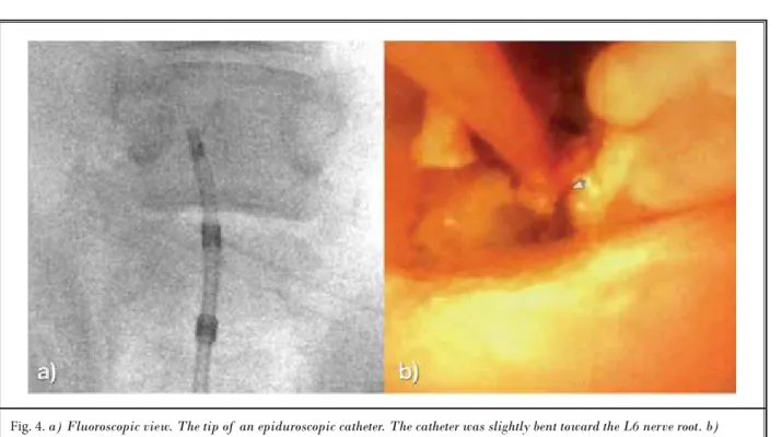
After topical anesthesia, the skin just above the sacral hiatus was incised and subcutaneous dissection was made to expose the sacral hiatus. After the sacral hiatus was exposed, the sacrococcygeal ligament was removed, and the rongeur was used to remove the surrounding bone and open the sacral hiatus more widely. An epidural, steerable, fiber-optic catheter system was introduced into the caudal region through the sacral hiatus. The tip of the catheter was advanced toward the L4-5 level, and the direction of the cath-eter was changed in the epidural space using the ro-bot. Then, the position of the catheter was confirmed through fluoroscopic and epiduroscopic images (Fig. 4). After the experiment, the pigs were allowed to re-cover after suturing, and after 2 weeks of observation, the pigs showed no signs of nerve damage or paralysis due to catheter insertion. The animals were returned to their home pen, and then the behavior of each indi-vidual pig was recorded for 1 month. Observations on appearance of the incision, behavioral characteristics, eating, drinking, urination, defecation, temperature, Adverse events, such as cancer and cataracts, are
commonly reported to be associated with a physi-cian’s exposure to radiation that occurs while steering the catheter during x-ray imaging (10-12). In addition, physicians wear lead aprons and thyroid shields during the procedure. These heavy aprons may cause arthritis and intervertebral disc diseases in physicians (13). Ad-ditionally, these tough working conditions may reduce the precision of the procedure.
Therefore, we developed a robot-controlled epi-duroscopic system to address the occupational hazards related to fluoroscope-guided spinal interventions, and to enhance the degree of precision of these procedures. Recently, various robotic devices have been intro-duced to enhance the performance of spinal surgeries, as well as to provide protection from the occupational hazards that are associated with the procedures (14-17). However, to the best of our knowledge, there has been no report on the use of a robotic system for epiduros-copy. The purpose of this study was to evaluate the fea-sibility of such a system through animal experiments.
M
ethodRobotic System
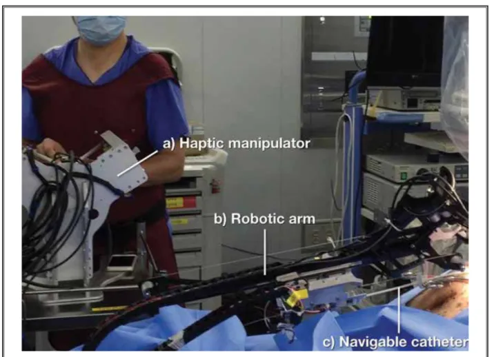
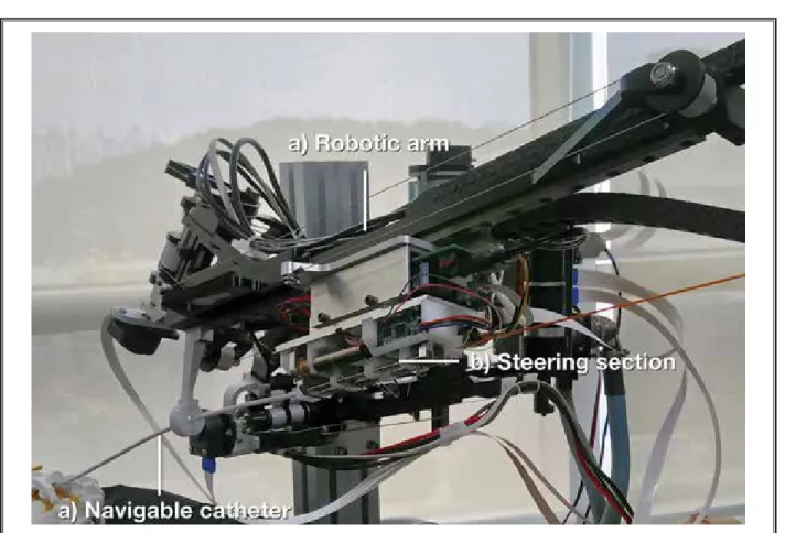
The robotic system consists of a navigable cath-eter with a built-in camera, a robotic arm moving the catheter, and a manipulator with haptic control (Fig. 1). The outer diameter of the catheter was 3 mm. The diameter of the working channel was 1.2 mm. The catheter was made of medical polyurethane. The distal 1 cm part of the catheter was made to be softer than the remaining part so as to be bent easily. The steer-ing section was located at the end of the catheter and consisted of 2 wire-driving motors. The steering sec-tion on the robotic arm is shown in Fig. 2. Inside the catheter, there are 4 metal wires for steering the distal part of the catheter. The catheter was flexed vertically or horizontally by using 2 DC motors which pulled and pushed on a pair of metal wires. The robotic arm, which controlled the steering section, was fixed on the side rail of the operating table. The robotic arm was built with a research surgical robot platform developed by the University of Washington (Raven Surgical Robotic System, CITRIS, Berkley, CA, USA). The robotic control program was based on an open-source robot-operating system (Robot Operating System, Open Source Robotics Foundation, CA, USA).
The robotic arm had a total of 3 degrees of free-dom. The first 2 axes moved the remote center of
rota-Fig. 1. Animal experiment using the robotic epiduroscope system. A physician is shown operating a) the haptic manipulator which
moves b) the mounted robotic arm equipped with c) the navigable epiduroscopic catheter.
pulse and respiration were carefully analyzed. The 2 primary endpoints were technical success and clinical success.
Technical success was defined as successful intraspi-nal advancement and target achievement by the robot-ic system, without conversion to manual operation. The duration of radiation exposure was measured. Clinical success was defined as the absence of major tions within 1 week of the procedure. Major complica-tions were defined as pain behavior, gait disturbance, voiding difficulties, and wound problems.
R
esultsBefore the animal experiment too place, a physician was trained to manipulate the robotic epiduroscope using the haptic control. After each training session, feedback was obtained from the physician in order to improve the shape of the tip of the epiduroscope and the image quality of the camera. A protocol for robotic
epiduroscopy was also developed. Robot-controlled epiduroscopy was performed twice on each pig.
All robotic epiduroscopic procedures were per-formed via trans sacral access, with 15-gauge Tuohy needles. A mean time of 17.5 ± 0.7 minutes was taken from catheter insertion to target achievement. X-ray exposure occurred for a mean of 13.5 ± 1.6 minutes during the procedure.
In all trials, advancement of the epiduroscope pro-ceeded uneventfully, without any nerve injury or dural perforation. We confirmed that the catheter could be precisely entered into the desired site. In all procedures, the tip of the catheter was delivered to the target, and the intervertebral discs were successfully visualized. No trial was converted to manual operation. There were no surgi-cal complications related to the use of the robotic system. Technical and clinical outcomes are detailed in Table 1. The primary endpoints were achieved in 100% of trials.
Pain Physician: September/October 2018: 21:E565-E571
Fig. 2. The robotic arm of the robot-assisted epiduroscopy system. a) The epiduroscope catheter was connected to b) the steering
section which was installed on c) the robotic arm.
d
iscussionThis study demonstrated the feasibility of robot-assisted epiduroscopy. In the 2 pigs treated with 4 tri-als, there were no treatment-related complications or adverse events. The primary endpoints were achieved in 100% of trials. There was no case of conversion to traditional manual operation. This study showed that the robotic system is safe and effective in the advance-ment and steering of the epiduroscope into the epi-dural space. Compared to conventional procedures, less time was required in x-ray to visualize the position of the epiduroscope. The robotic procedure can be used to visualize pain generators and to treat epidural patholo-gies, such as direct decompression, thermal annulo-plasty, epidural adhesiolysis, and precise drug delivery.
While new implants and surgical procedures are of-ten tested on human cadavers, the availability of fresh frozen human cadaver is very limited. The pig spine has been regarded as the most representative model for the human spine. Busscher et al (18) reported that the
pig spine is quite similar to the human spine in pedicle width and height, spinal canal width and depth, and transverse process length in all spinal regions. They concluded that the pig spine could be used in stud-ies testing new implants and surgical techniques (18). Pleticha et al (19) reported that the pig epidural space, demarcated by the ligamentum flavum dorsally and the dura mater ventrally, was filled with abundant adipose tissue in the pig. In the present study, we confirmed that the pig spine is appropriate for examining human epiduroscopy in terms of comparable canal size and redundant epidural space.
As the current practice of pain physician evolves into performing a higher volume of procedures, pain physicians have raised safety concerns, advocating for reducing radiation exposure to both patients and op-erators and for making interventions more ergonomic (10-12,20). A recent observational report raised con-cerns regarding a possible association between career-long exposure to radiation in interventional cardiolo-gists with the development of left-sided brain tumors
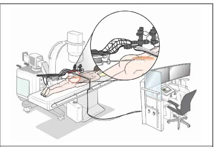
Fig. 3. A schematic diagram of the robot-assisted epiduroscopy system. A patient is lying on a surgical table onto which the robotic
system is installed. The operation is performed under fluoroscopic guidance. A physician is sitting on a chair at a radiation-blocked console. He is operating the epiduroscopic system remotely with a manipulator. However, this is a schematic illustration. In this study, the console is not developed yet.
(21). Robotic epiduroscopy has the potential to address this issue (Fig. 3). In the current study, we measured the duration of radiation exposure. During the procedure, epiduroscopy requires fluoroscopy to confirm the site of the scope tip. Komiya et al (22) reported that the average fluoroscopic time of epiduroscopy was 9.5 min-utes. However, the procedure time of vertebroplasty, a procedure similar to epiduroscopy, was reported to be 27.7 minutes (23). In our series, conventional epiduros-copy took about 21.3 minutes (not published yet). We found a significant decrease in radiation exposure dur-ing robotic epiduroscopy, with a reduction of almost 37% compared to a personal investigation regarding conventional epiduroscopy. The difference is possibly due to differences in skills or methods. If the robotic system can shorten the procedure time with increasing accuracy in the future, the patient’s exposure to radia-tion will also be significantly decreased.
No matter how skilled a physician is, if the robotic system helps, epiduroscopy will be more precise. The ability to remotely control precision instruments will significantly improve the outcome of epiduroscopy. Compared with other minimally invasive surgery, robot-assisted surgery allowsthe surgeon to manipulate the surgical tool precisely and to see the surgical site more clearly (16,24,25). In addition, physicians no longer need to stand throughout the epiduroscopy and do not get tired as quickly. Hand tremors can also be filtered out by the robot’s software (16,24,25).
Another major issue associated with epiduroscopy is occupational hazard, such as arthritis or a slipped disc due to long periods of added weight-bearing. Long hours donning a heavy lead apron while standing may adversely affect interventional physicians, result-ing in reduced performance and loss of productivity (12,26,27). Although no measurement of operator
com-Pain Physician: September/October 2018: 21:E565-E571
Table 1. The details and outcomes of the robot-controlled epiduroscopic system.
Trials Access Target Procedural Time (min) Approach Success Fluoroscopy time (min) Complications
1 Transsacral L4-5 18.3 Yes 15.1 None
2 Transsacral L4-5 17.2 Yes 13.8 None
3 Transsacral L5-6 17.9 Yes 14.3 None
4 Transsacral L5-6 16.5 Yes 10.9 None
Average 17.5 ± 0.7 13.5 ± 1.6
Fig. 4. a) Fluoroscopic view. The tip of an epiduroscopic catheter. The catheter was slightly bent toward the L6 nerve root. b)
Epiduroscopic view. The posterior annulus is seen ventrally. A laser fiber is located in the left upper corner.
fort was used in this study, the benefits of the system are intuitive. Sitting at the shielded interventional cockpit without the need for a heavy lead apron will minimize back discomfort, allowing the operator to focus on the procedure without being distracted by the physical strain. This will make physicians more ergonomically comfortable and thus, enhance their capabilities. In addition, one advantage of using the robot-assisted method is that the physician does not have to be pres-ent, but can be anywhere in the world, leading to the possibility of remote surgery.
This is the first report demonstrating the feasibility of robot-assisted epiduroscopy. The limitation of this study was that the robot-assisted epiduroscopic proce-dures were performed on pigs, and not in humans. Pig
experiments cannot fully account for use in humans, which have a bigger anatomical profile and different pain behavior, which may cause different results. While the current study only investigated the successful place-ment of the epiduroscopic catheter to a target lesion, the ability of epiduroscopy to treat spinal diseases may further increase the usefulness of the robot-assisted epiduroscopy system. This will be addressed in a future study.
c
onclusionThe robot-assisted epiduroscopy system was effec-tive in reaching target lesions in a swine model. Further research will address the safety and effectiveness of this procedure in a larger sample of patients.
R
efeRences1. Manchikanti L, Boswell MV, Rivera JJ, Brandon DE, , Pampati V, Damron KS, McManus CD, Wilson SR. A random-ized, controlled trial of spinal endoscop-ic adhesiolysis in chronendoscop-ic refractory low back and lower extremity pain. BMC
An-esthesiol 2005; 5:10.
2. Hayek SM, Helm S, Benyamin RM, Singh V, Bryce DA, Smith HS. Effective-ness of spinal endoscopic adhesiolysis in post lumbar surgery syndrome: A systematic review. Pain Physician 2009; 12:419-435.
3. Epstein J, Adler R. Laser-assisted per-cutaneous endoscopic neurolysis. Pain
Physician 2000; 3:43-45.
4. Takeshima N, Miyakawa H, Okuda K, Hattori S, Hagiwara S, Takatani J, No-guchi T. Evaluation of the therapeutic results of epiduroscopic adhesiolysis for failed back surgery syndrome. Br J
An-aesth 2009; 102:400-407.
5. Lee SH, Lee SH, Lim KT. Trans-sacral epiduroscopic laser decompression for symptomatic lumbar disc herniation: A preliminary case series. Photomedicine
and Laser Surgery 2016; 34:121-129.
6. Manchikanti L, Pampati V, Bakhit CE, Pakanati RR. Non-endoscopic and en-doscopic adhesiolysis in post-lumbar laminectomy syndrome: A one-year out-come study and cost effectiveness analy-sis. Pain Physician 1999; 2:52-58. 7. Richter EO, Abramova MV, Cantu F,
De-Andres J, Lierz P, Manchiaro P, Van Buy-ten J-P, Kim J-D, Jang J-H, Jung G-H, Kim J-Y, Jang S-J, Salgado H, Salgado P, Alo KM. {AU: need name of article}
Euro-pean Journal of Pain Supplements 2011;
5:401-407.
8. De Andrés J, Reina MA, Machés F, De Sola RG. Epidural fat: Considerations for minimally invasive spinal injection and surgical therapies. JNR 2011; 1:45-53. 9. Moon BJ, Lee HY, Kim KN, Yi S, Ha
Y, Yoon DH, Shin DA. Experimental evaluation of percutaneous lumbar la-ser disc decompression using a 1414 nm Nd:YAG laser. Pain Physician 2015; 18:E1091-E1099.
10. Hadelsberg UP, Harel R. Hazards of
ionizing radiation and its impact on spine surgery. World Neurosurgery 2016; 92:353-359.
11. Ahn Y, Kim C-H, Lee JH, Lee SH, Kim J-S. Radiation exposure to the surgeon during percutaneous endoscopic lumbar discectomy: A prospective study. Spine 2013; 38:617-625.
12. Plastaras C, Appasamy M, Sayeed Y, McLaughlin C, Charles J, Joshi A, Ma-cron D, Pukenas B. Fluoroscopy proce-dure and equipment changes to reduce staff radiation exposure in the interven-tional spine suite. Pain Physician 2013; 16:E731-E738.
13. Rayner S. Radiation hazards in hospital: A cultural analysis of occupational risk perception. RAIN 1984; 60:10.
14. Chenin L, Peltier J, Lefranc M. Mini-mally invasive transforaminal lumbar interbody fusion with the ROSATM Spine robot and intraoperative flat-panel CT guidance. Acta Neurochir (Wien) 2016; 158:1125-1128.
15. Yang MS, Hong JW, Lee SK, Lee EJ, Kim SH. Clinical management and outcome of 36 invasive prolactinomas treated with dopamine agonist. J Neurooncol 2011; 104:195-204.
16. Yang MS, Kim KN, Yoon DH, Pennant W, Ha Y. Robot-assisted resection of para-spinal schwannoma. J Korean Med Sci 2011; 26:150-153.
17. Stüer C, Ringel F, Stoffel M, Reinke A, Behr M, Meyer B. Robotic technology in spine surgery: Current applications and future developments. Acta Neurochir
Suppl 2011; 109:241-245.
18. Busscher I, Ploegmakers JJW, Verkerke GJ, Veldhuizen AG. Comparative ana-tomical dimensions of the complete hu-man and porcine spine. Eur Spine J 2010; 19:1104-1114.
19. Pleticha J, Maus TP, Jeng-Singh C, Marsh MP, Al-Saiegh F, Christner JA, Lee KH, Beutler AS. Pig lumbar spine anatomy and imaging-guided lateral lumbar puncture: A new large animal model for intrathecal drug delivery. J Neurosci
Method 2013; 216:10-15.
20. Weisz G, Metzger DC, Caputo RP, Del-gado JA, Marshall JJ, Vetrovec GW, Re-isman M, Waksman R, Granada JF, No-vack V, Moses JW, Carrozza JP. Safety and feasibility of robotic percutane-ous coronary intervention. JAC 2013; 61:1596-1600.
21. Roguin A, Goldstein J, Bar O. Brain tu-mours among interventional cardiolo-gists: A cause for alarm? Report of four new cases from two cities and a review of the literature. Euro Intervention 2012; 7:1081-1086.
22. Komiya K, Igarashi T, Suzuki H, Hira-bayashi Y, Waechter J, Seo N. In vitro study of patient’s and physician’s radia-tion exposure in the performance of epi-duroscopy. Reg Anesth Pain Med 2008; 33:98-101.
23. Fitousi NT, Efstathopoulos EP, Delis HB, Kottou S, Kelekis AD, Panayiotakis GS. Patient and staff dosimetry in ver-tebroplasty. Spine 2006; 31:E884-E889. Discussioin E890.
24. Yang MS, Yoon DH, Kim KN, Kim H, Yang JW, Yi S, Lee JYK, Jung WJ, Rha KH, Ha Y. Robot-assisted anterior lum-bar interbody fusion in a Swine model in vivo test of the da vinci surgical-as-sisted spinal surgery system. Spine 2011; 36:E139-E143.
25. Kim MJ, Ha Y, Yang MS, Yoon DH, Kim KN, Kim H, Yang JW, Lee JYK, Yi S, Jung WJ, Rha KH. Robot-assisted anterior lumbar interbody fusion (ALIF) using retroperitoneal approach. Acta Neurochir
(Wien) 2009; 152:675-679.
26. Botwin KP, Fuoco GS, Torres FM, Gru-ber RD, Bouchlas CC, Castellanos R, Rao S. Radiation exposure to the spi-nal interventiospi-nalist performing lum-bar discography. Pain Physician 2003; 6:295-300.
27. Goldstein JA, Balter S, Cowley M, Hodg-son J, Klein LW, Interventional Com-mittee of the Society of Cardiovascular Interventions. Occupational hazards of interventional cardiologists: Prevalence of orthopedic health problems in con-temporary practice. Catheter Cardiovasc