http://crossmark.crossref.org/dialog/?doi=10.14474/ptrs.2020.9.3.209&domain=pdf&date_stamp=2020-9-25
Received: 31 August, 2020 Revised: 17 September, 2020 Accepted: 17 September, 2020 Corresponding author: Wan-Hee Lee (ORCID https://orcid.org/0000-0001-8030-4853)
Department of Physical Therapy, College of Health Science and Social Welfare, Sahmyook University, 815 Hwarang-ro, Nowon-gu, Seoul 01795, Republic of Korea Tel: 82-2-3399-1633 Fax: 82-2-3399-1639 E-mail: [email protected]
This is an Open-Access article distributed under the terms of the Creative Commons Attribution Non-Commercial License (http://creativecommons.org/licenses/ by-nc/4.0) which permits unrestricted non-commercial use, distribution, and reproduction in any medium, provided the original work is properly cited.
Copyright © 2020 Korean Academy of Physical Therapy Rehabilitation Science https://doi.org/10.14474/ptrs.2020.9.3.209
pISSN 2287-7576 eISSN 2287-7584
Phys Ther Rehabil Sci 2020, 9 (3), 209-214 www.jptrs.org
The immediate effectiveness of mobile game-based
instruction with an extracorporeal biofeedback device for
an exercise program to improve pelvic floor muscle
contraction in healthy subjects
Hyo Jeong Kanga , Mi-Hwa Kima , Ji Hye Hwangb , Wan-Hee Leec
aDepartment of Physical Therapy, Cheongam College, Suncheon, Republic of Korea
bDepartment of Physical and Rehabilitation Medicine, Samsung Medical Center, Sungkyunkwan University School of Medicine, Seoul,
Republic of Korea
cDepartment of Physical Therapy, College of Health Science and Social Welfare, Sahmyook University, Seoul, Republic of Korea
Objective: Using biofeedback in instructing pelvic floor muscle (PFM) activation is a great method to provide information on
muscle contraction. This study aimed to determine the immediate effectiveness of a mobile game-based instruction with an ex-tracorporeal biofeedback device (EBD) to improve PFM contraction in healthy subjects.
Design: Cross-sectional study.
Methods: Sixteen healthy subjects (4 men and 12 nulliparous women; age, 31±5 years) were enrolled. The subjects were
ran-domly categorized into two groups, those who were to receive instructions on how to contract the PFM by using the EBD (n=7) and those who were to undergo a transabdominal ultrasonography (TAUS; n=9) with biofeedback. PFM function was measured as the displacement (mm) of PFM elevation by using the TAUS before and after the instructions in each group.
Results: The EBD and TAUS groups showed a significant increase in the displacement of PFM elevation after the instructions
from 5.93±4.03 mm to 7.62±3.77 mm and from 5.27±3.39 mm to 7.47±2.79 mm, respectively (p<0.05). No significant differences were found between the two groups.
Conclusions: The results of this study indicated that instructions for PFM contraction using the EBD and TAUS showed an
im-mediate effect; however, no significant difference in effectiveness was observed between the two instruction methods. Therefore, not only can the TAUS method be used but the EBD method can also be used as a PFM instruction method for noninvasive procedures. However, further studies are needed to demonstrate the effectiveness of training and exercise on larger sample sizes that includes patient populations with PFM dysfunction.
Key Words: Extracorporeal biofeedback, Game-based education, Pelvic floor muscle, Transabdominal ultrasonography
Introduction
The pelvic floor muscle (PFM) are a collection of muscles and ligaments that form the pelvic hammock below the pel-vis, supporting the bladder, rectum and uterus (in woman). The function of the PFM are to maintain continence as part of the urinary and anal sphincters, in addition to contract in
cooperation with the deep abdominal muscles and dia-phragm to assist in maintaining intra-abdominal pressure. Moreover, it performs various functions such as maintaining pelvic stability, posture control, and providing breathing assistance. Malfunctions due to PFM weakness or impair-ment disrupt the mechanism that counteracts the increase in abdominal pressure and can lead to incontinence, pelvic
or-gan prolapse, and pelvic and lower back pains [1]. Independent and repetitive contraction of the PFM for the prevention and treatment of pelvic floor dysfunction was re-ported in 1948 by Arnold Kegel, who argued that the recov-ery rate for various type of incontinence was 84% [2]. However, the isolated voluntary PFM contraction is difficult to maintain because the muscle is not located within a visible range and is attached to immovable joints.
Voluntary contraction of the PFM is performed accurately at less than 50% of the time when training and exercises are performed with verbally guided instructions [3]. Caagbay et al. [4] reported that verbal explanations using leaflets on how to contract the PFM were ineffective in subjects with low literacy, limited resources, and or those living in poor settings. The biofeedback method is effective and useful for promoting awareness and control of PFM contraction. It provides information on muscle contraction in real time, and for this reason, it is used as patient education in regards to voluntary PFM contraction [5]. The visual biofeedback re-search by Engel et al. [6] in 1974 for effective education and training of PFM contraction is still being studied for its method and effectiveness 40 years later. The main ap-proaches to visual feedback include the use of a perine-ometer, manometry, electromyography, and real-time ultra-sonography [7]. Most methods require insertion of a probe or an electrode in the vagina or rectum to monitor muscle ac-tivity and squeeze pressures exerted by the PFM. This may cause patient reproach or rejection, and may interfere with receiving instruction and training, and it is limited in appli-cation when surgery or treatment is performed on or around the insertion site [8].
Therefore, noninvasive visual feedback methods such as transperineal ultrasonography (TPUS) and transabdominal ultrasonography (TAUS) have been experimented accord-ing to a recent study, because they can inspect the PFM through contact with the body surface without inserting a probe [9]. Instead of the TPUS, in which a probe is directly in contact with the perineal area, the TAUS is mainly used to confirm the degree of PFM contraction by imaging the base of the bladder by contact of the probe with the lower abdomen. However, the TAUS is also difficult because the patient is instructed to have a full bladder by drinking water an hour before the monitoring.
However, PFM contraction education, training, and re-habilitation using computer games [10], virtual reality games [11], and game therapy [12] have recently been pro-posed and gained increasing interest as noninvasive
methods. These methods are performed using visual feed-back, but most programs only train the PFM indirectly by contracting the muscles of the torso that are related to the PFM. Lee et al. [8] published a study, which showed that symptoms and muscle contraction were improved by game-based training using an extracorporeal biofeedback device (EBD) with a pressure sensor in patients with urinary incontinence. However, the limitation of the study was that the quantitative contractile force of the PFM was difficult to confirm using the Oxford scale, which evaluates the con-tractile force of the PFM on a 5-point scale by inserting the evaluator’s index finger into the vagina. Furthermore, it is a computer-based game therapy that cannot be used in a lim-ited space and is expensive. Game therapy using a mobile phone is useful as a home training tool because it is portable and inexpensive. However, only a few studies have been conducted on the use of mobile phone game therapy to edu-cate and train the PFM contraction so far.
Therefore, this study aimed to confirm the possibility of education and training for PFM contraction using a mobile game-based EBD with a pressure sensor that can visualize PFM contractions in healthy adults. This study was con-ducted to compare and analyze the immediate effectiveness of the therapy by performing functional tests of the PFM us-ing TAUS to provide education.
Methods
Participants
The study was conducted with 16 healthy adults who met the selection criteria from among 22 volunteers who partici-pated in the experiment. We used the G*Power version 3.1.9.2 (Heinrich-Heine-Universität, Düsseldorf, Germany) to calculate the sample size of study subjects. According to the results of a previous study, the sample size calculated us-ing a test power (ρ) of 0.90 and the significance level of 0.05 was 22 [8]. An IRB-approved poster that explained the purpose and method of this study was used to recruit the subjects. The selection criteria were subjects aged 18 to 40 years, and those with normal weight as indicated by a body mass index (BMI) between 18.5 and 25.0 kg/m2. The ex-clusion criteria were subjects with urinary and fecal incon-tinence who have symptoms of constipation for the past 3 months or more, those who had undergone surgical surgery within the last year; those who have been diagnosed as hav-ing neurological, mental, and cardiovascular diseases, and respiratory musculoskeletal disorders; and pregnant and
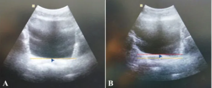
Figure 1. Pelvic floor muscle (PFM) function test using the trans-abdominal ultrasound images. (A) Rest. (B) PFM contraction.
Figure 2. Education using a mobile game-based extracorporeal bi-ofeedback device (EBD). (A) Posture during the experiment. (B) Any kegel (Furun Healthcare).
postpartum women. All the subjects provided voluntary consent after hearing the explanation of the purpose and method of the study. The study was approved by the Institutional Review Board of the Institutional Bioethics Committee of Sahmyook University (IRB No. 2-1040781- AB-N-01-2016108HR).
Outcome assessments
The subjects were assessed with the PFM function test us-ing ultrasonography (3D TRUS Accuvix V10; Samsung Medison Co., Ltd., Seongnam, Korea) by TAUS at two time points: immediately before and after education of the PFM contraction. The distance of the movement of the bladder base was calculated at rest and during the PFM contraction for the PFM function test. The location of the base of the bladder was measured using a 3.5 Hz curved linear array ul-trasonographic transducer probe. To fill the bladder, it was emptied 1 hour before the test and filled by drinking 500 ml of water. The measurement was performed in a crook-lying supine position which is to bend the hip and knee joints by 60° in the supine position. The examiner placed the probe on the abdomen 5 cm above the pubic symphysis region of sub-jects, and tilted it at 15°-30°. The arrow indicated the lowest point of the base of the bladder at rest and the image was captured. Then, the examiner marked with an arrow again to indicate the bladder base at the highest maximum displace-ment when the participant performed maximum PFM contraction. The probe was not moved during the testing procedure (Figure 1) [9].
Research procedure
The subjects were divided randomly into two groups ac-cording to the PFM contraction method used as follows: the mobile game-based EBD group and the TAUS group. Before the experiment, all subjects received full planations and understood the information of the
ex-perimental procedure and how to contract the PFM. To conduct the PFM contraction visual feedback educa-tion using a mobile game-based EBD (Any kegel; Furun Healthcare, Wonju, Korea), an application was installed in a mobile phone (Galaxy S7; Samsung, Suwon, Korea). At first, the subjects wore thin clothes to be able to feel the force more effectively and then sat on the center of the pressure sensor of the EBD, which is located on a chair with the hip and knee joint at approximately 90° of flexion. A pressure sensor was placed between the buttocks, positioned between the anus and the urethra, to contact the PFM. Calibration was performed while the subject sat comfortably on the pressure sensor without contracting the PFM. After the full ex-planation of the game method was provided to the subject, the game was played for 8 minutes (2 minutes/set, total 3 sets, each set with a 30 second rest) while looking at the mo-bile phone located 1m in front of the subject and the subject was educated on how to contract and relax the PFM through visual feedback. The method involved touching a coin while drawing a parabolic line and the balloon floating on the screen continuously that moved from left to right, rising up according to the degree of PFM contraction, and descending when the PFM relaxed (Figure 2).
Visual feedback education using the TAUS was con-ducted under the same conditions as the PFM function test. At this time, the subject could see the ultrasonography image through the monitor. The subject received information to as-sist in understanding the ultrasonography image, which was the basal surface of the risen bladder when the PFM con-tracted, and at the descended position with the PFM relaxed. Thus, the subject was then instructed to relax for 5 seconds after a maximum contraction of 10 seconds, and visual
feed-Table 2. Comparison of pre- and post- instruction of PFM functions in terms of displacement (in millimeters) of PFM elevation
between the two groups (N=16)
PFM functions TAUS group (n=9) EBD group (n=7) p-value
Pre-instruction 5.27 (3.39) 5.93 (4.03)
Post-instruction 7.47 (2.79) 7.62 (3.77)
Difference (post-pre) 2.20 (1.67) 1.68 (1.26) 0.529
p-value 0.008 0.028
Values are presented as mean (SD).
PFM: pelvic floor muscle, TAUS: transabdominal ultrasonography, EBD: extracorporeal biofeedback device.
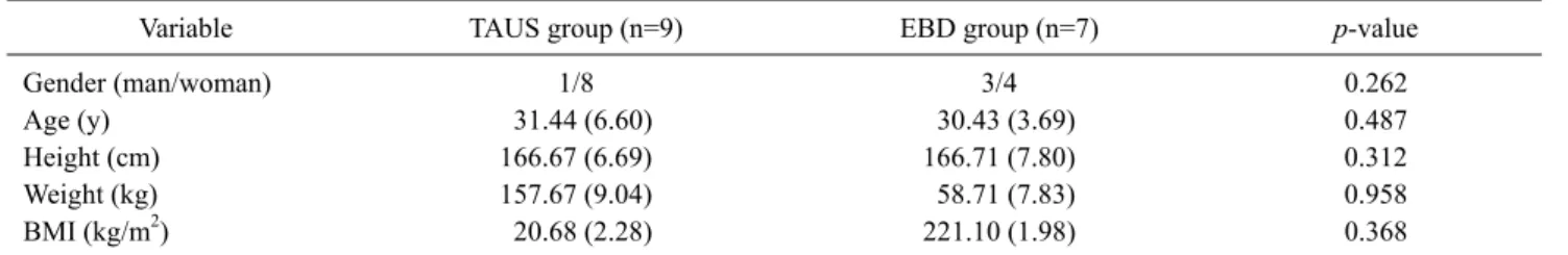
Table 1. General characteristics of the subjects (N=16)
Variable TAUS group (n=9) EBD group (n=7) p-value
Gender (man/woman) 1/8 3/4 0.262
Age (y) 31.44 (6.60) 30.43 (3.69) 0.487
Height (cm) 166.67 (6.69) 166.71 (7.80) 0.312
Weight (kg) 157.67 (9.04) 58.71 (7.83) 0.958
BMI (kg/m2) 20.68 (2.28) 221.10 (1.98) 0.368
Values are presented as number only or mean (SD).
TAUS: transabdominal ultrasonography, EBD: extracorporeal biofeedback device, BMI: body mass index. back training was conducted for 8 minutes (10 times/set,
to-tal 3 sets) [9]. Data processing
In this study, statistical analysis was performed using IBM SPSS Statistics for Windows, Version 22.0 (IBM Co., Armonk, NY, USA), and the general characteristics of the subjects were analyzed using descriptive statistics, Chi square test, and Mann-Whitney U-test. The Wilcoxon sign-ed-rank test was performed to confirm the pre- and post-group differences, and the Mann-Whitney U-test was performed to confirm the difference in PFM function be-tween the two groups. All statistical significance levels (α) were set to p<0.05.
Results
No significant differences were found between the two groups in terms of age, height, weight, and BMI (Table 1). The displacement of the PFM elevation significantly in-creased after the instructions from 5.93±4.03 mm to 7.62±3.77 mm (p<0.05) in the EBD education group, with a mean increase rate of 38% (range, 4%-80%), and from 5.27±3.39 mm to 7.47±2.79 mm (p<0.05), with an increase rate of 75% (range, 4%-180%) in the TAUS education group. No significant differences in PFM elevation
dis-placement was found between the two teaching methods af-ter the instructions (Table 2).
Discussion
Voluntary contraction of the PFM controls urine, gas, and bowel movements by tightening the vagina, urethra, and rec-tum, respectively. However, performing accurate voluntary contractions is difficult for patients with conditions such as urinary and fecal incontinence, and for healthy people. In some cases, symptoms may not improve or may actually worsen by increasing the abdominal pressure through in-correct training methods for PFM contraction. A visual feed-back training method is used to recognize and educate on PFM contractions. It is a useful method for visually confirm-ing muscle contraction usconfirm-ing ultrasonography techniques such as TAUS and TPUS, which do not involve directly in-serting an ultrasonography probe in the vagina or rectum. However, the application of this method is not easy clin-ically because it requires a long preparation time and much effort before implementation. Therefore, nowadays, a game-based intervention method with an EBD is being de-veloped for patients to induce voluntary contraction of the PFM, with the use of technological developments such as computers, mobile phones, and internet of things.
of using the TAUS and mobile game-based education using the EBD to confirm the possibility of education and training for voluntary PFM contraction in healthy adults. This study showed that the PFM contractile force in the education group using the TAUS was significantly increased from 5.27±3.39 mm to 7.47±2.79 mm, with a mean rate of change of 75%. Dietz et al. [12] reported that among women who were able to contract their PFM voluntarily, 57% could not correctly contract the PFM after training for 5 minutes using the TPUS [12]. This difference is thought to be because of the fact that this study was conducted in healthy adults with-out symptoms rather than in a disease group whose volun-tary PFM contractility was decreased before education.
The EBD education group showed a significant increase in displacement of the PFM elevation from 5.93±4.03 mm to 7.62±3.77 mm (p<0.05), with a mean rate of change of 38%. Silva et al. [13] applied game therapy for contraction train-ing of the PFM and abdominal muscles in young women and reported that coactivation of the PFM using electro-myography increased significantly from 127.27 to 147.84. According to the results of this study, PEM contraction education using the EBD and TAUS showed immediate ef-fectiveness, which did not significantly differ between the two education methods. Therefore, these results demon-strate the possibility of using not only the method using the TAUS but also that method using the EBD as a non-insertion method of PFM contraction education.
Low effectiveness of PFM contraction education and training conducted through oral explanations has been re-ported [3], Caagbay et al. [4] did not use leaflet verbal ex-planations in subjects with low literacy level. For effective education and training of the PFM, the visual feedback method introduced by Engel et al. [6] in 1974 has been used continuously on the subjects to investigate its procedures and effects even after 40 years.
In this study, an EBD based on mobile games was used to confirm the effectiveness of PFM contraction education. The EBD and TAUS methods did not show statistically sig-nificant difference, showing the potential usefulness of the EBD method for PFM contraction. However, the EBD method was shown to be less effective than the TAUS meth-od using visual feedback for education. The limitations of this study include the small sample size, and the fact that it was conducted in healthy adults. Also, PFM contraction ed-ucation was conducted with 8 minutes of training time in this study. A previous study showed an effect with 5 minutes of visual feedback training [12], but there are still a lack of
re-search on effective training time. Therefore, future studies are needed to compare effective training and education protocols. In addition, further studies are needed to verify the suitable methods of education in subjects with urinary and fecal incontinence, genitourinary cancer treatment, and surgical diseases, which cause functional abnormalities of the PFM.
In conclusion, in this study, mobile game-based in-struction with an EBD was demonstrated to have potential usefulness as an education and training method for volun-tary PFM contraction in healthy subjects.
Conflict of Interest
The authors declared no potential conflicts of interest with respect to the authorship and/or publication of this article.
References
1. Bø K, Berghmans B, Mørkved S, Van Kampen M. Evidence- based physical therapy for the pelvic floor: bridging science and clinical practice. 2nd ed. Philadelphia (PA): Churchill Livingstone; 2014.
2. Kegel AH. Progressive resistance exercise in the functional re-storation of the perineal muscles. Am J Obstet Gynecol 1948;56: 238-48.
3. Bump RC, Hurt WG, Fantl JA, Wyman JF. Assessment of Kegel pelvic muscle exercise performance after brief verbal instruction. Am J Obstet Gynecol 1991;165:322-7; discussion 327-9. 4. Caagbay DM, Black K, Dangal G, Raynes-Greenow C. Can a
leaflet with brief verbal instruction teach Nepali women how to correctly contract their pelvic floor muscles? J Nepal Health Res Counc 2017;15:105-9.
5. Ibrahim IK, Hameed MM, Taher EM, Shaheen EM, Elsawy MS. Efficacy of biofeedback-assisted pelvic floor muscle training in females with pelvic floor dysfunction. Alexandria J Med 2015; 51:137-42.
6. Engel BT, Nikoomanesh P, Schuster MM. Operant conditioning of rectosphincteric responses in the treatment of fecal incon-tinence. N Engl J Med 1974;290:646-9.
7. Scott KM. Pelvic floor rehabilitation in the treatment of fecal incontinence. Clin Colon Rectal Surg 2014;27:99-105. 8. Lee HN, Lee SY, Lee YS, Han JY, Choo MS, Lee KS. Pelvic floor
muscle training using an extracorporeal biofeedback device for female stress urinary incontinence. Int Urogynecol J 2013;24: 831-8.
9. Ariail A, Sears T, Hampton E. Use of transabdominal ultrasound imaging in retraining the pelvic-floor muscles of a woman postpartum. Phys Ther 2008;88:1208-17.
10. McKenna PH, Herndon CD, Connery S, Ferrer FA. Pelvic floor muscle retraining for pediatric voiding dysfunction using inter-active computer games. J Urol 1999;162(3 Pt 2):1056-62;
dis-cussion 1062-3.
11. Botelho S, Martinho NM, Silva VR, Marques J, Carvalho LC, Riccetto C. Virtual reality: a proposal for pelvic floor muscle training. Int Urogynecol J 2015;26:1709-12.
12. Dietz HP, Wilson PD, Clarke B. The use of perineal ultrasound to quantify levator activity and teach pelvic floor muscle exercises.
Int Urogynecol J Pelvic Floor Dysfunct 2001;12:166-8; dis-cussion 168-9.
13. Silva VR, Riccetto CL, Martinho NM, Marques J, Carvalho LC, Botelho S. Training through gametherapy promotes coactivation of the pelvic floor and abdominal muscles in young women, nul-liparous and continents. Int Braz J Urol 2016;42:779-86.