A Thesis
For the Degree of Master of Veterinary Medicine
Effects of Fucoidan on Survival
and Hematopoietic Recovery
in Irradiated Mice
Jee-young Lee
Department of Veterinary Medicine
GRADUATE SCHOOL, CHEJU NATIONAL UNIVERSITY
Effects of Fucoidan on Survival and Hematopoietic
Recovery in Irradiated Mice
Jeeyoung Lee
(Supervised by professor Taekyun Shin)
A thesis submitted in partial fulfillment of the requirement for the degree of Master of Veterinary Medicine
2006. 10.
This thesis has been examined and approved
Thesis director, Youngheun Jee, Prof. of Veterinary Medicine
Hong-gu Joo, Prof. of Veterinary Medicine
Taekyun Shin, Prof. of Veterinary Medicine
2006. 12.
Abstract
To evaluate the possible in vivo radioprotection conferred by fucoidan, the survival rate and hematopoietic changes in mice subjected to total body irradiation were investigated. BALB/c mice were administered 10, 50, or 100 mg of fucoidan/kg body wt i.p. before exposure to 9 Gy of gamma radiation, and then were monitored for 30 days. To see the effect of fucoidan on the radiation-induced damage to peripheral blood leukocytes, the number of blood leukocytes was counted in mice treated with fucoidan 4 h post-irradiation (6 Gy). The changes in bone marrow viability and the endogenous hematopoietic spleen colony
formation after irradiation (6 Gy) were measured at 9 days in mice given fucoidan (10 and 100 mg/kg body wt, i.p.). The fucoidan pretreatment improved the survival rate of irradiated mice; 67% (6/9) of the mice treated with 100 mg/kg of fucoidan survived until day 30 after exposure to 9 Gy of gamma radiation, whereas most of the mice treated with less fucoidan died before day 15. This effect accompanied significant increases in peripheral blood leukocytes, endogenous spleen colonies, and bone marrow cells in irradiated mice pretreated with fucoidan, compared with control mice. Pretreatment with fucoidan before irradiation plays an important role in the survival of irradiated mice, possibly by protecting
endogenous spleen colonies. The molecular mechanism of the radioprotection conferred by fucoidan remains to be determined.
CONTENTS
I. Introduction --- 1
II. Materials and Methods --- 3
III. Results --- 6
IV. Discussion --- 14
I. Introduction
Radiation is used to treat various malignancies [4]. However, a severe side effect of radiotherapy is that it damages normal cells. The hematopoietic system and hematocytes are sensitive to low doses of radiation [14]. Radioprotective agents are administered before exposure to ionizing radiation to reduce the damaging effects, including radiation-induced lethality [14]. The efficacy of many synthetic and natural agents at protecting against radiation injuries has been investigated in recent years [9].
Fucoidans are a group of sulfated fucose-containing polysaccharides that are derived from marine brown algae including Fucus vesiculosus [1]. Many studies have shown that fucoidans have a wide spectrum of activity in biological systems [10]. Fucoidans have potent anti-thrombotic activity [1]. Another outstanding physiological function of fucoidans is their anti-inflammatory properties via anti-complement [2] and anti-leukocyte migration effects [7].
In vivo, fucoidan blocks leukocyte rolling in a dose-dependent manner, interfering
with various inflammatory responses in several animal models [3, 6, 11]. In addition, it was reported that fucoidan increased circulating mature white blood cells and hematopoietic
2
progenitor/stem cells in mice and nonhuman primates [11].
Little is known of the biological effects of fucoidan in vivo in animals that have been irradiated. Therefore, this study investigated the effects of fucoidan on mice given a sublethal dose of radiation.
II. Materials and Methods
1. Animals and experiments
Sixty female BALB/c mice (Orient Bio, Gyunggi-do, Korea), 6 to 8 weeks old, were used in this experiment. Fucoidan (Sigma, St. Louis, MO, USA) was dissolved in phosphate-buffered saline (PBS) and administered intraperitoneally (i.p.) 24 and 1 h before irradiation. Control animals were given PBS instead, at the same times.
After treatment, the mice were placed in a specially designed, well-ventilated acrylic container and subjected to whole-body radiation with 6 or 9 Gy in a single fraction using a
60Co γ-ray source (10,000 Ci; Co-60 Irradiation Facility, Applied Radiological Science
Research Institute, Cheju National University, Korea).
2. Survival assays
Survival was monitored daily and reported as the percentage of animals surviving 30 days after 9 Gy irradiation. Each treatment group (irradiation plus vehicle or 10, 50, or 100 mg fucoidan /kg body weight) consisted of nine mice. The data are expressed as the percent survival.
4
3. Hematologic examination
Four hours following irradiation (6 Gy), 1 ml of blood was collected from the hearts of mice treated with nothing (n = 3), fucoidan only (n = 3), irradiation only (n = 3), and irradiation plus fucoidan (100 mg/kg body weight) (n = 3) into tubes containing
ethylenediaminetetraacetic acid (EDTA) before sacrifice. Total leukocytes were counted microscopically with a hemocytometer.
4. Hematopoietic stem cell assays
To test the effect of fucoidan after irradiation, the number of bone marrow cells was counted 9 days after 6 Gy irradiation. Each treatment group (normal control plus vehicle, irradiation plus vehicle, and irradiation plus fucoidan 10 or 100 mg/kg) consisted of five mice. Bone marrow cells were obtained from anesthetized mice by aseptic isolation of the femurs from which the marrow was flushed with Hank’s balanced salt solution (HBSS) using a 25-gauge needle. The cells were suspended in HBSS and counted with a
hemocytometer. The results are expressed as the number of live bone marrow cells per 106
cells/femur.
To test the effect of fucoidan on endogenous hematopoietic spleen colony formation, the number of spleen colonies was counted using the method of Till and McCulloch [12] on
day 9 after radiation. The mice were sacrificed; their spleens were removed and fixed in Bouin’s fixative for 24 h. The number of macroscopic spleen colonies was then counted.
5. Histological examination
For histological examination, femurs (n=5/each group) from mice (normal control plus vehicle, irradiation plus vehicle, and irradiation plus fucoidan 10 or 100 mg/kg) were isolated and fixed in 4% paraformaldehyde in phosphate buffer, decalcified in the
decalcification solution (formic acid-sodium citrate method) [8] for five days and processed for paraffin embedding. Paraffin sections were stained with hematoxylin and eosin.
6. Statistical analysis
The results are presented as the mean ± S.E. The results were analyzed statistically using one-way analysis of variance (ANOVA) followed by the post-hoc Student-Newman-Keuls procedure for multiple comparisons. In all cases, p < 0.05 was considered significant.
6
III. Results
1. Survival rate of mice after irradiation
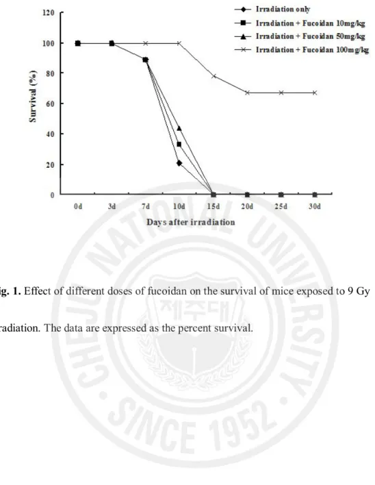
Of the control mice given 9 Gy irradiation, 78% (7/9) died by day 10, and all died before 15 days post-irradiation (Fig. 1). The mortality of the irradiated mice pretreated with fucoidan 10 mg/kg was 67% (6/9) at day 10, and 100% by day 15. The mortality of the mice pretreated with fucoidan 50 mg/kg was delayed: 56% (5/9) died at day 10, and the remainder (4/9) all died by day 15. The mortality of the irradiated mice treated with 100 mg
fucoidan/kg was significantly reduced compared with that of the other groups. Only 22% (2/9) of the mice died before day 15, and 67% (6/9) remained alive at day 30. These results suggest that fucoidan decreases the radiation-induced mortality.
Fig. 1. Effect of different doses of fucoidan on the survival of mice exposed to 9 Gy
8
2. Hematologic examination
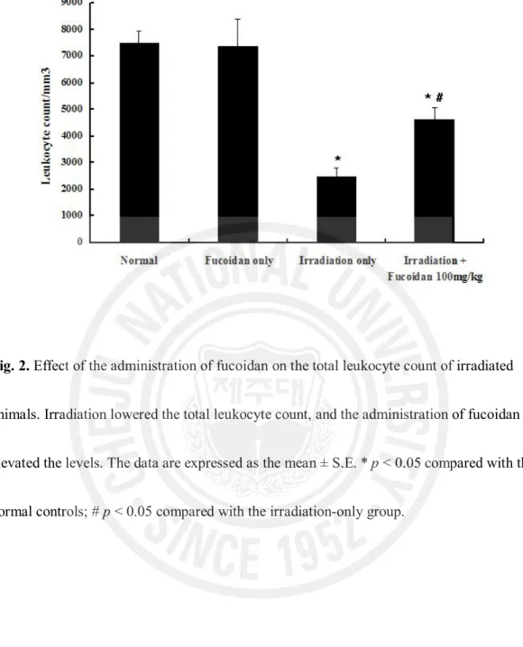
To see the effect of fucoidan on the radiation-induced damage to peripheral blood leukocytes, the blood leukocytes were counted in untreated mice and in mice treated with irradiation only, fucoidan only, and irradiation plus fucoidan (100 mg/kg).
Total body radiation significantly lowered the total leukocyte count (Fig. 2). The number of peripheral blood leukocytes in the irradiation-only group (2,483 ± 324/mL) was significantly lower than in the fucoidan-only (7,367 ± 1024/mL) and normal control (7,500 ± 425/mL) groups (both p < 0.01). The peripheral blood leukocytes in the mice treated with irradiation plus 100 mg/kg of fucoidan were significantly increased compared with the number in the irradiation-only group (4,617 ± 451/mL vs. 2,483 ± 324/mL; p < 0.05).
Fig. 2. Effect of the administration of fucoidan on the total leukocyte count of irradiated
animals. Irradiation lowered the total leukocyte count, and the administration of fucoidan elevated the levels. The data are expressed as the mean ± S.E. * p < 0.05 compared with the normal controls; # p < 0.05 compared with the irradiation-only group.
10
3. Effect of fucoidan on bone marrow nucleated cells
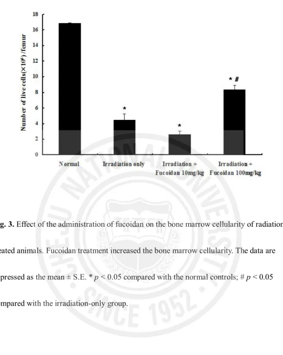
Bone marrow viability (Fig. 3) was significantly lower in the irradiation-only group (4.46 ± 0.79 ´ 106 cells/femur) than in the normal control group (16.88 ± 0.06 ´ 106
cells/femur, p < 0.001). In the irradiation plus 100 mg fucoidan/kg group, bone marrow viability was significantly higher compared with that in the irradiation-only group (8.36 ± 0.59 ´ 106 vs. 4.46 ± 0.79 ´ 106 cells/femur, p < 0.001). However, there was no
significant difference in bone marrow viability between the irradiation-only and irradiation plus 10 mg fucoidan/kg groups (4.46 ± 0.79 ´ 106 vs. 2.65 ± 0.94 ´ 106 cells/femur,
p > 0.05).
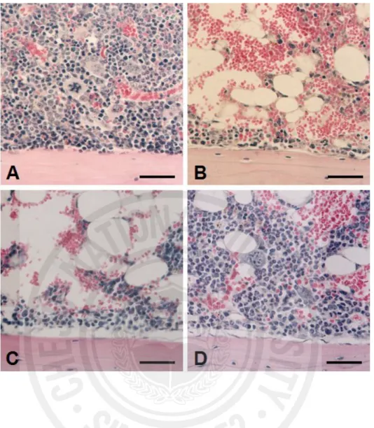
Histological examination of bone marrow from irradiated mice showed marked hypocellularity (Fig. 4B) in comparison to normal cellularity in untreated control mice (Fig. 4A). Fucoidan (100 mg/kg) administration elevated the lowered cellularity of bone marrow after irradiaton (Fig. 4D). This suggests that the higher dose of fucoidan (100 mg/kg) effectively protects the bone marrow cells of mice exposed to radiation.
Fig. 3. Effect of the administration of fucoidan on the bone marrow cellularity of
radiation-treated animals. Fucoidan treatment increased the bone marrow cellularity. The data are expressed as the mean ± S.E. * p < 0.05 compared with the normal controls; # p < 0.05 compared with the irradiation-only group.
12
Fig. 4. Histological findings in bone marrow of the normal control (A), irradiation-only (B),
irradiation plus 10 mg fucoidan/kg (C), and irradiation plus 100 mg fucoidan/kg groups (D). The scale bars represent 40 μm. Panels A–D stained with hematoxylin-eosin.
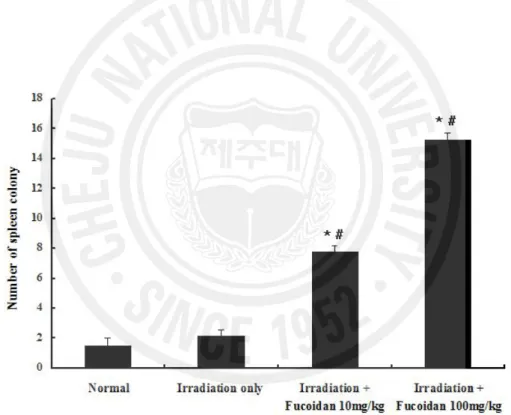
4. Effect of fucoidan on endogenous hematopoietic spleen colony formation
The results for spleen colonies (Fig. 5) largely matched those for bone marrow cells. Few spleen colonies were found in the spleens of mice given sub-lethal irradiation. The number of spleen colonies was increased significantly in the mice that received 10 (7.8 ± 0.37) or 100 (15.25 ± 0.48) mg fucoidan/kg compared with the mice exposed to irradiation only (2.2 ± 0.37) and the normal controls (1.5 ± 0.5) (p < 0.001).
Fig. 5. Effect of the administration of fucoidan on endogenous hematopoietic spleen colony
formation in irradiated animals. The data are expressed as the mean ± S.E. * p < 0.05 compared with the normal controls; # p < 0.05 compared with the irradiation-only group.
14
IV. Discussion
Our study indicates that fucoidan at a dose of 100 mg/kg provided the highest protection against radiation-induced mortality. Moreover, our data show that the pre-irradiation administration of fucoidan stimulated the recovery of total leukocytes and reduced the decrease in bone marrow nucleated cells induced by radiation. Therefore, fucoidan administration appears to protect both circulating blood cells and progenitor cells from irradiation. The enhanced spleen colony counts in the fucoidan-treated irradiated mice indicates that fucoidan protects stem cells or stimulates the proliferation of surviving cells. These results suggest that the increase in the 30-day survival is attributable to the protection afforded by fucoidan to the stem cell compartments of the bone marrow and spleen, which continue to supply the requisite number of cells in survivors.
It was reported that fucoidan elevated circulating white blood cells and mobilized hematopoietic progenitor/stem cells within hours [11]. The cells mobilized by fucoidan included radioprotective cells and probably also the cells required for long-term repopulation [11]. Therefore, the protective effect on leukocytes and bone marrow cells after irradiation seen in our study is supported by these roles of fucoidan.
Fucoidan is a ligand for the macrophage scavenging receptor 1 (MSR1), which increases the level of tumor necrosis factor-α and interleukin-1 secretion [5]. It was recently reported that fucoidan inhibited the lipopolysaccharide-mediated expression of inducible nitric oxide synthase by blocking activator protein 1 activation in macrophages [13].
Recently, fucoidan was proposed to prevent concanavalin A-induced liver injury through the production of endogenous interleukin-10 by Kupffer cells, and these effects were mediated by MSR1 [10]. These anti-inflammatory roles of fucoidan may be another important mechanism of its radioprotective efficacy.
Taken all into considerations, we postulated that fucoidan administration before irradiation has a dose-dependent effect on increasing the survival of irradiated mice by stimulating hematopoiesis. The results suggest that fucoidan is useful for radioprotection.
16
V. References
1. Berteau O, Mulloy B. Sulfated fucans, fresh perspectives: structures, functions, and biological properties of sulfated fucans and an overview of enzymes active toward this class of polysaccharide. Glycobiology 2003, 13, 29-40.
2. Blondin C, Chaubet F, Nardella A, Sinquin C, Jozefonvicz J. Relationships between chemical characteristics and anticomplementary activity of fucans. Biomaterials 1996, 17, 597-603.
3. Granert C, Raud J, Xie X, Lindquist L, Lindbom L. Inhibition of leukocyte rolling with polysaccharide fucoidin prevents pleocytosis in experimental meningitis in the rabbit. J Clin Invest 1994, 93, 929-936.
4. Hari Kumar KB, Sabu MC, Lima PS, Kuttan R. Modulation of haematopoetic system and antioxidant enzymes by Emblica officinalis gaertn and its protective role against gamma-radiation induced damages in mice. J Radiat Res (Tokyo) 2004, 45, 549-555. 5. Hsu HY, Chiu SL, Wen MH, Chen KY, Hua KF. Ligands of macrophage scavenger
receptor induce cytokine expression via differential modulation of protein kinase signaling pathways. J Biol Chem 2001, 276, 28719-28730.
6. Ley K, Linnemann G, Meinen M, Stoolman LM, Gaehtgens P. Fucoidin, but not yeast polyphosphomannan PPME, inhibits leukocyte rolling in venules of the rat mesentery. Blood 1993, 81, 177-185.
7. Linnemann G, Reinhart K, Parade U, Philipp A, Pfister W, Straube E, Karzai W. The effects of inhibiting leukocyte migration with fucoidin in a rat peritonitis model. Intensive Care Med 2000, 26, 1540-1546.
8. Luna LG. Manual of histologic staining methods of the armed forced institute of pathology. 3rd ed. pp. 6-8, McGraw-Hill Book Company, New York, 1968.
9. Nair CK, Parida DK, Nomura T. Radioprotectors in radiotherapy. J Radiat Res (Tokyo) 2001, 42, 21-37.
10. Saito A, Yoneda M, Yokohama S, Okada M, Haneda M, Nakamura K. Fucoidan prevents concanavalin A-induced liver injury through induction of endogenous IL-10 in mice. Hepatol Res 2006, 35, 190-198.
11. Sweeney EA, Priestley GV, Nakamoto B, Collins RG, Beaudet AL,
Papayannopoulou T. Mobilization of stem/progenitor cells by sulfated
polysaccharides does not require selectin presence. Proc Natl Acad Sci U S A 2000, 97, 6544-6549.
18
12. Till JE, Mcculloch EA. A direct measurement of the radiation sensitivity of normal mouse bone marrow cells. Radiat Res 1961, 14, 213-222.
13. Yang JW, Yoon SY, Oh SJ, Kim SK, Kang KW. Bifunctional effects of fucoidan on the expression of inducible nitric oxide synthase. Biochem Biophys Res Commun 2006, 21, 345-350.
14. Zhou Y, Mi MT. Genistein stimulates hematopoiesis and increases survival in irradiated mice. J Radiat Res (Tokyo) 2005, 46, 425-433.