Molecular cloning of clathrin assembly protein gene (rCALM)
and its differential expression to AP180 in rat brain
Hyung-Lae Kim1,3 and Sunhee Cho Lee2
1Department of Biochemistry and 2Department of Physiology, College of Medicine, Ewha Womans University,
Seoul 158-056, Korea
3Corresponding author: Tel, 82-2-650-5727; Fax, 82-2-650-5791, E-mail, [email protected]
Accepted 10 November 1999
Abbreviations: CCV, clathrin-coated vesicle; CALM, clathrin assem-bly protein lymphoid myeloid; PLD, phospholipase D; RT-PCR, reverse transcriptase polymerase chain reaction; AP, adaptor pro-tein
Abstract
Binding of clathrin assembly protein to clathrin triskelia induces their assembly into clathrin-coated vesicle (CCV) in neurons. The clathrin assembly protein gene (rCALM) was cloned from rat brain cDNA library. rCALM deduced 69 kD molecule has overall 73% amino acid homology compared with that of AP180 protein. The N-terminal domain, where amino acid sequences are very similar with AP180, harbours binding sites for clathrin and inositides, as well as possible phosphorylation sites, but the proline rich C-terminal domain is different from that of AP180. The mRNA expression of rCALM and AP180 by in situ hybridization histochemistry revealed that the rCALM mRNA was more intensely expressed than that of AP180, and the distribution patterns were different from each other. These results suggest that the rCALM mediates the assem-bly of clathrin in neural and supporting cells of brain, and regulates the clathrin coated-vesicle formation through phosphorylation and inositide metabolism.
Keywords: clathrin-coated vesicle, AP180, CALM, gene
cloning, expression
Introduction
Clathrin-coated vesicles (CCV) are involved in pathways of receptor-mediated intracellular transport and transfer of proteins from trans-Golgi network to pre-lysosomal
compartment and recycling of synaptic vesicles (Pearse and Robins, 1990; Keen, 1990). The CCV coat is formed by polymerization of triskelion-shaped clathrin molecules into lattice of clathrin cage, and is catalyzed by the assembly proteins. The major coat protein is a clathrin, which consists of triskelion having three identical 190 kD heavy chains and three 23-27 kD light chains. Coated vesicle also contains one or more of the assembly proteins (Robinson, 1994). The assembly proteins are adaptors, and are believed to link receptors to the clathrin network. The assembly proteins all participate in promoting an assembly of clathrin triskelia into artificial clathrin cage that resemble coated vesicle. To date, six different adaptor proteins (AP) are known: AP-1, AP-2, AP-3, AP4, AP180, CALM, and auxillin. The AP-1, AP-2, AP-3, and AP-4 are tetramer, whereas AP180, CALM, and auxillin are monomer.
AP-1 and AP-2 adaptors mediate clathrin assembly at the trans-Golgi network and plasma membrane, respec-tively, and play a role in selecting cargo protein in CCVs (Marsh and McMahon, 1999). AP-3 plays a role in traffick-ing from trans-Golgi network to the lysosome (Simpson et al., 1997). The function of AP-4 associated with trans-Golgi network was not characterized precisely (Dell'Angelica et al., 1999). AP180 is well studied because it is mono-meric protein and neuron specific (Kondury and Roland, 1988; Zhou et al., 1992). AP180 was first discovered as a specific coat component of clathrincoated vesicles from neural tissue and as a phosphoprotein (Stephan et al., 1990). The native protein was shown to associate with clathrin triskelia on equimolar basis and induces clathrin assembly into a homogenous population of 60-70 nm coats (Zhou et al., 1993). AP180 is phosphorylated at serine residues (Morris et al., 1990). The disassembly of clathrin from the vesicle coat was promoted by the phosphorylation of AP's (Wilde and Brodsky, 1996). Two known primary functional properties of the AP180 are clathrin assembling and high affinity for specific inositol polyphosphates (Morris et al., 1993; Ye et al., 1995; Norris et al., 1995). The CALM gene was cloned from human leukemia cell line, and was shown to be homologous to the monomeric clathrin adaptor of AP180. But the mole-cular property of the gene was not characterized. To understand the regulation of CCV formation at a mole-cular level, I have cloned and characterized the clathrin assembly protein gene from rat brain. In this study, the primary structure of CALM gene (rCALM) from the rat brain cDNA libraries and the differential expression between two monomeric adaptor proteins, AP180 and rCALM, in adult rat brain are presented.
Rat brain cDNA library was purchased from Stratagene. T7 sequencing kit was from US Biochemicals. Exo III deletion kit and Wizard miniprep kit were obtained from Promega. Restriction endonucleases were from Boerhinger Manheim. Nitrocellulose transfer membranes (BA85, 0.45
µm) were from Schleicher and Schuell. Other chemicals were the highest purity available.
Cloning of rCALM gene from rat cDNA library
The rCALM gene was isolated from rat cDNA library using oligonucleotides, 5'-AGC CAG GTT GGC TGT GTA-3', which was designed from the third transmem-brane segment of the glutamate receptor 1. About 7.0
×105 plaques were screened with the 32P end-labelled oligonucleotide probe. The plaques were transferred onto nitrocellulose membranes, immobilized, and hybridized with hybridization solution containing the probe. The membranes were washed with 0.2x SSC/ 0.1% SDS for 10 min three times at room temperature and followed by at 37oC for 10 min. The signal was visualized by exposure
onto X-OMAT film overnight. The positive plaques were picked, and the second screening was performed as described above. The resulting two plaques, G12 and G18, were cultured and their phage DNA s were isolated. The clones were digested with Eco RI, shown to have 1.2 and 2.3 kb insert. Restriction mapping was carried out to make deletion mutant for the sequencing.
Sequencing and sequence analysis of cDNA
Basically the deletion mutants were prepared as pre-viously described (Kim et al., 1998). Briefly, the 5'-/3'-overhang DNA was made, and was digested with Exo III nuclease followed by S1 nuclease. The unidirectional deletion DNA was ligated and transformation was carried out. The plasmids DNA from the deletion clones were prepared by Wizard miniprep kit (Promega). The template DNA for the sequencing was prepared by alkali denatu-ration-neutralization of the double-stranded plasmid. The sequencing was carried out by using the Sequenase v.2.0 sequencing kit (Amersham). The sequencing data from the deletion mutants were analyzed by using the MacVector program from IBI Co.
Reverse transcription (RT) and PCR
Total RNA was isolated from a 3 month-old rat cerebrum by using the acid guanidium phenol method (Chomczynski and Cacchi, 1987). Approximately 5 µg of total RNA was incubated at 37oC for 60 min in 50 µl volume
containing 10 units of MMLV reverse transcriptase, 50 mM Tris-HCl, pH 8.3, 75 mM KCl, 3 mM MgCl2, 0.5 mM
dNTP, 0.1µg of random hexamer primers. After incu-bation, 10 µl of the first-strand reaction mixture was
35 cycles (10 sec at 94oC, 10 sec at 50oC, and 0.5 min
at 72oC) in Thermocycler Model 9600 (Perkin Elmer). The PCR products were separated on 3% agarose (Agarose : NuSieve = 2 : 1) gel. To verify the alternative splicing, the PCR products were cloned into T-vector and the sequencing was carried out as previously described.
In situ hybridization histochemistry
The hybridization probe for rCALM was prepared from pGEM plasmid containing 615 bp fragment of rCALM cDNA (Pst I and Eco RI fragment, nucleotide residues 1095-1700) by transcribing with appropriate RNA poly-merases using a Riboprobe System (Promega Co.) in the presence of α-[35S]UTP (1000-1500 Ci/mmol, New
England Nuclear). The 645 bp-sized AP180 cDNA (nucleotide residues 2222-2866) was prepared by RT PCR of rat brain cDNA and labelled with [35S]UTP as
above. In situ hybridization histochemistry was perform-ed essentially as previously describperform-ed (Kim et al., 1992). Briefly, frozen rat brain sections (12 µm thick) were cut, thaw-mounted onto gelatin- coated slides. The sections were fixed in 4% paraformaldehyde, treated with 0.25% acetic anhydride in 0.1 M triethanolamine/0.9% NaCl (pH 8.0) to reduce nonspecific hybridization due to electro-static force, dehydrated and defatted in ethanol and chloroform, and finally air-dried. The sections were hybridized overnight at 53oC with 5×10⁶ cpm of labeled
RNA probe per slide. Then the sections were washed in 2x SSC, treated with RNase A (20 mg/ml, Boehringer-Mannheim) for 30 min at 37oC and washed sequentially
for 60 min in 2x SSC at 50oC, 60 min in 0.2x SSC at
55oC, and 60 min in 0.2x SSC at 60oC. After drying, the slides were processed for film (β-max Hyperfilm, Amer-shampharmacia) autoradiography by exposing 4 days.
Results and Discussion
Primary structure of clathrin assembly protein from rat (rCALM)
The clathrin assembly protein gene was found from the rat brain cDNA library by low stringency screening using the oligonucleotide designed from the third transmem-brane segment of the glutamate receptor 1. Among 48 cDNA clones, two clones, G12 and G18, were shown to have high homology with N-terminal region of AP180 and CALM when they were searched with blast program of NCBI at NIH. The size of G12 and G18 were 1.2 and 2.3 kb respectively. G18 encompasses a full coding region sequences from putative translation initiation
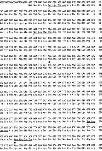
methionine (ATG) at position 30 through termination codon (TAA) located at position 1946 (Figure 1). The 3'-untranslated region (3'-UTR) terminates with a polyA tail preceded by an AAUAAA polyadenylation signal. Analysis of the deduced amino acid sequences of the rCALM did not reveal a signal sequences, or any regions of extensive hydrophobicity. G18 contains a full open reading frame encoding the clathrin assembly protein (short form of rCALM) with 597 amino acids. Because the nucleotide sequence of rCALM shows 129 bp-short comparing to that of CALM, PCR was carried out to determine whether alternative splicing exists in rCALM. The rCALM with 640 amino acids was shown to have an alterntive splicing variant with 129 nucleotide insert at the middle of the transcript (long form of rCALM). Estimated molecular masses of short form and long form of rCALM are 63,792 and 69,280, respectively.
The primary structure of the rCALM, shows a remark-able homology to the murine and rat clathrin assembly protein AP180 (Zhou et al., 1992; Morris et al., 1993;
Zhou et al., 1993) (Figure 2). The homology ranges from 97% to 28% in different portions of the protein. The most striking homology of more than 95% is found between the first 289 amino acids of rCALM and that of AP180. The rCALM protein was 256 amino acid short compared to AP180, the midlle part of which is missing in rCALM. The amino acid sequences of the C-terminal half of the rCALM is different from that of AP180. The overall pI values of the rCALM and AP180 were 8.7 and 4.6, respectively. The missing 250 amino acids which correspond to the acidic middle region of AP180 caused the pI value of rCALM alkaline. AP180 is interrupted by acidic and proline rich middle domains.
The distribution of charged amino acids, proline and alanine suggest a two-domain structure of rCALM in contrast to the three-domain structure of AP180. The N-terminal region of 340 amino acids is highly charged and predominantly basic. Twenty five percent of the amino acids in the region is charged, 14% of which is basic amino acids. Especially the first 289 residues are
Figure 1. Complete nucleotide and deduced amino acid sequence of the rat CALM. Numbers refer to nucleotide and amino acid positions. Beneath the nucleotide sequence is the deduced amino acid sequence coded for by the open reading frame between nucleotide 24 and 2048. The alternatively spliced region is shown as bold. The arrows indicate the primers (ASU and ASD) to verify the alternatively splicing sequences. The possible phophorylation sites are; casein kinase II (S-X-X-D/E, shown as open circles), cAMP-dependent proein kinase A (R-R-X-S, closed circle), protein kinase C (S/T-X-R/K, open square), Ca2+/ calmodulin-dependent protein kinase (R-X-X-S/T, closed square). The above sequence has been deposited in the GenBank database under the accession number AF041373 (short form) and AF041374 (long form).
almost identical to the corresponding sequences of AP180 from rat. Functional studies of the AP180 have shown that a clathrin-binding domain resides in the 33 kD N-terminal portion of the protein. This 33 kD region is thought to have a globular structure and consists mainly of helices (Ye and Lafer, 1995). Although this N-terminal domain is able to bind clathrin triskelia, it is unable to assemble them into clathrin cages and bind to preassembled cages. The assembling function resides in the 58 kD C-terminal region of the AP180. The high conservation of N-half amino acid of the rCALM suggests that the protein has similar clathrin binding properties. Another important function of the 33 kD N-terminal region is its high affinity binding of both inositol hexakisphosphate and diphosphoinositol pentakisphosphate (Norris et al., 1995; Ye et al., 1995). Binding of these ligands inhibits the ability of AP180 to assemble clathrin into cages (Ye et al., 1995). The binding site for the inositide was suggested to be KKK at N-terminal side, that is well conserved in rCALM and AP180. Thus, inositol poly-phosphates and phosphatidyl inositides may regulate clathrin-coated vesicle assembly and/or disassembly by means of the monomeric clathrin assembly proteins,
rCALM and AP180. Prasad (1995) reported that myelin basic protein would be a novel clathrin assesmbly protein. The protein has 23% of charged amino acids, and 30 out of 38 charged amino acids are basic. The highly charged and basic character of N-half of the rCALM is similar with that of myelin basic protein.
Analysis of deduced amino acid composition revealed an unusually large number of proline residues (45 prolines /640 residues). Most (36 out of 45) of the prolines are localized at the C-terminal half of the rCALM, where the proline levels approach 12.5%, while the proline com-position is just 1.6% at the N-terminal half. In the case of AP180, which has three domain structures, the middle and C-terminal domain is rich in proline (15%). There are multiple PXXP amino acid sequences, which is a known ligand for SH3 domain. Amphiphysin and other proteins containing SH3 domains would participate in the regulation of the clathrin cage foramtion. The polyproline could be the ligand for the WW domain. Although the typical sequence of XPPPXY was not found in the rCALM, there are XPPX sequences that could bind WW domain. Considering the role of WW domain containing protein, such as dystrophin, in the
Figure 2. Amino acid sequence comparison between the rat CALM and the human CALM and rat AP180. There is high degree of homology(more than 90%) between the genes from amino acids 1 to 289. The interrupted dots show the very different region, and the low degree of identity (around 30%) was shown at the C-terminal region. Differences between the sequences are indicated as small letter. The dashed line shows the missing of the amino acids and asterisk shows the translation-termination of amino acid.
interaction between cytoskeleton and membrane structure (Sudol, 1996), the rCALM and AP180 are important in the transport of vesicle through the bridge-formation between cytoskeleton and CCV. The possible function of the marked different region of the C-terminal half could be an interaction site(s) with proteins in the plasma membrane, possibly contributing to the regulation of the endocytotic activity and receptor turnover of the cell.
There are eight possible phosphorylation sites; three casein protein kinase II and protein kinase C, one cAMP-dependent protein kinase A, and one Ca2+/ calmodulin kinase. Except one protein kinase C site, most of the kinase substrate sites are located on the N-terminal half of the protein. where prolines and alanines are rare. The AP180 protein was known as a neuron-specific phosphoprotein (Keen and Black, 1986). High degree of homology of rCALM with AP180 suggested that the rCALM would be phosphorylated in vivo. The major role of AP180 would be the formation of uniform-sized vesicle in the nervous system (McMahon, 1999). Considering the ubiquitous expression of the rCALM and inhibitory activity of phosphorylated AP-1 and AP-2 (Wilde and Brodsky, 1996) on the formation of clathrin cage, the phosphorylation of the rCALM would be involved in the regulation of the uniform-sized clathrin cage formation in non-nervous systems.
Alternatively splicing
To determine the differential expression of the alternatively splicing variants, RT-PCR was carried out. The difference between the long and short form of rCALM was 129 nucleotide insertion or deletion by alternatively splicing. The short form was shown as 203 bp-sized PCR product, that was observed in most of the tissues examined (Figure 3). The 332 bp PCR product, the long form of the rCALM, was not observed in heart, lung, and pancreas. The rCALM might have a complementary role in the formation of uniform-sized CCV in neural and non-neural tissues. But there was no functional domains so far known in the alternatively spliced segment. The
exact functional role of the differential expression remains to be studied.
Expression in adult rat brain
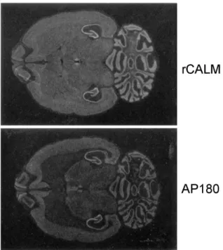
The expression pattern of the rCALM gene was deter-mined in adult rat brain by in situ hybridization histo-chemistry. The rCALM riboprobe was 615 bp-sized Pst I and Eco RI fragment (nucleotide residues 1,095-1700) from G18 cDNA, which encodes a unique sequence compared to that of other types of assembly proteins, in order to avoid a cross reaction with other assembly protein mRNAs. The riboprobe of AP180 was also designed from the unique 3'-end lower one third sequences. To establish the specificity of the labelled probe, control experiments were conducted as follows. First, a control hybridization using a sense probe resulted in autoradiograms virtually devoid of signal. Second, hybridization signal from the tissue was abolished by addition of RNase in the hybridization solution. Figure 4 shows the transverse section of the adult rat brain. The expression of the rCALM mRNA was more intense than that of AP180 and the distribution pattern was different each other. The rCALM mRNA was widely expressed throughout the brain. High levels of rCALM were found on the hippocampus, dentate gyrus, medial habenula nucleus, and cerebellar granule cells. Relatively high levels of expression were observed in olfactory bulb, and cerebral cortex. Moderate densities were in the
Figure 3. Differential expression of the clathrin assembly protein transcript in various tissues of rat. The oligonucleotides used for PCR are indicated by arrows in Figure 1. The alternative splicing sites were amplified from the mRNA of adult rat tissues by using RT PCR as described in ‘Materials and Methods’, and analyzed on 3% agarose (Agarose: NuSieve = 2 : 1) gel. The left lane indicates ΦX174 Hae III size marker. The sizes of the upper and lower band are 332 bp and 203 bp respectively. The tissues from the left are: cr, cerebrum, cl; cerebellum, ht; heart, in; intestine, lv; liver, lu; lung, kd; kidney, sp; spleen, pn; pancrease, ts; testis, th; thymus, sg; salivary gland, sm; skeletal muscle.
Figure 4. Localization of rCALM and AP180 in adult rat brain by in situ hybridization. Both figures show the negative film image of the expression of rCALM (shown upper) and AP180 (lower) in the transverse section of adult rat brain. In situ hybridization was carried out as described in ‘Materials and Methods’.
caudate-putamen. From this different distribution pattern, it may be suggested that these two monomeric clathrin assembly proteins function differently; AP180 would work as a neuronal synaptic vesicle assembly protein, and rCALM might assemble endocytic vesicles in neural and supporting cells in brain.
Acknowledgement
I thank Hye-Sun Jung at Dept. of Anatomy, Korea University for her excellent technical assistance of in situ hybridization. This study was supported by the Grant of the Korea Research Fund, Basic Medical Science (997-021-F0019).
References
Chomczynski, P. and Sacchi, N. (1987) Singl-step method of RNA isolation by acid guanidium thiocyanate-phenol-chloroform extraction. Anal. Biochem. 162: 156-159
Dell'Angelica, E. C., Mullins, C. and Bonifacino, J. S. (1999) AP-4, a novel protein complex related to clathrin adaptors. J. Biol. Chem. 274: 7278-7285
Keen, J. H. and Black, M. B. (1986) The phosphorylation of coated membrane proteins in intact neurons. J. Cell Biol. 102: 1325-1333
Keen, J. H. (1990) Clathrin associated assembly and disas-sembly proteins. Ann. Rev. Biochem.. 59: 415-438
Kim, H.-L., Chang, Y.-J., Lee, S.-M. and Hong, Y.-S. (1998) Genomic structure of the regulatory region of the voltage-gated calcium channel 1D. Exp. Mol. Med. 30: 246-251
Kim, H.-L., Kim, H., Lee, P., King, R. G. and Chin, H. (1992) Rat brain expressed an alternatively spliced form of the dihydropyrdine-sensitive L-type calcium channel a2 subunit. Proc. Nat'l Acad. Sci. USA. 89: 3251-3255
Kondury, P. and Roland, E. L. (1988) Molecular characteri-zation of the AP180 coated vesicle assembly protein. Biochem. 27: 6098-6104
Marsh, M. and McMahon, H. T. (1999) The structural era of endocytosis. Science 285: 215-219
Ungewickell, E. (1993) Clathrin assembly protein AP 180: primary structure, domain organization and identification of a clathrin binding site. The EMBO J. 12: 667-675
Norris, F. A., Ungewickell, E. and Majerus, P. W. (1995) Inositol hexakisphosphate binds to clathrin assembly protein 3(AP-3/AP180) and inhibits clathrin cage assembly in vitro. J. Biol. Chem. 270: 214-217
Pearse, B. M. and Robins, M. S. (1990) Clathrin,adaptors, and sorting. Ann. Rev. Cell. Biol. 6: 151-171
Prasad, K., Barouch, W., Martin, B. M., Greene, L. E. and Eisenberg, E. (1995) Purification of new clathrin assembly protein from bovine brain coated vesicles and its identification as myelin basic protein. J. Biol. Chem. 270: 30551-30556 Robinson, M. S. (1994) The role of clathrin, adaptors, and dynamin in endocytosis. Curr. Opin. Cell. Biol. 6: 538-544 Simpson, F., Peden, A. A., Christopoulou, L. and Robinson, M. S. (1997) Characterization of the adaptor-related protein complex, AP-3. J. Cell Biol. 137: 835-845
Stephen, A. M., Annette, M. and Ungewickell, E. (1990) Anaysis of 100-180 kD phosphoprotein in clathrin-coated vesicles from bovine brain. J. Biol. Chem. 265: 3354-3357
Sudol, M. (1996) The WW domain binds polyproline and is involved in human diseases. Exp. Mol. Med. 28: 65-69 Wilde, A. and Brodsky, F. M. (1996) In vivo phophorylation of adaptors regulates their interaction with clathrin. J. Cell Biol. 135: 635-645
Ye, W., Ali, N., Bembenek, M. E., Shears, S. B. and Lafer, E. M. (1995) Inhibition of clathrin assembly by high affinity binding of specific inositol polyphosphate to the synapse-specific clathrin assembly protein AP-3. J. Biol. Chem. 270: 1564-1568
Ye, W. and Lafer, E. M. (1995) Clathrin binding and assembly activities of expressed domains of the synapse-specific clathrin assembly protein AP-3. J. Biol. Chem. 270: 10933-10939 Zhou, S., Sousa, R. and Lafer, E. M. (1992) Characterization of a novel synapse-specific protein II. cDNA cloning and sequence analysis of F1-20 protein. J. Neurochem. 12: 2114-2155
Zhou, S., Tannery, N. H. and Lafer, E. M. (1993) The synapse-specific phosphoprotein F1-20 is identical to the clathrin assembly protein AP-3. J. Biol. Chem. 268: 12655-12662