www.kaom.org
ISSN 2288-9272 J Oral Med Pain 2014;39(1):22-25 http://dx.doi.org/10.14476/jomp.2014.39.1.22
Synovial Chondromatosis in Temporomandibular Joint
A-Young Chung, Jung-Hun Hong, Jeong-Seung Kwon, Hyung-Joon Ahn, Jong-Hoon Choi, Seong-Taek Kim
Department of Orofacial Pain and Oral Medicine, College of Dentistry, Yonsei University, Seoul, Korea
Received January 10, 2014 Revised January 23, 2014 Accepted February 5, 2014
Synovial chondromatosis (SC) in temporomandibular joint is a rare benign disorder charac-terized by cartilaginous metaplasia of the mesenchymal remnants of the synovial tissue. The etiology of the disease is unclear but may be associated with trauma, overuse, local infection, and embryologic disturbance. SC does not spontaneously resolve and respond to nonsurgical treatment. SC should be differentially diagnosed with other temporomandibular joint disorders such as arthralgia or osteoarthritis because surgery should be done for managing it. However, primary diagnosis of SC is not easy because of nonspecific symptoms and signs. For the pa-tients with unsuccessful conservative treatment response, especially accompanied by crepitus, preauricular swelling or posterior open bite, computed tomography/cone-beam computed to-mography or magnetic resonance imaging should to be performed to exclude SC. We discussed the importance of the early diagnosis and surgical treatment of SC from this case.
Key Words: Arthroscopy; Chondromatosis, synovial; Joint loose bodies; Malocclusion;
Maxil-lofacial surgery; Temporomandibular joint
Correspondence to: Seong-Taek Kim
Department of Orofacial Pain and Oral Medicine, Dental Hospital, College of Dentistry, Yonsei University, 50, Yonsei-ro, Seodaemun-gu, Seoul 120-752, Korea
Tel: +82-2-2228-3110 Fax: +82-2-393-5673 E-mail: [email protected]
mm, including the vertical incisal overlap, and (4) crepitus. Clinical diagnosis is made mainly based on imaging such as the following: (1) magnetic resonance imaging (MRI): multiple chondroid nodules, joint effusion, and amorphous iso-intensity signal tissues within the joint space and cap-sule, (2) computed tomography (CT)/cone-beam CT (CBCT): loose calcified bodies in the soft tissues of the TMJ.
Histologic examination confirming cartilaginous meta-plasia makes the definitive diagnosis of SC.
The disease may be related to malocclusion; progressive ipsilateral posterior open bite.1) In the cases with
intracrani-al invasion, headache can be often complained.2) Small
car-tilaginous nodules with lack of calcification or ossification are easy to be overlooked on CT.2)
CASE REPORT
A 48-year-old female presented with pain on the left pre-auricular area when chewing. She had been experiencing crepitus sound and pain on the left TMJ region for about 1 year, and had no specific medical history except for being a
INTRODUCTION
Synovial chondromatosis (SC) in temporomandibular joint (TMJ) is a rare benign disorder characterized by car-tilaginous metaplasia of the mesenchymal remnants of the synovial tissue.1)
It is usually confined to the joint space, mainly the superior joint space, but it can occasionally ex-tend beyond the joint capsule into the cranium, parotid gland, infratemporal fossa, or external auditory meatus.2)
The etiology of the disease is unclear; the development of these pathologies may be associated with trauma, overuse, local infection, and embryologic disturbance.2)
The American Academy of Orofacial Pain has published the diagnostic criteria for SC.1)
First of all, the patient has the history of at least one of the following: (1) report of preauricular swelling, (2) ar-thralgia, (3) progressive limitation in mouth opening, and (4) joint noise in the past month.
In addition, at least one of the following must be proved by examination: (1) preauricular swelling, (2) arthralgia, (3) maximum assisted opening (passive stretch) less than 40
Case Report
JOMP
Journal of Oral Medicine and Pain
23 A-Young Chung, et al. Synovial Chondromatosis in Temporomandibular Joint
www.kaom.org
on the left TMJ and myofascial pain on the left masseter and both SCM, trapezius, and splenius capitis muscles. The patient was instructed precautions for temporomandibular disorder and treated with pharmacologic and physical ther-apy. After 2 months, the pain disappeared and then treat-ment was finished.
One year and a half later, she revisited for the same com-plaint. She said that the pain had recurred 6 months af-ter the end of treatment and splint therapy, conducted at a local clinic, had failed due to discomfort associated with wearing the appliance.
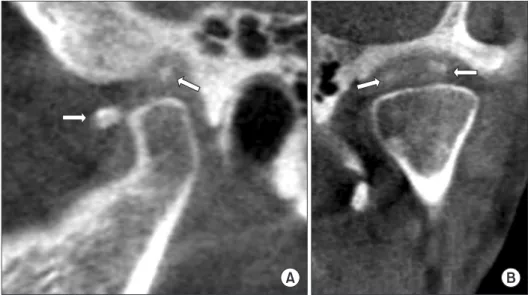
Active mouth opening was 45 mm and she presented with pain on her left TMJ during opening and eccentric move-ment. Tenderness was observed when palpating on both TMJ, masseter, SCM, trapezius, and splenius capitis muscles. The occlusion remained unchanged, but crepitus sound was detected on the left TMJ. CBCT was taken, and sagittal and frontal images showed bony flattening of the superior por-tions of the both condyle heads and small high attenuated bony materials around the left condyle (Fig. 3). As the re-sult, a presumptive radiological diagnosis was made as SC.
After 8 weeks of pharmacologic and physical treatments, hepatitis B carrier.
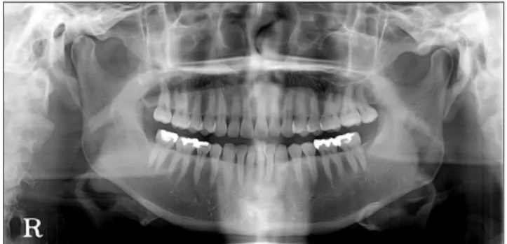
She reported pain on the left TMJ during active mouth opening (50 mm), laterotrusive movement (5 mm) and pro-trusive movement (7 mm). Physical examination revealed tenderness to palpation on the left TMJ and masseter, and both sternocleidomastoid (SCM), trapezius, and splenius capitis muscles. The occlusion was stable on the both side evenly and the sound of the TMJ was not detected at the clinical examination. There were no significant patholog-ic bony change on the panorampatholog-ic and transcranial radio-graphic view (Figs. 1, 2). She was diagnosed with arthralgia
Fig. 1. The panoramic radiography at first visit.
Fig. 2. Transcranial radiography at first visit. (A) Closing, left temporomandibular joint (TMJ), (B) opening, left TMJ, (C) closing, right TMJ, (D) opening, right TMJ.
24 J Oral Med Pain Vol. 39 No. 1, March 2014
www.kaom.org
Open TMJ surgery is recommended as the therapy of choice for SC.3,7)
It removes loose bodies greater than 3 mm.3) Additional procedure, such as synovectomy which
removes affected active synovial membrane with synovial metaplastic activity is performed most frequently. It is of-ten accompanied by diskectomy, and, less frequently, by condylectomy.7)
Postoperative follow-up examination is suggested ini-tially at 3-6 month intervals in the first year, and later once per year.2,4)
Once the loose bodies have been removed, the recurrence rate seems to be very low.7)
Primary diagnosis of SC is not easy because of low in-cidence in TMJ and nonspecific symptoms and signs. SC should be differentially diagnosed with other TMJ disorders such as arthralgia or osteoarthritis because surgery should be done for managing it. It must be suspected if the patient reports preauricular swelling, crepitus or posterior openbite or the symptom is not relieved and progressive despite ap-propriate conservative treatment. It may not be detected by conventional X-rays in the case of lack of calcification or ossification.
In this case, the patient revisited for the recurrence of the same symptom. If a CT scan had been conducted at the first visit, diagnosis for SC would have been made earlier. In ad-dition, surgical approach should have been considered right after the clinical diagnosis of SC.
In conclusion, for the patients with unsuccessful conser-vative treatment response, especially accompanied by crepi-tus, preauricular swelling or posterior open bite, CT/CBCT or MRI should to be performed to exclude SC. MRI may be the patient complained of occlusion change, and clinical
examination identified light contacts of the left second pre-molar, and first and second molars. The patient’s signs and symptom repeated waxing and waning during conservative treatment including occlusal stabilization splint therapy.
The patient underwent follow-up CBCT 1 year later, and no significant change was observed.
Surgical removal of the mass via a preauricular approach was performed under general anesthesia at the department of oral and maxillofacial surgery 1 year and 10 months lat-er since hlat-er revisitation. Histopathological assessment con-firmed SC. After the surgery, subjective pain and joint noise improved, but anterior openbite was observed, likely result-ing from degenerative joint disease.
So far, the occlusion has been recovering without pain recurrence.
DISCUSSION
SC is a benign disease, but does not spontaneously re-solve nor respond to nonsurgical treatment.2-5)
Furthermore, it has potential to invade the intracranial structure and recur.2,4) Therefore, surgical intervention is needed as
following.
Arthroscopy is less invasive technique that is useful for the primary treatment of small loose bodies less than 3 mm, confined to the superior joint space.2,6) Success rate is no
better than 55%.3) Arthroscopy has limitation and
dif-ficulty to retrieve the larger intra-articular loose bodies completely.2,6)
Fig. 3. Cone-beam computed tomo-graphic images after 1 year and a half. (A) Sagittal view, (B) coronal view. Bony flattening of the superior portions of the both condyle heads and small high attenuated bony materials (arrows) around the left condyle were shown.
25 A-Young Chung, et al. Synovial Chondromatosis in Temporomandibular Joint
www.kaom.org dibular joint. Oral Surg Oral Med Oral Pathol Oral Radiol Endod 2010;109:441-448.
3. Shah SB, Ramanojam S, Gadre PK, Gadre KS. Synovial chondro-matosis of temporomandibular joint: journey through 25 decades and a case report. J Oral Maxillofac Surg 2011;69:2795-2814. 4. Lim SW, Jeon SJ, Choi SS, Choi KH. Synovial chondromatosis in
the temporomandibular joint: a case with typical imaging fea-tures and pathological findings. Br J Radiol 2011;84:e213-e216. 5. Sink J, Bell B, Mesa H. Synovial chondromatosis of the
temporo-mandibular joint: clinical, cytologic, histologic, radiologic, thera-peutic aspects, and differential diagnosis of an uncommon lesion. Oral Surg Oral Med Oral Pathol Oral Radiol 2014;117:e269-e274. 6. Cai XY, Yang C, Chen MJ, et al. Arthroscopic management for
sy-novial chondromatosis of the temporomandibular joint: a retro-spective review of 33 cases. J Oral Maxillofac Surg 2012;70:2106-2113.
7. Guarda-Nardini L, Piccotti F, Ferronato G, Manfredini D. Synovial chondromatosis of the temporomandibular joint: a case descrip-tion with systematic literature review. Int J Oral Maxillofac Surg 2010;39:745-755.
useful for detecting the nodules in the early stages of for-mation, before ossification.7)
For the treatment of SC which is needed surgical intervention, early diagnosis is important to promote adequate therapy and improve the prognosis.
CONFLICT OF INTEREST
No potential conflict of interest relevant to this article was reported.
REFERENCES
1. de Leeuw R, Klasser GD. Orofacial pain: guidelines for assess-ment, diagnosis, and management. 5th ed. Chicago: Quintessence Publishing Co.; 2013. pp. 145.
2. Meng J, Guo C, Yi B, Zhao Y, Luo H, Ma X. Clinical and radiologic findings of synovial chondromatosis affecting the