저작자표시-비영리-변경금지 2.0 대한민국 이용자는 아래의 조건을 따르는 경우에 한하여 자유롭게 l 이 저작물을 복제, 배포, 전송, 전시, 공연 및 방송할 수 있습니다. 다음과 같은 조건을 따라야 합니다: l 귀하는, 이 저작물의 재이용이나 배포의 경우, 이 저작물에 적용된 이용허락조건 을 명확하게 나타내어야 합니다. l 저작권자로부터 별도의 허가를 받으면 이러한 조건들은 적용되지 않습니다. 저작권법에 따른 이용자의 권리는 위의 내용에 의하여 영향을 받지 않습니다. 이것은 이용허락규약(Legal Code)을 이해하기 쉽게 요약한 것입니다. Disclaimer 저작자표시. 귀하는 원저작자를 표시하여야 합니다. 비영리. 귀하는 이 저작물을 영리 목적으로 이용할 수 없습니다. 변경금지. 귀하는 이 저작물을 개작, 변형 또는 가공할 수 없습니다.
Micro-computed tomographic evaluation of bone healing
following dental extraction in beagle dogs treated with
zoledronic acid
Seung-Won Chung
Micro-computed tomographic evaluation of bone healing
following dental extraction in beagle dogs treated with
zoledronic acid
Directed by Professor Young-Soo Jung
A Doctoral Dissertation
Submitted to the Department of Dentistry
and the Graduate School of Yonsei University
in partial fulfillment of the
requirements for the degree of
Doctor of Philosophy in Dental Science
Seung-Won Chung
December 2016
감사의 글
먼저 이 모든 것이 가능하게 하신 하나님의 은혜에 감사 드립니다. 제가 논문을 완성할 수 있도록 세심한 지도로 이끌어 주신 정영수 지도 교수님께 진심으로 감사의 말씀을 드립니다. 또한 논문이 완성될 때까지 지속적인 관심과 도움을 주신 정한성 교수님, 김형준 교수님, 박원서 교수님, 그리고 정휘동 교수님께도 깊이 감사 드립니다. 그리고 제가 한 사람의 구강악안면외과 의사로 성장할 수 있도록 지도해 주신 이충국 교수님, 박형식 교수님, 차인호 교수님, 이상휘 교수님, 강정완 교수님, 허종기 교수님, 그리고 남웅 교수님께도 깊은 감사의 말씀을 드립니다. 항상 사랑과 믿음으로 지켜봐 주시고 지원해 주시는 아버지, 어머니, 그리고 저를 믿고 응원해 주시는 할머니, 장인어른, 장모님께도 깊은 감사의 마음을 전합니다. 마지막으로 늘 곁에서 힘이 되어주는 사랑하는 아내와 딸 아인이에게도 고마움을 전합니다. 2016년 12월 정승원TABLE OF CONTENTS
TABLE OF CONTENTS ··· ⅰ LIST OF FIGURES ··· ⅱ ABSTRACT ··· ⅲ
I. INTRODUCTION ···1
II. MATERIALS AND METHODS ···4
Pilot study ···4 Experimental design ···7 Macroscopic assessment ···8 Micro-CT analysis ···8 Histologic analysis ··· 12 Statistical analysis ··· 12 III. RESULTS··· 13 Macroscopic assessment ··· 13
Micro-CT and histologic images ··· 15
Micro-CT analysis ··· 23
IV. DISCUSSION··· 27
LIST OF FIGURES
Figure 1. Macroscopic assessment of the extraction sites in the pilot study ···6
Figure 2. Region of interest ···9
Figure 3. Macroscopic assessment of the extraction sites in the CON animal ··· 13
Figure 4. Macroscopic assessment of the extraction sites in the ZOL animal ··· 14
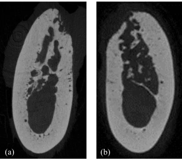
Figure 5. Representative micro-CT images of the extraction socket in the CON animal ··· 15
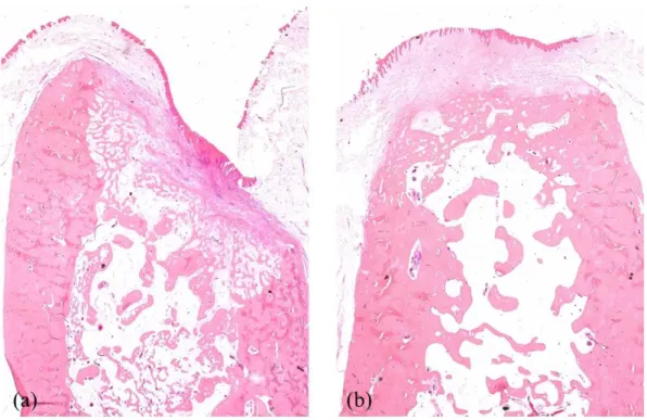
Figure 6. Representative histologic images of the extraction socket in the CON animal ··· 16
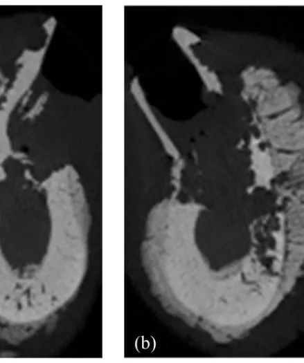
Figure 7. Representative micro-CT images of the septal bone in the CON animal ··· 17
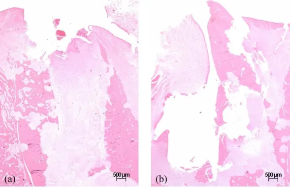
Figure 8. Representative histologic images of the septal bone in the CON animal ··· 18
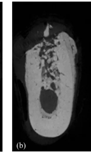
Figure 9. Representative micro-CT images of the extraction socket in the ZOL animal ··· 19
Figure 10. Representative histologic images of the extraction socket in the ZOL animal ··· 20
Figure 11. Representative micro-CT images of the septal bone in the ZOL animal ··· 21
Figure 12. Representative histologic images of the septal bone in the ZOL animal ··· 22
Figure 13. Bone microarchitectural morphometric analysis of the extraction sockets ··· 24
Figure 14. Bone microarchitectural morphometric analysis of the septal bone ··· 25
ABSTRACT
Micro-computed tomographic evaluation of bone healing
following dental extraction in beagle dogs treated with
zoledronic acid
Seung-Won Chung
Department of Dentistry
The Graduate School, Yonsei University
(Directed by Professor Young-Soo Jung, DDS, PhD)
Medication-related osteonecrosis of the jaws (MRONJ) is a rare but potentially severe side effect of long-term use of antiresorptive and antiangiogenic agents. However, the etiopathology of this condition is still not fully understood. In order to establish the pathogenesis of this disease, a reliable and reproducible animal model is required. The purpose of this study was to induce clinical features of MRONJ after tooth extraction in
1. In the experimental animal, extraction sites showed exposed bone with impaired wound healing at both post-extraction 1 month and 2 months.
2. Extraction sockets of the experimental animal showed significantly lower values of bone volume fraction, trabecular number, and trabecular thickness than those of the control animal at both post-extraction 1 month and 2 months.
3. Septal bone of the experimental animal showed significantly lower values of bone volume fraction than those of the control animal at both post-extraction 1 month and 2 months.
4. Cortical bone of the experimental animal showed significantly lower values of cortical area fraction, higher values of cortical porosity and average pore volume than those of the control animal at both post-extraction 1 month and 2 months. 5. In the experimental animal, new bone formation was noted outside buccal and
lingual cortex at both post-extraction 1 month and 2 months.
Although the sample size of this study is limited, this study introduces beagle dog as a promising large animal model for MRONJ. From this model, microarchitectural morphometric changes of bone healing after tooth extraction were evaluated. This animal model can be used for further research in the fields of MRONJ etiopathology and treatment.
Micro-computed tomographic evaluation of bone healing
following dental extraction in beagle dogs treated with
zoledronic acid
Seung-Won Chung
Department of Dental Science The Graduate School, Yonsei University
(Directed by Professor Young-Soo Jung, DDS, PhD)
I. INTRODUCTION
Bisphosphonates have been widely used for the treatment of osteoporosis and other various bone disease including skeletal complications induced by cancer metastasis by inhibiting bone turnover. (Delmas, 2005; Hatoum et al., 2008; Sambrook and Cooper, 2006) However, osteonecrosis of the jaws (ONJ), a rare but potentially severe side effect
jaws (BRONJ) in 2007 and updated it in 2009. (Advisory Task Force on Bisphosphonate-Related Ostenonecrosis of the Jaws and Maxillofacial, 2007; Ruggiero et al., 2009) With growing number of ONJ associated with other antiresorptive and antiangiogenic agents, AAOMS proposed the term medication-related osteonecrosis of the jaw (MRONJ) in 2014. (Ruggiero et al., 2014)
The current definition of MRONJ is based on its clinical characteristics and medical history; exposed bone or bone that can be probed through an intraoral or extraoral fistula in the maxillofacial region that has persisted for more than eight weeks, with current or previous treatment with antiresorptive or antiangiogenic agents, and absence of radiation therapy history to the jaws or obvious metastatic disease to the jaws. (Ruggiero et al., 2014) This definition, however, does not include pathogenesis or histopathology. (Pautke et al., 2010)
Despite the efforts to establish the pathogenesis of this disease, the etiopathology of this condition is not yet fully understood. (Allen and Burr, 2009; Otto et al., 2010; Ruggiero et al., 2014; Subramanian et al., 2011) Although MRONJ has been reported to occur spontaneously, dental extraction has been considered as a common risk factor that increases the incidence of the disease. (Ruggiero et al., 2009; Sonis et al., 2009) There have been various studies to develop clinical features of MRONJ in small animal models, such as mice and rodents. (Barba-Recreo et al., 2014; Biasotto et al., 2010; Hokugo et al.,
2010; Lopez-Jornet et al., 2010; Maahs et al., 2011; Sharma et al., 2013; Sonis et al., 2009) However, these small animal models have a crucial limitation due to anatomical divergence to human that they lack intracortical remodeling. (Kubek et al., 2010; Lelovas et al., 2008) Larger animal models, such as dogs, minipigs, and sheep, are considered more appropriate for the study of MRONJ because the nature of the disease affects cortical bone as well as trabecular bone. (Allen et al., 2013; Jee and Yao, 2001) Especially, canine bones have similar bone turnover rates to human bones. (Aerssens et al., 1998; Allen and Burr, 2008)
Recent studies on developing a large animal model for MRONJ have shown variable success rates. (Pautke et al., 2012; Voss et al., 2015; Voss et al., 2016) Although beagle dog is an established large animal model for investigating skeletal changes, reliable induction of exposed bone has not yet been developed. (Allen et al., 2013; Allen et al., 2010; Allen et al., 2011)
The purpose of this study was to induce clinical features of MRONJ after tooth extraction in zoledronate treated beagle dogs, and analyze bone healing by using micro-computed tomography (micro-CT).
II. MATERIALS AND METHODS
All animal procedures were performed under protocols approved by the institutional animal research ethics committee at CRONEX Co., Ltd (CRONEX-IACUC: 201506003).
1. Pilot study
One skeletally mature male beagle dog (9-month-old) with weight of 11.9kg was used in the pilot study. Following 1 week of acclimatization, the animal received zoledronic acid (Zometa®) intravenously at a dose of 0.067mg/kg every 10 days. Zoledronic acid was dissolved in saline and administered in a 10mL volume by using a 20-gauge catheter in the cephalic vein over 15-minute period under sedation. This dosage was determined according to the following factors; (1) oncological zoledronic dose in human is 0.067mg/kg/month (Clemons et al., 2006) and (2) relatively higher bone turnover rate in dogs (Cardaropoli et al., 2003). Therefore, the frequency of injection was about three times as frequently as it is used clinically.
After 3 months of injection, the animal underwent dental extraction of the lower left premolar 2, 3, 4 and molar 1 under sedation as a surgical intervention. Zoledronic acid injection was continued every 10 days for 1 month, and the animal underwent dental extraction of the lower right premolar 2, 3, 4 and molar 1. This study design was to allow
two different post-extraction times (post-extraction 1 month and 2 months) to be evaluated in the same animal.
For dental extractions, the animal was anesthetized using Propofol (8mg/kg) intravenously and intubated with continuous isofluorane during the surgery. After intraoral regional block of the inferior alveolar nerve, the crown of the tooth was split in the middle using high speed dental drill and each of the two roots was removed using dental forceps. After extraction, the extraction sockets were left open without any sutures. 1 month later, the animal was sacrificed for macroscopic evaluation of both extraction sites. However, there was absence of exposed bone on both extraction sites. (Fig. 1)
Fig. 1. Macroscopic assessment of the extraction sites in the pilot study. (a) Right mandible (post-extraction 1 month) showing a small inflammatory lesion, and (b) left mandible (post-extraction 2 months) showing complete mucosal coverage.
2. Experimental design
Two skeletally mature male beagle dogs (10-month-old) with weight of 12.7 and 12.9kg were used in the main study. Following 1 week of acclimatization, the animals were assigned to untreated control (CON; n=1) and zoledronic acid treated (ZOL; n=1) group. ZOL animal was administered with the same dose of zoledronic acid (Zometa®) intravenously every 10 days for 6 months, totalizing 18 administrations before extraction. CON animal was not treated during the experimental period.
After 6 months, all animals underwent dental extraction of the left lower premolar 2, 3, 4 and molar 1. 1 month later, all animals underwent extraction of the right lower premolar 2, 3, 4 and molar 1.
1 month after the extraction of the right premolars and molar, the animals were euthanized by intravenous administration of sodium pentobarbital. The right and left mandibles were dissected and placed in 10% formalin solution.
3. Macroscopic assessment
Clinical analysis was performed to verify the presence of exposed bone or fistula and photographs were taken for documentation.
4. Micro-CT analysis
The extraction sites of both the right and left hemi-mandibles were isolated and scanned by using high resolution micro-CT scanner SkyScan1173 (Bruker-microCT, Kartuizersweg 3B 2550 Kontich, Belgium). The tomographic images were acquired at energy settings of 90 kV and 88μA, integration time of 500ms, filtration by 1.0mm aluminum, beam hardening correction 40%, and 25μm pixel size. The cross-section reconstruction was made by using NreconⓇsoftware (ver. 1.6.9.8).
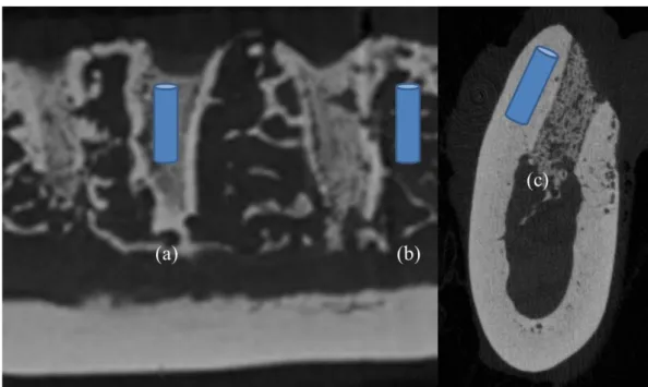
Comparing original 2D images with the segmented micro-CT scan images, a proper threshold was selected (82~255 mg HA/cm3). The cylindrical region of interest (ROI) was selected and measured with CT Analyzer (ver. 1.15.4.0) software.
Microarchitectural morphometric properties of the specimen were evaluated within the following ROIs; (1) center of the extraction sockets of each root, (2) center of the septal bone between the roots, and (3) Buccal and lingual cortical bone of each root. (Fig. 2)
Fig. 2. Region of interest. (a) Center of the extraction socket (b) Center of the septal bone (c) Buccal and lingual cortical bone
(1) Trabecular bone morphology indices included were as follows;
Total volume (TV, mm3) - Volume of the region of interest
Bone volume (BV, mm3) - Volume of the region segmented as trabecular bone Bone volume fraction (BV/TV, %) - Ratio of the segmented bone volume to the total
volume of the ROI
Trabecular number (Tb.N, 1/mm) - average number of trabeculae per unit length Trabecular thickness (Tb.Th, mm) - Mean thickness of trabeculae, assessed using
direct 3D methods
Trabecular separation (Tb.Sp, mm) - Mean distance between trabeculae, assessed using direct 3D methods
(2) Cortical bone morphology indices included were as follows;
Total cross-sectional area (Tt.Ar, mm2) - total cross-sectional area inside the periosteal envelope
Cortical volume (Ct.V, mm3) - Volume of cortical bone compartment
Cortical bone area (Ct.Ar, mm2) - Cortical volume (Ct.V) ÷ (number of slices × slice thickness
Total pore volume (Po.V, mm3) - Total volume occupied by cortical pores in the region of interest
Pore number (Po.N, n) - Number of cortical pores in a given volume
Cortical area fraction (Ct.Ar/Tt.Ar, %) - Ratio of cortical cross-sectional area to total cross-sectional area
Cortical porosity (Ct.Po, %) - Pore volume divided by the total cortical compartment volume
Average pore volume (Po.V/Po.N, mm3) - Total pore volume divided by the pore number
5. Histologic analysis
The extraction sockets, adjacent septal bone and cortical bone of the hemi-mandibles were processed for histologic analysis. Specimens were fixed with 4% phosphate-buffered saline formalin for 2 weeks and decalcified in 10% EDTA (pH 7.2) at 4°C for 28 days and immersed in paraffin. The samples were then embedded in paraffin and cut into 4-μm-thick sections. The sections were deparaffinized, rehydrated, and stained with hematoxylin and eosin (H-E).
6. Statistical analysis
Statistical analysis was carried out using SPSS 20.0 (IBM Corp., Armonk, NY, USA). Microarchitectural properties were compared between CON and ZOL animals using two-sample t tests. Difference in microarchitectural properties was compared between time (post-extraction 1 month and 2 months) within each group using paired t tests. A p-value of less than 0.05 was considered statistically significant.
III. RESULTS
1. Macroscopic assessment
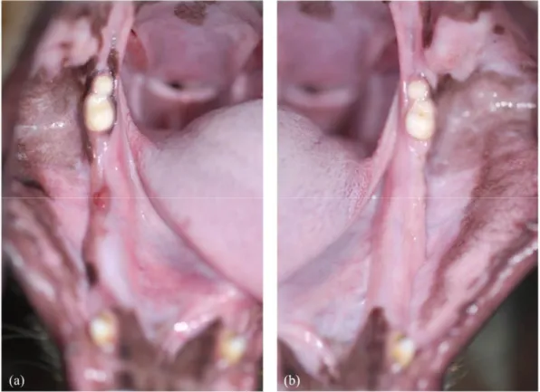
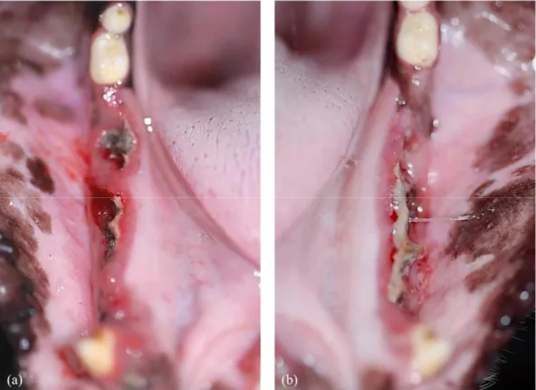
CON animal presented normal healing process and did not exhibit exposed bone or fistula at both post-extraction 1 month (CON1M) and 2 months (CON2M). (Fig. 3)
ZOL animal exhibited impaired wound healing and exposed bone at all extraction sites at both post-extraction 1 month (ZOL1M) and 2 months (ZOL2M). (Fig. 4)
Fig. 4. Macroscopic assessment of the extraction sites in the ZOL animal. (a) ZOL1M (b) ZOL2M showing impaired wound healing with exposed bone.
2. Micro-CT and histologic images
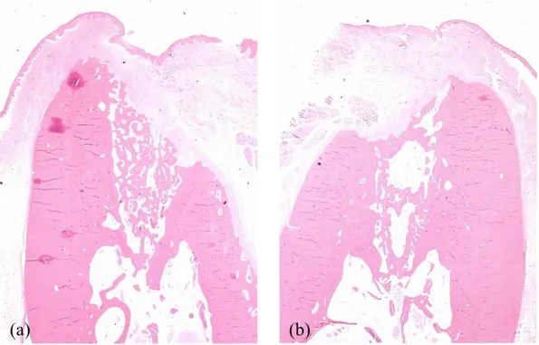
Extraction sockets of the CON animal demonstrated newly formed bone with normal trabecular pattern at both post-extraction 1 month and 2 months. (Fig. 5, 6)
Fig. 6. Representative histologic images of the extraction socket in the CON animal (x1.25). (a) CON1M (b) CON2M showing sufficient bone healing with normal trabecular pattern and epithelial coverage.
Septal bones of the CON animal showed normal trabecular pattern with intact cortical bone. (Fig. 7, 8)
Fig. 7. Representative micro-CT images of the septal bone in the CON animal. (a) CON1M (b) CON2M showing normal trabecular pattern.
Fig. 8. Representative histologic images of the septal bone in the CON animal (x1.25). (a) CON1M (b) CON2M showing normal trabecular pattern and epithelial coverage.
Extraction sockets of the ZOL animal presented insufficient amount of bone filling with extensive destruction of the pre-existing trabecular and cortical bone and sequestrum formation, and intense periosteal reaction around the outer cortex at both post-extraction 1 month and 2 months. (Fig. 9, 10)
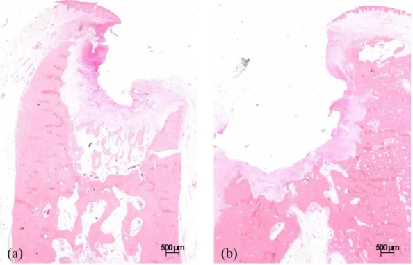
Fig. 10. Representative histologic images of the extraction socket in the ZOL animal (x1.25). (a) ZOL1M (b) ZOL2M showing insufficient bone filling, extensive destruction of the trabecular and cortical bone with sequestrum formation and intense periosteal reaction.
Septal bones of the ZOL animal showed destruction of the trabecular pattern and cortical bone with sequestrum formation with slight periosteal reaction and mucosal inflammation at both post-extraction 1 month and 2 months. (Fig. 11, 12)
Fig. 11. Representative micro-CT images of the septal bone in the ZOL animal. (a) ZOL1M (b) ZOL2M showing destruction of the trabecular and cortical bone with
Fig. 12. Representative histologic images of the septal bone in the ZOL animal (x1.25). (a) ZOL1M (b) ZOL2M destruction of the trabecular and cortical bone with severe mucosal inflammation.
3. Micro-CT analysis
(1) Extraction sockets
Bone microarchitectural morphometric assessment of the extraction sockets of each root revealed significantly lower values of bone volume fraction (BV/BT) in ZOL animal compared to that of the CON animal at both post-extraction 1 month and 2 months, suggesting severely compromised socket healing. ZOL animal presented deteriorated architectures showing significantly lower values of trabecular number (Tb.N) and trabecular thickness (Tb.Th), and significantly higher values of trabecular separation (Tb.Sp ) than those of CON animal at both post-extraction 1 month and 2 months. In ZOL animal, BV/BT and Tb.N decreased significantly from post-extraction 1 month to 2 months.
Fig. 13. Bone microarchitectural morphometric assessment of the extraction sockets. Abbreviations; BV/BT, bone volume fraction; Tb.N, trabecular number; Tb.Th, trabecular thickness; Tb.Sp, trabecular separation. Column and bar indicate mean and standard error, respectively. * indicates P<0.05 between CON and ZOL at the same post-extraction period; ** indicates P<0.05 between post-post-extraction 1 month and 2 months in the same group.
(2) Septal bone
Bone microarchitectural morphometric assessment of the septal bone revealed significantly lower values of BV/BT in ZOL animal compared to that of CON animal at both post-extraction 1 month and 2 months. There was no significant difference between the values of Tb.N, Tb.Th and Tb.Sp between ZOL and CON group.
(3) Cortical bone
Bone microarchitectural morphometric assessment of the cortical bone revealed significantly lower values cortical area fraction (Ct.Ar/Tt.Ar) and higher values of cortical porosity (Ct.Po) and average pore volume (Po.V/Po.N) in ZOL animal compared to those of CON animal at both post-extraction 1 month and 2 months.
Fig. 15. Bone microarchitectural morphometric assessment of the cortical bone. Abbreviations; Ct.Ar/Tt.Ar, cortical area fraction; Ct.Po, cortical porosity; Po.V/Po.N, average pore volume. * indicates P<0.05 between CON and ZOL at the same post-extraction period.
III. DISCUSSION
This study was conducted to induce clinical features of MRONJ after tooth extraction in zoledronate treated beagle dog, and evaluate the microarchitectural changes of bone healing using micro-CT. The results showed that zoledronic acid (0.067mg/kg) administrated approximately 3 times more frequently than the oncological use for 6 months successfully induced exposed bone at post-extraction 1 month and 2 months with insufficient bone fill in the extraction sockets and destruction of adjacent trabecular bone and cortical bone.
Until now, various animal models of MRONJ have been reported with most of them being rodents. (Barba-Recreo et al., 2014; Biasotto et al., 2010; Hokugo et al., 2010; Lopez-Jornet et al., 2010; Maahs et al., 2011; Sharma et al., 2013; Sonis et al., 2009) However, a potential drawback to the use of rodent models for MRONJ is the lack of Haversian remodeling in the rat skeleton. In humans, increased Haversian remodeling is the main cause of cortical porosity, but rodents lack a well-developed Haversian remodeling system. (Kubek et al., 2010)
reported compromised bone healing of extraction sites in only one of six zoledronic acid treated beagle dogs, after intravenous infusion of zoledronic acid (0.067mg/kg) every 2 weeks for 3 months. (Allen et al., 2011) Allen et al. also reported absence of exposed bone following dental extraction in beagle dogs treated after intravenous infusion of zoledronic acid (0.067mg/kg) every 2 weeks for 8 months. (Allen et al., 2013) Likewise, Huja et al. reported absence of ONJ lesion at surgical site in zoledronic acid treated beagle dogs after monthly infusion of 0.1mg/kg for 4 months. (Huja et al., 2011) From the variability of these study designs, it could be concluded that the frequency or the duration of zoledronic acid administration was insufficient to develop MRONJ model in beagle dogs.
However, there have been reports on successful inductions of MRONJ in other animals. Peutke et al. reported exposed bone following dental extraction in zoledronic acid treated minipigs after intravenous infusion of zoledronic acid (0.05mg/kg) every 1 week for 6 months. (Pautke et al., 2012) Also, Voss et al. reported spontaneous development of exposed bone following dental extraction in sheep after intravenous infusion of higher dose of zoledronic acid (0.075mg/kg) every 3 weeks for 4 months. (Voss et al., 2015) This may be attributed to the different mechanism of chewing of these animals from the beagle dogs.
This study attempted to compensate for the more rapid bone metabolism of beagle dogs. In the pilot study, zoledronic acid (0.067mg/kg) was administrated approximately 3 times more frequently than the oncological used in human. However, this frequency for 3 months duration was insufficient to develop spontaneous exposed bone after tooth extraction in a beagle dog. Although a small inflammatory lesion was present at the extraction site at postoperative 1 month, complete mucosal healing occurred at postoperative 2 months. Therefore, duration of zoledronic acid administration was increased with the same frequency. As a result, exposed bone was developed in ZOL animal at both at post-extraction 1 month and 2 months, while CON animal showed complete mucosal coverage at post-extraction 1 month and 2 months. Previous report on normal epithelial closure after tooth extraction in beagle dogs was within 14 days. (Cardaropoli et al., 2003)
In ZOL animal, the mean BV/TV of the extraction socket was 5% and 3% at post-extraction 1 month and 2 months respectively. This was significantly lower compared to the mean BV/TV of 50% and 51% in CON animal, showing significantly lower amount of bone fill in the extraction socket and severely compromised socket healing of the ZOL animal. Also, Tb.N and Tb.Th was significantly lower and Tb.Sp was significantly higher in ZOL animal compare to those of CON animal, implying deteriorated architectures of
The mean BV/TV of the septal bone in ZOL animal was 17% and 15% at post-extraction 1 month and 2 months respectively. This also was significantly lower compared to the mean BV/TV of 28% and 26% in CON animal, suggesting destruction of the trabecular bone outside the extraction socket. ZOL animal also presented lower values of mean Tb.N and Tb.Th and higher values of mean Tb.Sp of the septal bone compared to those of the CON animal, although they were not statistically significant.
ZOL animal also showed reduction of Ct.Ar/Tt.Ar, increase in Ct.Po and Po.V/Po.N at both post-extraction 1M and 2M period, compared with CON animal, demonstrating cortical disruption. ZOL animal also presented intense periosteal bone formation on both buccal and lingual surface of the cortical bone. Periosteal reaction was also reported in the study by Allen, despite the absence of exposed bone following dental extraction. (Allen et al., 2013) Periosteal reaction also suggests the innate high bone healing potential of the beagle dogs, which makes it more time consuming to develop an MRONJ model.
Recently, there have been attempts to develop an animal model of MRONJ with precedent osteoporosis. (Kim et al., 2015; Voss et al., 2016) Further study is required to establish a beagle dog model that includes osteopenic changes prior to bisphosphonate treatment in order to further evaluate the pathogenesis of this condition.
IV. Conclusion
Medication-related osteonecrosis of the jaws (MRONJ) is a rare but potentially severe side effect of long-term use of antiresorptive and antiangiogenic agents. However, the etiopathology of this condition is still not fully understood. In order to establish the pathogenesis of this disease, a reliable and reproducible animal model is required. The purpose of this study was to induce clinical features of MRONJ after tooth extraction in zoledronate treated beagle dogs and analyze bone healing using micro-CT. The results are as follows;
1. In the experimental animal, extraction sites showed exposed bone with impaired wound healing at both post-extraction 1 month and 2 months.
2. Extraction sockets of the experimental animal showed significantly lower values of bone volume fraction, trabecular number, and trabecular thickness than those of the control animal at both post-extraction 1 month and 2 months.
3. Septal bone of the experimental animal showed significantly lower values of bone volume fraction than those of the control animal at both post-extraction 1 month and 2 months.
5. In the experimental animal, new bone formation noted outside buccal and lingual cortex at both post-extraction 1 month and 2 months.
Although the sample size of this study is limited, this study introduces beagle dog as a promising large animal model for MRONJ. From this model, microarchitectural morphometric changes of bone healing after tooth extraction were evaluated. This animal model can be used for further research in the fields of MRONJ etiopathology and treatment.
V. References
Advisory Task Force on Bisphosphonate-Related Ostenonecrosis of the Jaws AAoO, Maxillofacial S: American Association of Oral and Maxillofacial Surgeons position paper on bisphosphonate-related osteonecrosis of the jaws. J Oral Maxillofac Surg 65(3): 369-376, 2007.
Aerssens J, Boonen S, Lowet G, Dequeker J: Interspecies differences in bone composition, density, and quality: potential implications for in vivo bone research. Endocrinology 139(2): 663-670, 1998.
Allen MR, Burr DB: Mandible matrix necrosis in beagle dogs after 3 years of daily oral bisphosphonate treatment. J Oral Maxillofac Surg 66(5): 987-994, 2008.
Allen MR, Burr DB: The pathogenesis of bisphosphonate-related osteonecrosis of the jaw: so many hypotheses, so few data. J Oral Maxillofac Surg 67(5 Suppl): 61-70, 2009.
Allen MR, Chu TM, Ruggiero SL: Absence of exposed bone following dental extraction in beagle dogs treated with 9 months of high-dose zoledronic acid combined with dexamethasone. J Oral Maxillofac Surg 71(6): 1017-1026, 2013.
Allen MR, Kubek DJ, Burr DB: Cancer treatment dosing regimens of zoledronic acid result in near-complete suppression of mandible intracortical bone remodeling in beagle dogs. J Bone Miner Res 25(1): 98-105, 2010.
Allen MR, Kubek DJ, Burr DB, Ruggiero SL, Chu TM: Compromised osseous healing of dental extraction sites in zoledronic acid-treated dogs. Osteoporos Int 22(2): 693-702, 2011.
Biasotto M, Chiandussi S, Zacchigna S, Moimas S, Dore F, Pozzato G, et al.: A novel animal model to study non-spontaneous bisphosphonates osteonecrosis of jaw. J Oral Pathol Med 39(5): 390-396, 2010.
Cardaropoli G, Araujo M, Lindhe J: Dynamics of bone tissue formation in tooth extraction sites. An experimental study in dogs. J Clin Periodontol 30(9): 809-818, 2003.
Clemons MJ, Dranitsaris G, Ooi WS, Yogendran G, Sukovic T, Wong BY, et al.: Phase II trial evaluating the palliative benefit of second-line zoledronic acid in breast cancer patients with either a skeletal-related event or progressive bone metastases despite first-line bisphosphonate therapy. J Clin Oncol 24(30): 4895-4900, 2006.
Delmas PD: The use of bisphosphonates in the treatment of osteoporosis. Curr Opin Rheumatol 17(4): 462-466, 2005.
Hatoum HT, Lin SJ, Smith MR, Barghout V, Lipton A: Zoledronic acid and skeletal complications in patients with solid tumors and bone metastases: analysis of a national medical claims database. Cancer 113(6): 1438-1445, 2008.
Hokugo A, Christensen R, Chung EM, Sung EC, Felsenfeld AL, Sayre JW, et al.: Increased prevalence of bisphosphonate-related osteonecrosis of the jaw with vitamin D deficiency in rats. J Bone Miner Res 25(6): 1337-1349, 2010.
Huja SS, Mason A, Fenell CE, Mo X, Hueni S, D'Atri AM, et al.: Effects of short-term zoledronic acid treatment on bone remodeling and healing at surgical sites in the maxilla and mandible of aged dogs. J Oral Maxillofac Surg 69(2): 418-427, 2011.
Jee WS, Yao W: Overview: animal models of osteopenia and osteoporosis. J Musculoskelet Neuronal Interact 1(3): 193-207, 2001.
Kim JW, Tatad JC, Landayan ME, Kim SJ, Kim MR: Animal model for medication-related osteonecrosis of the jaw with precedent metabolic bone disease. Bone 81: 442-448, 2015.
Kubek DJ, Burr DB, Allen MR: Ovariectomy stimulates and bisphosphonates inhibit intracortical remodeling in the mouse mandible. Orthod Craniofac Res 13(4): 214-222, 2010.
Lelovas PP, Xanthos TT, Thoma SE, Lyritis GP, Dontas IA: The laboratory rat as an animal model for osteoporosis research. Comp Med 58(5): 424-430, 2008.
Lopez-Jornet P, Camacho-Alonso F, Molina-Minano F, Gomez-Garcia F, Vicente-Ortega V: An experimental study of bisphosphonate-induced jaws osteonecrosis in Sprague-Dawley rats. J Oral Pathol Med 39(9): 697-702, 2010.
Maahs MP, Azambuja AA, Campos MM, Salum FG, Cherubini K: Association between bisphosphonates and jaw osteonecrosis: a study in Wistar rats. Head Neck 33(2): 199-207, 2011.
Marx RE: Pamidronate (Aredia) and zoledronate (Zometa) induced avascular necrosis of the jaws: a growing epidemic. J Oral Maxillofac Surg 61(9): 1115-1117, 2003.
Otto S, Hafner S, Mast G, Tischer T, Volkmer E, Schieker M, et al.: Bisphosphonate-related osteonecrosis of the jaw: is pH the missing part in the pathogenesis puzzle? J Oral Maxillofac Surg 68(5): 1158-1161, 2010.
Pautke C, Bauer F, Bissinger O, Tischer T, Kreutzer K, Steiner T, et al.: Tetracycline bone fluorescence: a valuable marker for osteonecrosis characterization and therapy. J Oral Maxillofac Surg 68(1): 125-129, 2010.
Pautke C, Kreutzer K, Weitz J, Knodler M, Munzel D, Wexel G, et al.: Bisphosphonate related osteonecrosis of the jaw: A minipig large animal model. Bone 51(3): 592-599, 2012. Ruggiero SL, Dodson TB, Assael LA, Landesberg R, Marx RE, Mehrotra B, et al.: American
Ruggiero SL, Dodson TB, Fantasia J, Goodday R, Aghaloo T, Mehrotra B, et al.: American Association of Oral and Maxillofacial Surgeons position paper on medication-related osteonecrosis of the jaw--2014 update. J Oral Maxillofac Surg 72(10): 1938-1956, 2014. Sambrook P, Cooper C: Osteoporosis. Lancet 367(9527): 2010-2018, 2006.
Sharma D, Hamlet S, Petcu E, Ivanovski S: Animal models for bisphosphonate-related osteonecrosis of the jaws--an appraisal. Oral Dis 19(8): 747-754, 2013.
Sonis ST, Watkins BA, Lyng GD, Lerman MA, Anderson KC: Bony changes in the jaws of rats treated with zoledronic acid and dexamethasone before dental extractions mimic bisphosphonate-related osteonecrosis in cancer patients. Oral Oncol 45(2): 164-172, 2009. Subramanian G, Cohen HV, Quek SY: A model for the pathogenesis of bisphosphonate-associated osteonecrosis of the jaw and teriparatide's potential role in its resolution. Oral Surg Oral Med Oral Pathol Oral Radiol Endod 112(6): 744-753, 2011.
Voss PJ, Stoddart M, Ziebart T, Zeiter S, Nelson K, Bittermann G, et al.: Zoledronate induces osteonecrosis of the jaw in sheep. J Craniomaxillofac Surg 43(7): 1133-1138, 2015. Voss PJ, Stoddart MJ, Bernstein A, Schmelzeisen R, Nelson K, Stadelmann V, et al.: Zoledronate
induces bisphosphonate-related osteonecrosis of the jaw in osteopenic sheep. Clin Oral Investig 20(1): 31-38, 2016.
ABSTRACT (IN KOREAN)
Micro-CT를 이용한 비스포스포네이트가 투여된
비글견의 발치 후 골 치유 분석
<지도교수 정 영 수>
연세대학교 대학원 치의학과
정 승 원
약제 관련 턱뼈 괴사(Medication-related osteonecrosis of the jaw, MRONJ)는 비스포스포네이트, RANKL antibody 계열의 골흡수억제제 및 혈관신생억제제의 장기간 사용과 관련되어 드물게 발생하지만 때로는 심각하게 나타날 수 있는 합병증이다. 턱뼈에서만 특이적으로 괴사가
아직까지 만족할만한 대형 동물모델은 개발되지 않은 상태이다. 이번 연구에서는 비스포스포네이트가 투여된 비글견에서 발치 후 MRONJ를 유도하고, 골 치유 양상을 micro-CT를 이용하여 분석하여 다음과 같은 결과를 얻었다 1. 대조군에서는 발치 후 1개월, 2개월에서 발치와 주변의 정상적인 연조직 치유가 관찰되었으나, 실험군에서는 발치 후 1개월, 2개월에서 모두 발치와 주변의 골노출이 관찰되었다. 2. 실험군에서는 대조군에 비해 발치 후 1개월, 2개월에서 발치와의 bone volume fraction, trabecular number, 그리고 trabecular thickness가 유의하게 낮게 나타났다.
3. 실험군에서는 대조군에 비해 발치 후 1개월, 2개월에서 치간 해면골의 bone volume fraction이 유의하게 낮게 나타났다.
4. 실험군에서는 대조군에 비해 발치 후 1개월, 2개월에서 피질골의 cortical area fraction이 유의하게 낮게, cortical porosity와 average pore volume은 유의하게 높게 나타났다.
5. 실험군에서는 발치 후 1개월, 2개월에서 협설측 피질골 외측에 신생골 형성이 관찰되었다.
이번 연구를 통해 Zoledronic acid가 투여된 비글견에서 발치 후 1개월부터 임상적으로 MRONJ의 정의에 부합하는 결과를 얻을 수 있었으며, 미세구조학적으로 심하게 손상된 발치와 주변 해면골 및 피질골의 골 구조를 확인하였다. 이 모델이 MRONJ의 병태생리 규명 및 치료 개발을 위한 연구에 사용될 수 있을 것으로 평가된다.