Review Article
Toxicol. Res. Vol. 35, No. 4, pp. 331-341 (2019) https://doi.org/10.5487/TR.2019.35.4.331
Open Access
Toxicological Research
Molecular Markers in Sex Differences in Cancer
Ji Yoon Shin, Hee Jin Jung and Aree Moon
Duksung Innovative Drug Center, College of Pharmacy, Duksung Women’s University, Seoul, Korea
Abstract
Cancer is one of the common causes of death with a high degree of mortality, worldwide. In many types of can-cers, if not all, sex-biased disparities have been observed. In these cancan-cers, an individual’s sex has been shown to be one of the crucial factors underlying the incidence and mortality of cancer. Accumulating evidence suggests that differentially expressed genes and proteins may contribute to sex-biased differences in male and female can-cers. Therefore, identification of these molecular differences is important for early diagnosis of cancer, prediction of cancer prognosis, and determination of response to specific therapies. In the present review, we summarize the differentially expressed genes and proteins in several cancers including bladder, colorectal, liver, lung, and non-small cell lung cancers as well as renal clear cell carcinoma, and head and neck squamous cell carcinoma. The sex-biased molecular differences were identified via proteomics, genomics, and big data analysis. The identified molecules represent potential candidates as sex-specific cancer biomarkers. Our study provides molecular insights into the impact of sex on cancers, suggesting strategies for sex-biased therapy against certain types of cancers. Key words: Sex difference, Cancer, Sex hormone, Chemotherapy
INTRODUCTION
Cancer is one of the most common diseases with an extremely high mortality rate (1). Several types of cancer show sex-biased tendencies in mortality and incidence (2-4). Lung cancer shows the highest incidence and mortal-ity in males, and ranks third in incidence and second in mortality among females (5). The incidence and mortality of liver cancer in males are significantly higher than in females (6). The incidence of bladder cancer is three- to four-fold higher in males than in females (7). However, females are diagnosed with more advanced disease and worse prognosis after treatment compared with males (8,9). Data obtained from Korea and Japan indicate that colorectal cancer is not only associated with the highest mortality in females over 65 years of age, but also has a
significantly higher incidence and mortality rate in females than in males over the age of 65 years (10-12). In renal clear cell carcinoma, the incidence rate was twice as com-mon in males than in females, and males showed a lower survival rate (13). Studies investigating the role of gender and incidence or mortality in head and neck squamous cell carcinoma had no significant differences by sex (14).
Sex differences in cancer are presumably induced by sex hormones, especially estrogen (15). Sex hormones are known to modulate gene expression in many cancers (16). Sexually dimorphic expression of molecules (gene or pro-tein) are referred to as ‘sex-biased’ (17-19). Sex-biased disparities in responsiveness and side effects to anticancer drugs have also been observed (reviewed in 16). For instance, the clearance of 5-fluorouracil (FU) is higher in males than in females (20,21). Females treated with 5-FU showed higher levels of toxicity such as nausea, vomiting, alope-cia and leukopenia than males (21,22). The influence of gender on drug metabolism may contribute at least in part, to the sex-specific differences in susceptibility to antican-cer drugs (23-27).
Mounting evidence suggests that genetic and molecular differences between male and female cancers may contrib-ute to sex-biased disparities in mortality and incidence of cancers (28). For instance, male patients with lung adeno-Correspondence to: Aree Moon, Duksung Innovative Drug
Cen-ter, College of Pharmacy, Duksung, Women’s University, 33, 144-gil, Samyang-ro, Dobong-gu, Seoul 01369, Korea
Email: [email protected]
This is an Open-Access article distributed under the terms of the Creative Commons Attribution Non-Commercial License (http:// creativecommons.org/licenses/by-nc/3.0) which permits unre-stricted non-commercial use, distribution, and reproduction in any medium, provided the original work is properly cited.
carcinoma showed 1.636-fold higher frequency of genetic alterations than female patients, and greater genetic alter-ations were related to worse overall survival in males (29). At the genetic/molecular level, gene polymorphism and differential expression have been recognized as key fac-tors for sex-biased differences in cancer incidence between males and females (16). However, the molecular differ-ences between male and female cancer patients have yet to be elucidated. A comprehensive characterization of sex-biased molecular differences in cancer patients was con-ducted by analyzing the high-throughput molecular data obtained from The Cancer Genome Atlas (TCGA) (18).
Proteomics/genomics is one of the most efficient ap-proaches to study the expression of proteins and genes (30). Currently, big data analytics of patient information is widely used in molecular studies. In this review, we have summarized the genes and proteins associated with sex-dependent expression via approaches such as proteomics, genomics, and big data analytics in bladder, colorectal, liver, lung and non-small cell lung cancers, renal clear cell and neck squamous cell carcinoma. All these cancers except renal clear cell and head and neck squamous cell carcinoma show sex-biased incidence and mortality. This review facili-tates the design of an efficient strategy for sex-specific
can-cer therapy.
BLADDER CANCER
The incidence of bladder cancer is four times higher in females than in males (31,32). Studies investigated the association between the risk of bladder cancer and hypometh-ylation of long interspersed nuclear element-1 (LINE-1) (33). The findings suggest a lower frequency of methyla-tion of LINE-1 in females than in males. Hypomethyla-tion of LINE-1 may be an effective biomarker for the risk of bladder cancer in females. The lowest degree of LINE-1 methylation in females indicates a 2.5-fold significantly higher risk of bladder cancer. In males, however, there was no correlation between hypomethylation of LINE-1 and risk of bladder cancer (33). Sex-biased molecules in bladder cancer are listed in Table 1.
COLORECTAL CANCER
A comparison of 54 colorectal cancer biopsies and mRNAs of adjacent normal mucosal tissues by RT-PCR analysis revealed a significant overexpression of twist family basic helix-loop-helix transcription factor 1 (TWIST1) in colorec-Table 1. Sex-biased molecular differences in cancers
Bladder cancer
Name Gender Method Reference Remarks
Male Female
LINE1
(Methylation) −
Quantitative Bisulfite
Pyrosequencing 33
Hypomethylation of LINE1 in females indicates higher risk of bladder cancer. No relation in male.
−: Low frequency of methylation related to bladder cancer
Colorectal cancer
Name Gender Method Reference Remarks
Male Female
ERα +
Immunoblot assay 42
Significantly increased in tumor tissue than normal tissue of male.
No alteration in female.
ERβ − −− Significantly decreased in tumor tissue of
female than male.
K-ras
(Point mutations)
−
(younger male) Sequence-specific
Oligonucleotide probes 41
More frequent mutations in older male than older female. + (older male) p16INK4a (Methylation) + Methylation-specific PCR analysis 44 TWIST1 + RT-PCR analysis 34
+: High expression/point mutation/methylation related to colorectal cancer −: Low expression/point mutation related to colorectal cancer
Table 1. Continued
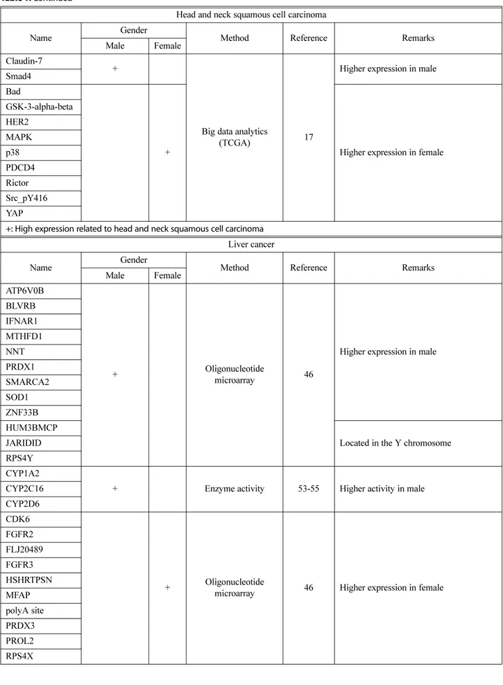
Head and neck squamous cell carcinoma
Name Gender Method Reference Remarks
Male Female
Claudin-7
+
Big data analytics
(TCGA) 17
Higher expression in male Smad4
Bad
+ Higher expression in female
GSK-3-alpha-beta HER2 MAPK p38 PDCD4 Rictor Src_pY416 YAP
+: High expression related to head and neck squamous cell carcinoma Liver cancer
Name Gender Method Reference Remarks
Male Female
ATP6V0B
+ Oligonucleotide
microarray 46
Higher expression in male BLVRB IFNAR1 MTHFD1 NNT PRDX1 SMARCA2 SOD1 ZNF33B HUM3BMCP
Located in the Y chromosome JARIDID
RPS4Y CYP1A2
+ Enzyme activity 53-55 Higher activity in male
CYP2C16 CYP2D6 CDK6
+ Oligonucleotide
microarray 46 Higher expression in female FGFR2 FLJ20489 FGFR3 HSHRTPSN MFAP polyA site PRDX3 PROL2 RPS4X
Table 1. Continued
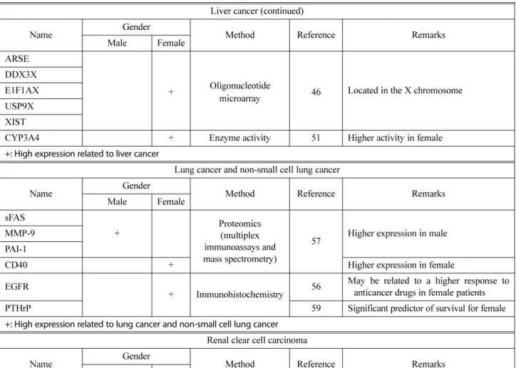
Liver cancer (continued)
Name Gender Method Reference Remarks
Male Female
ARSE
+ Oligonucleotide
microarray 46 Located in the X chromosome DDX3X
E1F1AX USP9X XIST
CYP3A4 + Enzyme activity 51 Higher activity in female
+: High expression related to liver cancer
Lung cancer and non-small cell lung cancer
Name Gender Method Reference Remarks
Male Female sFAS + Proteomics (multiplex immunoassays and mass spectrometry)
57 Higher expression in male MMP-9
PAI-1
CD40 + Higher expression in female
EGFR
+ Immunohistochemistry 56
May be related to a higher response to anticancer drugs in female patients
PTHrP 59 Significant predictor of survival for female
+: High expression related to lung cancer and non-small cell lung cancer Renal clear cell carcinoma
Name Gender Method Reference Remarks
Male Female
AR
+
Big data analytics
(TCGA) 17
Higher expression in male ARHJ IRS1 JAB1 PAI-1 PKC-alpha Transglutaminase VEGFR2 VHL 4E-BP1
+ Higher expression in female
Akt DJ-1 NDRG1 p38 PTEN Src VEGFR2 VHL
tal cancer samples compared with benign colon mucosa and a significant increase in patients with nodal invasion (34). TWIST1 is involved in the regulation of epithelial-mesenchymal transition (EMT), inducing cancer progres-sion and metastasis, in various cancers (35-37). Notably, the expression of TWIST1 mRNA was higher in males than in females, suggesting the possibility of differential transcriptional regulation of TWIST1 by sex hormones (34).
In early stage of colorectal carcinogenesis, KRAS muta-tions were observed in 27-43% of patients (38-40). DNA from 251 primary tumors of Norwegian patients with col-orectal carcinoma was analyzed using sequence-specific oligonucleotide probes for the presence of k-ras point mutations at codons 12 and 13 (41). Male patients younger than 40 years carried fewer k-ras mutations in colon tumors compared with female patients (41). This finding sug-gested dominance of ras-independent pathways of colon cancer in younger males (41). In contrast, older males car-ried a higher number of k-ras mutations than older females (41).
The expression of ERα and ERβ protein showed no sex differences between normal colon mucosal tissues; how-ever, significant differences were observed between tumor tissues (42). The ERα protein expression was significantly increased in tumor tissues of male patients compared with normal tissues (42). By contrast, the ERα level did not dif-fer between tumor tissues and normal tissues of female patients (42). The level of ERβ protein and mRNA was significantly reduced in both male and female patients diag-nosed with colon cancer, although a greater decrease was observed in males (42,43). The ERβ level was decreased in poorly differentiated tumors of males (42).
Colorectal cancer associated with the proximal colon in females may show a different disease subtype (44). In 120 sporadic colorectal cancers, methylation-specific PCR was performed to determine whether the methylation of the CpG island in the 5' region of the p16INK4a tumor suppres-sor gene was associated with sex or other clinicopathologi-cal characteristics. In female patients, methylation-positive cancer was 8.8-fold higher than in male patients, and meth-ylation of p16INK4a was associated with poorly differenti-ated tumors. Female patients with p16INK4a methylation may represent an important database of molecular alterations associated with sporadic colorectal cancers (44). Sex-biased molecular differences in colorectal cancer are listed in Table 1.
HEAD AND NECK SQUAMOUS
CELL CARCINOMA
Abundant signals associated with sex-biased protein expres-sion in head and neck squamous cell carcinoma were detected via TCGA database analysis (18). In patients with head and neck squamous cell carcinoma, 12 of 15
sex-biased proteins were identified, and the expression of Yes associated protein (YAP), programmed cell death 4 (PDCD4), glycogen synthase kinase 3 (GSK3-α/β), mitogen-acti-vated protein kinase (MAPK), Src, p38, human epidermal growth factor receptor 2 (HER2), B cell leukemia/lym-phoma (BCL2) associated agonist of cell death (Bad), and Rictor were upregulated in females whereas Claudin-7 and Smad4 were upregulated in males (18). SRC plays an important role in regulating a variety of cellular signal transduction pathways. The SRC kinase pathways are fre-quently activated in many carcinomas, especially metastatic diseases (45). In head and neck squamous cell carcinoma, SRC kinase may be the key molecule that shows a poten-tial to be a female-specific prognosis marker. Sex-biased molecular differences in head and neck squamous cell car-cinoma are listed in Table 1.
LIVER CANCER
In hepatocellular carcinoma, 27 genes were identified from male (n = 34) and female (n = 16) hepatocellular car-cinoma patient tissue mRNAs, which showed sex-specific differential expression (46). Among these genes, 12 showed higher and 15 lower levels of expression in males com-pared with females. Among the 12 genes expressed higher in male samples, interferon (α and β) receptor 1 (IFNAR1), ATPase H+ transporting V0 subunit b (ATP6V0B), biliv-erdin reductase B (BLVRB), zinc finger protein 33B (ZNF33B), nicotinamide nucleotide transhydrogenase (NNT), methylenetetrahydrofolate dehydrogenase, cyclohy-drolase and formyltetrahydrofolate synthetase 1 (MTHFD1), superoxide dismutase 1 (SOD1), SWI/SNF related, matrix associated, actin dependent regulator of chromatin, sub-family a, member 2 (SMARCA2) and peroxide peroxidase 1 (PRDX1) were located on the autosome while ribo-somal protein S4 Y-linked 1 (RPS4Y), lysine demethylase 5D (JARIDID), and HUM3BMCP were located on the Y chromosome. PRDX1 and SOD1 were highly expressed in male hepatocellular carcinoma samples (46). In hepato-cellular carcinoma, PRDX1 may be a key molecule for sex-biased mortality and incidence. PRDX, a major com-ponent of antioxidant enzymes, removes reactive oxygen species (47). PRDX1 is a versatile molecule that regulates cell growth, differentiation, cell death and tumor suppres-sion (48,49). However, the 15 genes with a higher expres-sion in female samples include cyclin dependent kinase 6 (CDK6), PROL2, FLJ20489, fibroblast growth factor receptor 2 (FGFR2), microfibrillar-associated protein 2 (MFAP), peroxiredoxin 3 (PRDX3), fibroblast growth fac-tor recepfac-tor 3 (FGFR3), polyA site, and HSHRTPSN located on the autosome, and inactive X specific transcripts (XIST), ubiquitin specific peptidase 9 X-linked (USP9X), E1F1AX, ribosomal protein S4 X-linked (RPS4X), arylsulfatase E (ARSE), and DEAD-box helicase 3 X-linked (DDX3X)
on the X chromosome (46).
Sex differences in the gene expression of drug metabo-lizing enzymes and transporters result in differences in drug absorption, distribution, metabolism and excretion, possi-bly affecting drug efficacy and adverse reactions (50). Cyto-chrome P450 family 3 subfamily A member 4 (CYP3A4) is an enzyme that is responsible for the metabolism of over 50% of all therapeutic drugs (51,52). Higher CYP3A4 activity was observed in females than in males; by con-trast, higher activities of cytochrome P450 family 1 sub-family A member 2 (CYP1A2), cytochrome P450 2C16 (CYP2C16), cytochrome P450 family 2 subfamily D mem-ber 6 (CYP2D6) and cytochrome P450 family 2 subfam-ily E member 1 (CYP2E1) were detected in males than in females (53-55). The analysis of enzymatic activity in human liver microsomes and hepatocytes revealed no sig-nificant differences in hepatic CYP3A4 activity between males and females (50). However, in primary hepato-cytes, a two-fold higher activity of CYP3A4 was observed in females than in males (51). Sex-biased molecular differ-ences in liver cancer are listed in Table 1.
LUNG CANCER AND NON-SMALL CELL
LUNG CANCER
Epidermal growth factor receptor (EGFR), the most important therapeutic target in lung cancer, was specifi-cally expressed in females (18). Erlotinib and gefitinib are the major anti-cancer drugs that target EGFR pathway in lung cancer (56). Female patients showed higher response to erlotinib than male patients (56). This higher response among female patients may be related to a higher expres-sion of EGFR observed in females (18,56).
Sex-specific biomarkers were identified in non-small cell lung cancer, using multiplex immunoassays and mass spectrometry (57). In males, soluble Fas (sFAS), matrix metalloproteinase-9 (MMP-9), and plasminogen activator inhibitor-1 (PAI-1) were strongly predictive biomarkers, whereas soluble cluster of differentiation 40 (sCD40) was prognostic for cancers in females (57). The factors under-lying these gender-biased differences in non-small cell lung cancer are not clear. Studies to date indicate that these pro-teins play different roles in male and female patients due to endocrine differences.
Parathyroid hormone-related protein (PTHrP) is upregu-lated in tumors with skeletal metastasis and commonly expressed in non-small cell lung carcinomas (58,59). Female patients diagnosed with non-small cell lung carcinoma with and without PTHrP showed a median survival of 55 and 22 months, respectively, whereas male survival was independent of PTHrP status for 38 months. PTHrP was suggested as a significant predictor of survival in female patients after adjusting for stage, age, and histology (59). Sex-biased molecular differences in lung cancer and
non-small cell lung cancer are listed in Table 1.
RENAL CLEAR CELL CARCINOMA
Using TCGA database analytics, sex-biased proteins from renal clear cell carcinoma were analyzed (18). In renal clear cell carcinoma, 18 out of 25 proteins were uplated in females. These include N-myc downstream regu-lated 1 (NDRG1), Akt, phosphatase and tensin homolog (PTEN), DJ-1, 4E binding protein 1 (4E-BP1), Src, and p38. The von Hippel-Lindau (VHL), phosphoribosylanthrani-late isomerase 1 (PAI-1), PKC-alpha, VEGFR2, androgen receptor (AR), ARHJ, insulin receptor substrate 1 (IRS1), C-Jun activation domain-binding protein-1 (JAB1) genes were upregulated in males (18). Sex-biased molecular dif-ferences in renal clear cell carcinoma are listed in Table 1.
SEX-BIASED MOLECULES IN ANIMAL CANCERS
Large portions of genome in animals are shown to be sex-specific. Sex-biased gene expression may increase the differences between female and male development, to facilitate adaptive sex differentiation in vivo (60-62). The genomic distribution of sex-biased genes (i.e., chromo-somal binding patterns) in animals also provides clues to the evolutionary process and the underlying genetic varia-tion in development according to sex (63,64). Mice and rats are frequently used for cancer research (65). In this review, we have summarized the sex-biased molecular differ-ences in animal cancers.
Recently, a Hras12V transgenic mouse model was gen-erated to demonstrate the development of male-biased hepatic tumorigenesis (66). The peritumor and tumoral tis-sues of male and female transgenic mice were compared with normal liver tissues of non-transgenic mice via pro-teomic analysis. During hepatic tumorigenesis, 19 proteins derived from males and 10 proteins from female mice showed sex-biased expression (66). The upregulated pro-teins in males were Rho GDP dissociation inhibitor alpha (ARHGDIA), fatty acid binding protein 1 (FABP1), nucle-ophosmin 1 (NPM1), ribosomal protein lateral stalk sub-unit P2 (RPLP2), dihydrodiol dehydrogenase (DHDH), ATP5D, farnesyl diphosphate synthase (FDPS), and ubiqui-lin 1 (UBQLN1). The proteins involved in DNA packing include Histone H4, Histone H2AA, and Histone H2AB. SFL2, protein disulfide isomerase family A member 6 (PDIA6), tropomyosin 1 (TPM1) were also upregulated in a male-specific manner (66). Female-specific proteins such as acyl-CoA oxidase 1 (ACOX1), adenosylhomocystein-ase (AHCY), betaine-homocysteine S-methyltransferadenosylhomocystein-ase 2 (BHMT2), glutathione S-transferase alpha 3 (GSTA3), basonuclin 2 (BNC2), calmodulin 1 (CALM1), and profil-ing 1 (PFN1) were downregulated (66). However, apoli-poprotein A4 (APOA4), eukaryotic translation elongation
factor 2 (Eef2), and heat shock protein family A member 8 (HSPA8) were upregulated in females. The metabolic cat-egory of proteins including aldehyde dehydrogenase 1 family member L1 (ALDH1L1), phosphoglycerate kinase 1 (PGK1), and sterol carrier protein 2 (SCP2) were down-regulated in males. Glutathione peroxidase 1 (GPX1), per-oxiredoxin 6 (PRDX6), cytochrome b5 type A (CYB5A),
and heat shock protein family E member 1 (HSPE1) showed a reversal of expression in males and females (66).
To study the effects of forkhead box A1 (FOXA1), a member of the forkhead class of DNA-binding proteins, on liver cancer production, liver tumors were induced in female and male controls and liver-specific FOXA1/2-deficient mice (67). These hepatocyte nuclear factors are Table 2. Sex-biased molecules in animal liver cancers
Gene name Gender Method Reference Remarks
Male Female
ARHGDIA
+
Proteomics (2D-Fluorescence difference gel electrophoresis), genetics, and immunoblot assay
66 No significance in female ATP5D DHDH FABP1 FDPS Histone H2AA Histone H2AB Histone H4 NPM1 PDIA6 RPLP2 SFL2 TPM1 UBQLN1 FOXA1 + Chromatin Immunoprecipitation (ChIP) assays 67 FOXA2 p53 + - Immunoblot assay, RT-PCR
analysis, and microarray 68 Higher expression in male Pten
Rb APOA4
+
Proteomics (2D-Fluorescence difference gel electrophoresis), genetics, and immunoblot assay
66 No significance in male Eef2 HSPA8 ALDH1L1 - No significance in female PGK1 SCP2 ACOX1 - No significance in male AHCY BHMT2 BNC2 CALM1 GSTA3 PFN1
+: High expression related to hepatocellular carcinomas in animals −: low expression related to hepatocellular carcinomas in animals
transcriptional activators of liver-specific transcripts such as albumin and transthyretin, and interact with chromatin as pioneering factors. After treatment with carcinogens, large and diverse tumors were detected in the females with FOXA1/2 deficiency, whereas tumor growth in male mutants was reduced compared with the control. Thus, the expres-sion of FOXA-specific genes in mice seems to be related to the prognosis of hepatocellular carcinoma. It was sug-gested that FOXA1 and FOXA2 may promote hepatocel-lular carcinoma in male mice, while protecting female mice against hepatocellular carcinoma (67).
The expression of tumor suppressor genes (PTEN, p53 and Rb) was down-regulated in the early stages of female glycine N-methyltransferase (Gnmt)-/- mice, but not in male mice (65). The GNMT mediating 1-carbon metabolism affects DNA methylation by controlling the ratio of S-ade-nosylmethionine to S-adenosylmorphine (68,69). These data suggest that Gnmt deficiency not only increases the expres-sion of tumor genes but also induces a decrease in the expression of other tumor suppressor genes in the early stages of tumorigenesis in female rats, which explains the higher risk of hepatocellular carcinoma in female Gnmt-/-mice (67). The sex-biased molecules are listed in Table 2.
CONCLUSIONS
We have summarized the molecules that are differentially expressed between males and females in bladder cancer, colorectal cancer, liver cancer, lung cancer, non-small cell lung cancer, and head and neck squamous cell carcinoma and renal clear cell carcinoma. The sex-biased molecular differences in cancers are listed in Table 1 and 2. Molecu-lar differences include sex-specific upregulation or down-regulation of proteins and mRNAs, frequency of gene methylation, and activity of enzymes.
Sex-specific cancer therapies are indicated according to the differential role played by sex-biased molecular expres-sion in oncology and pharmacology. Nevertheless, sex-specific follow-up clinical trials beyond sex hormone-spe-cific therapy remain at an early stage (70). Many clinical and pre-clinical results suggest that sex and gender differ-ences may affect drug-promoted pathological states such as drug digestion and drug dependence or addiction. Gen-der differences in drug pharmacodynamics and pharmaco-kinetics will also affect drug addiction, dependence and side effects (71,72). Efforts are needed to evaluate the clinical utility of sex-biased target therapies in a larger range of patient cohorts (70). There is still a lack of bio-logic relevance of cancer diagnosis, prognosis, severity, and prediction of response to treatment. Genomics studies showed a variation in the expression of autosomal genes between males and females (71). We believe that this vari-ation may affect sex-specific cancer prognosis. Metabolo-mics studies also revealed gender differences in metabolite
levels and their correlations with genetic markers (72). Mechanisms underlying the differential expression of molecules in male and female cancers remain to be eluci-dated. Further investigation into the association of these molecules with sex-specific incidence and mortality of cancers is also needed. Our review elucidates the molecu-lar differences to facilitate the identification of sex-spe-cific cancer biomarkers.
ACKNOWLEDGMENTS
This study was supported by the Duksung Women’s University Research Grant 2018.
CONFLICT OF INTEREST
The authors have no conflict of interest to disclose. Received July 3, 2019; Revised July 5, 2019; Accepted July 10, 2019
REFERENCES
1. Jemal, A., Bray, F., Center, M.M., Ferlay, J., Ward, E. and Forman, D. (2011) Global cancer statistics. CA Cancer J. Clin., 61, 69-90.
2. Cook, M.B., McGlynn, K.A., Devesa, S.S., Freedman, N.D. and Anderson, W.F. (2011) Sex disparities in cancer mortal-ity and survival. Cancer Epidemiol. Biomarkers Prev., 20, 1629-1637.
3. Siegel, R.L., Miller, K.D. and Jemal, A. (2016) Cancer sta-tistics. CA Cancer J. Clin., 66, 7-30.
4. Fitzmaurice, C., Allen, C., Barber, R.M., Barregard, L., Bhu-tta, Z.A., Brenner, H., Dicker, D.J., Chimed-Orchir, O., Dan-dona, R., DanDan-dona, L., Fleming, T., Forouzanfar, M.H., Hancock, J., Hay, R.J., Hunter-Merrill, R., Huynh, C., Hos-good, H.D., Johnson, C.O., Jonas, J.B., Khubchandani, J., Kumar, G.A., Kutz, M., Lan, Q., Larson, H.J., Liang, X., Lim, S.S., Lopez, A.D., MacIntyre, M.F., Marczak, L., Mar-quez, N., Mokdad, A.H., Pinho, C., Pourmalek, F., Salomon, J.A., Sanabria, J.R., Sandar, L., Sartorius, B., Schwartz, S.M., Shackelford, K.A., Shibuya, K., Stanaway, J., Steiner, C., Sun, J., Takahashi, K., Vollset, S.E., Vos, T., Wagner, J.A., Wang, H., Westerman, R., Zeeb, H., Zoeckler, L., Abd-Allah, F., Ahmed, M.B., Alabed, S., Alam, N.K., Aldhahri, S.F., Alem, G., Alemayohu, M.A., Ali, R., Al-Raddadi, R., Amare, A., Amoako, Y., Artaman, A., Asayesh, H., Atnafu, N., Awasthi, A., Saleem, H.B., Barac, A., Bedi, N., Bensenor, I., Berhane, A., Bernabé, E., Betsu, B., Binagwaho, A., Boneya, D., Campos-Nonato, I., Castañeda-Orjula, C., Catalá-López, F., Chiang, P., Chibueze, C., Chitheer, A., Choi, J.Y., Cowie, B., Damtew, S., das, N.J., Dey, S., Dharmaratne, S., Dhil-lon, P., Ding, E., Driscoll, T., Ekwueme, D., Endries, A.Y., Farvid, M., Farzadfar, F., Fernandes, J., Fischer, F., G/Hiwot, T.T., Gebru, A., Gopalani, S., Hailu, A., Horino, M., Horita, N., Husseini, A., Huybrechts, I., Inoue, M., Islami, F., Jakovl-jevic, M., James, S., Javanbakht, M., Jee, S.H., Kasaeian, A.,
Kedir, M.S., Khader, Y.S., Khang, Y.H., Kim, D., Leigh, J., Linn, S., Lunevicius, R., El Razek, H.M.A., Malekzadeh, R., Malta, D.C., Marcenes, W., Markos, D., Melaku, Y.A., Meles, K.G., Mendoza, W., Mengiste, D.T., Meretoja, T.J., Miller, T.R., Mohammad, K.A., Mohammadi, A., Moham-med, S., Moradi-Lakeh, M., Nagel, G., Nand, D., Le Nguyen, Q., Nolte, S., Ogbo, F.A., Oladimeji, K.E., Oren, E., Pa, M., Park, E.K., Pereira, D.M., Plass, D., Qorbani, M., Radfar, A., Rafay, A., Rahman, M., Rana, S.M., Søreide, K., Satpathy, M., Sawhney, M., Sepanlou, S.G., Shaikh, M.A., She, J., Shiue, I., Shore, H.R., Shrime, M.G., So, S., Soneji, S., Stathopoulou, V., Stroumpoulis, K., Sufiyan, M.B., Sykes, B.L., Tabarés-Seisdedos, R., Tadese, F., Tedla, B.A., Tessema, G.A., Thakur, J.S., Tran, B.X., Ukwaja, K.N., Uzochukwu, B.S.C., Vlassov, V.V., Weiderpass, E., Wubshet, T.M., Yebyo, H.G., Yimam, H.H., Yonemoto, N., Younis, M.Z., Yu, C., Zaidi, Z., Zaki, M.E.S., Zenebe, Z.M., Murray, C.J.L. and Naghavi, M. (2017) Global, regional, and national cancer incidence, mortality, years of life lost, years lived with dis-ability, and disability-adjusted life-years for 32 cancer groups, 1990 to 2015: a systematic analysis for the global burden of disease study. JAMA Oncol., 3, 524-548.
5. Bray, F., Ferlay, J., Soerjomataram, I., Siegel, R.L., Torre. L.A. and Jemal, A. (2018) Global cancer statistics 2018: GLOBOCAN estimates of incidence and mortality world-wide for 36 cancers in 185 countries. CA Cancer J. Clin., 68, 394-424.
6. Hefaiedh, R., Ennaifer, R., Romdhane, H., Ben, N.H., Arfa, N., Belhadj, N., Gharbi, L. and Khalfallah, T. (2013) Gen-der difference in patients with hepatocellular carcinoma. Tunis Med., 91, 505-508.
7. Dobruch, J., Daneshmand, S., Fisch, M., Lotan, Y., Noon, A.P., Resnick, M.J., Shariat, S.F., Zlotta, A.R. and Boorjian, S.A. (2016) Gender and bladder cancer: a collaborative review of etiology, biology, and outcomes. Eur. Urol., 69, 300-310.
8. Horstmann, M., Witthuhn, R., Falk, M. and Stenzl, A. (2008) Gender-specific differences in bladder cancer: a ret-rospective analysis. Gend. Med., 5, 385-394.
9. Shariat, S.F., Sfakianos, J.P., Droller, M.J., Karakiewicz, P.I., Meryn, S. and Bochner, B.H.. (2010) The effect of age and gender on bladder cancer: a critical review of the literature. BJU Int., 105, 300-308.
10. Jung, K.W., Won, Y.J., Kong, H.J., Oh, C.M., Cho, H., Lee, D.H. and Lee, K.H. (2015) Cancer statistics in Korea: inci-dence, mortality, survival, and prevalence in 2012. Cancer Res. Treat., 47, 127-141.
11. Matsuda, A., Matsuda, T., Shibata, A., Katanoda, K., Sobue, T. and Nishimoto, H. (2008) Cancer incidence and inci-dence rates in Japan in 2008: a study of 25 population-based cancer registries for the Monitoring of Cancer Incidence in Japan (MCIJ) project. Jpn. J. Clin. Oncol., 44, 388-396. 12. Ferlay, J., Ervik, M., Dikshit, R., Eser, S., Mathers, C.,
Rebelo, M., Parkin, D.M., Forman, D. and Bray, F. (2015) Cancer incidence and mortality worldwide: sources, meth-ods and major patterns in GLOBOCAN 2012. Int. J. Can-cer, 136, 359-386.
13. Stafford, H.S., Saltzstein, S.L., Shimasaki, S., Sanders, C., Downs, T.M. and Sadler, G.R. (2008) Racial/ethnic and
gen-der disparities in renal cell carcinoma incidence and sur-vival. J. Urol., 179, 1704-1708.
14. Roberts, J.C., Li, G., Reitzel, L.R., Wei, Q. and Sturgis, E.M. (2010) No evidence of sex-related survival disparities among head and neck cancer patients receiving similar mul-tidisciplinary care: a matched-pair analysis. Clin. Cancer Res., 16, 5019-5027.
15. Özdemir, B.C. and Dotto, G.P. (2019) Sex hormones and anticancer immunity. Clin. Cancer Res. doi: 10.1158/1078-0432.CCR-19-0137 [Epub ahead of print].
16. Kim, H.I., Lim, H. and Moon, A. (2018) Sex differences in cancer: epidemiology, genetics and therapy. Biomol. Ther. (Seoul), 26, 335-342.
17. Cui, C., Yang, W., Shi, J., Zhou, Y., Yang, J., Cui, Q. and Zhou, Y. (2018) Identification and analysis of human sex-biased microRNAs. Genomics Proteomics Bioinformatics, 16, 200-211.
18. Yuan, Y., Liu, L., Chen, H., Wang, Y., Xu, Y., Mao, H., Li, J., Mills, G.B., Shu, Y., Li, L. and Liang, H. (2016) Compre-hensive characterization of molecular differences in cancer between male and female patients. Cancer Cell., 29, 711-722.
19. Ellegren, H. and Parsch, J. (2007) The evolution of sex-biased genes and sex-sex-biased gene expression. Nat. Rev. Genet., 8, 689-698.
20. Milano, G., Etienne, M.C., Cassuto-Viguier, E., Thyss, A., Santini, J., Frenay, M., Renee, N., Schneider, M. and Demard, F. (1992) Influence of sex and age on fluorouracil clearance. J. Clin. Oncol., 10, 1171-1175.
21. Sloan, J.A., Goldberg, R.M., Sargent, D.J., Vargas-Chanes, D., Nair, S., Cha, S.S., Novotny, P.J., Poon, M.A., O’Con-nell, M.J. and Loprinzi, C.L. (2002) Women experience greater toxicity with fluorouracil-based chemotherapy for colorectal cancer. J. Clin. Oncol., 20, 1491-1498.
22. Lim, H., Kim, S.Y., Lee, E., Lee, S., Oh, S., Jung, J., Kim, K.S. and Moon, A. (2019) Sex-dependent adverse drug reac-tions to 5-fluorouracil in colorectal cancer. Biol. Pharm. Bull., 42, 594-600.
23. Giudicelli, J.F. and Tillement, J.P. (1977) Influence of sex on drug kinetics in man. Clin. Pharmacokinet., 2, 157-166. 24. Wilson, K. (1984) Sex-related differences in drug
disposi-tion in man. Clin. Pharmacokinet., 9, 189-202.
25. Bonate, P.L. (1991) Gender-related differences in xenobi-otic metabolism. J. Clin Pharmacol., 31, 684-690.
26. Harris, J.R., Tambs, K. and Magnus, P. (1995) Sex-specific effects for body mass index in the new Norwegian twin panel. Genet. Epidemiol., 12, 251-265.
27. Xie, Y., Miller, G.G., Cubitt, S.A., Soderlind, K.J., Allalunis-Turner, M.J. and Lown, J.W. (1997) Enediyne-lexitrop-sinDNA-targeted anticancer agents. Physicochemical and cytotoxic properties in human neoplastic cells in vitro, and intracellular distribution. Anticancer Drug Des., 12, 169-179.
28. Dorak, M.T. and Karpuzoglu, E. (2012) Gender differences in cancer susceptibility: an inadequately addressed issue. Front. Genet., 3, 268-276.
29. Xiao, D., Pan, H., Li, F., Wu, K., Zhang, X. and He, J. (2016) Analysis of ultra-deep targeted sequencing reveals mutation burden is associated with gender and clinical
out-come in lung adenocarcinoma. Oncotarget, 7, 22857-22864. 30. Baak, J.P., Path, F.R., Hermsen, M.A., Meijer, G., Schmidt,
J. and Janssen, E.A. (2003) Genomics and proteomics in cancer. Eur. J. Cancer, 39, 1199-1215.
31. Parkin, D.M. (2008) The global burden of urinary bladder cancer. Scand. J. Urol. Nephrol. Suppl., 218, 12-20. 32. American Cancer Society (2009) Cancer Facts & Figures
2009, American Cancer Society, Atlanta, pp. 1-5.
33. Wilhelm, C.S., Kelsey, K.T., Butler, R., Plaza, S., Gagne, L., Zens, M.S., Andrew, A.S., Morris, S., Nelson, H.H., Schned, A.R., Karagas, M.R. and Marsit, C.J. (2010) Implications of LINE1 methylation for bladder cancer risk in women. Clin. Cancer Res., 16, 1682-1689.
34. Valdés-Mora, F., Gómez del, P.T., Bandrés, E., Cejas, P., Ramírez de Molina, A., Pérez-Palacios, R., Gallego-Ortega, D., García-Cabezas, M.A., Casado, E., Larrauri, J., Nistal, M., González-Barón, M., García-Foncillas, J. and Lacal, J.C. (2009) TWIST1 overexpression is associated with nodal invasion and male sex in primary colorectal cancer. Ann. Surg. Oncol., 16, 78-87.
35. Van der Auwera, I., Bovie, C., Svensson, C., Trinh, X.B., Limame, R., van Dam, P., van Laere, S.J., van Marck, E.A., Dirix, L.Y. and Vermeulen, P.B. (2010) Quantitative methyl-ation profiling in tumor and matched morphologically nor-mal tissues from breast cancer patients. BMC Cancer, 10, 97.
36. Sasaki, K., Natsugoe, S., Ishigami, S., Matsumoto, M., Oku-mura, H., Setoyama, T., Uchikado, Y., Kita, Y., Tamotsu, K., Sakamoto, A., Owaki, T. and Aikou, T. (2009) Significance of Twist expression and its association with E-cadherin in esophageal squamous cell carcinoma. J. Exp. Clin. Cancer Res., 28, 158.
37. Mikheeva, S.A., Mikheev, A.M., Petit, A., Beyer, R., Oxford, R.G., Khorasani, L., Maxwell, J.P., Glackin, C.A., Wakimoto, H., Gonzalez-Herrero, I., Sanchez-Garcia, I., Silber, J.R., Horner, P.J. and Rostomily, R.C. (2010) TWIST1 promotes invasion through mesenchymal change in human glioblas-toma. Mol. Cancer, 9, 194.
38. De Roock, W., Piessevaux, H., De Schutter, J., Janssens, M., De Hertogh, G., Personeni, N., Biesmans, B., Van Laethem, J.L., Peeters, M., Humblet, Y., Van Cutsem, E. and Tejpar, S. (2008) KRAS wild-type state predicts survival and is associ-ated to early radiological response in metastatic colorectal cancer treated with cetuximab. Ann. Oncol., 19, 508-515. 39. Lièvre, A., Bachet, J.B., Le Corre, D., Boige, V., Landi, B.,
Emile, J.F., Côté, J.F., Tomasic, G., Penna, C., Ducreux, M., Rougier, P., Penault-Llorca, F. and Laurent-Puig, P. (2006) KRAS mutation status is predictive of response to cetux-imab therapy in colorectal cancer. Cancer Res., 66, 3992-3995.
40. Freeman, D.J., Juan, T., Reiner, M., Hecht, J.R., Meropol, N.J., Berlin, J., Mitchell, E., Sarosi, I., Radinsky, R. and Amado, R.G. (2008) Association of K-ras mutational status and clinical outcomes in patients with metastatic colorectal cancer receiving panitumumab alone. Clin. Colorectal Can-cer, 7, 184-190.
41. Breivik, J., Meling, G.I., Spurkland, A., Rognum, T.O. and Gaudernack, G. (1994) K-ras mutation in colorectal cancer: relations to patient age, sex and tumour location. Br. J.
Can-cer, 69, 367-371.
42. Nüssler, N.C., Reinbacher, K., Shanny, N., Schirmeier, A., Glanemann, M., Neuhaus, P., Nussler, A.K. and Kirschner, M. (2008) Sex-specific differences in the expression levels of estrogen receptor subtypes in colorectal cancer. Gend. Med., 5, 209-217.
43. Campbell-Thompson, M., Lynch, I.J. and Bhardwaj, B. (2001) Expression of estrogen receptor (ER) subtypes and ERbeta isoforms in colon cancer. Cancer Res., 61, 632-640. 44. Wiencke, J.K., Zheng, S., Lafuente, A., Lafuente, M.J.,
Grudzen, C., Wrensch, M.R., Miike, R., Ballesta, A. and Trias, M. (1999) Aberrant methylation of p16INK4a in ana-tomic and gender-specific subtypes of sporadic colorectal cancer. Cancer Epidemiol. Biomarkers Prev., 8, 501-506. 45. Dehm, S.M. and Bonham, K. (2004) SRC gene expression
in human cancer: the role of transcriptional activation. Bio-chem. Cell Biol., 82, 263-274.
46. Takemoto, N., Iizuka, N., Yamada-Okabe, H., Hamada, K., Tamesa, T., Okada, T., Hashimoto, K., Sakamoto, K., Takashima, M., Miyamoto, T., Uchimura, S., Hamamoto, Y. and Oka, M. (2005) Sex-based molecular profiling of hepati-tis C virus-related hepatocellular carcinoma. Int. J. Oncol., 26, 673-678.
47. Sun, Q.K., Zhu, J.Y., Wang, W., Lv, Y., Zhou, H.C., Yu, J.H., Xu, G.L., Ma, J.L., Zhong, W. and Jia, W.D. (2014) Diag-nostic and progDiag-nostic significance of peroxiredoxin 1 expression in human hepatocellular carcinoma. Med. Oncol., 31, 786-797.
48. Lehtonen, S.T., Svensk, A.M., Soini, Y., Pääkkö, P., Hir-vikoski, P., Kang, S.W., Säily, M. and Kinnula, V.L. (2004) Peroxiredoxins, a novel protein family in lung cancer. Int. J. Cancer, 111, 514-521.
49. Cha, M.K., Suh, K.H. and Kim, I.H. (2009) Overexpression of peroxiredoxin I and thioredoxin1 in human breast carci-noma. J. Exp. Clin. Cancer Res., 28, 93.
50. Yang, L., Li, Y., Hong, H., Chang, C.W., Guo, L.W., Lyn-Cook, B., Shi, L. and Ning, B. (2012) Sex differences in the expression of drug-metabolizing and transporter genes in human liver. J. Drug Metab. Toxicol., 3, 1000119.
51. Parkinson, A., Mudra, D.R., Johnson, C., Dwyer, A. and Carroll, K.M. (2004) The effects of gender, age, ethnicity, and liver cirrhosis on cytochrome P450 enzyme activity in human liver microsomes and inducibility in cultured human hepatocytes. Toxicol. Appl. Pharmacol., 199, 193-209. 52. Paris, P.L., Kupelian, P.A., Hall, J.M., Williams, T.L., Levin,
H., Klein, E.A., Casey, G. and Witte, J.S. (1999) Associa-tion between a CYP3A4 genetic variant and clinical presen-tation in African-American prostate cancer patients. Cancer Epidemiol. Biomarkers Prev., 8, 901-905.
53. Tanaka, E. (1999) Gender-related differences in pharmacoki-netics and their clinical significance. J. Clin. Pharm. Ther., 24, 339-346.
54. Scandlyn, M.J., Stuart, E.C. and Rosengren, R.J. (2008) Sex-specific differences in CYP450 isoforms in humans. Expert Opin. Drug Metab. Toxicol., 4, 413-424.
55. Ou-Yang, D.S., Huang, S.L., Wang, W., Xie, H.G., Xu, Z.H., Shu, Y. and Zhou, H.H. (2000) Phenotypic polymorphism and gender-related differences of CYP1A2 activity in a Chi-nese population. Br. J. Clin. Pharmacol., 49, 145-151.
56. Shepherd, F.A., Rodrigues Pereira, J., Ciuleanu, T., Tan, E.H., Hirsh, V., Thongprasert, S., Campos, D., Maoleekoon-piroj, S., Smylie, M., Martins, R., van Kooten, M., Dediu, M., Findlay, B., Tu, D., Johnston, D., Bezjak, A., Clark, G., Santabárbara, P. and Seymour, L.; National Cancer Institute of Canada Clinical Trials Group (2005) Erlotinib in previ-ously treated non-small-cell lung cancer. N. Engl. J. Med., 353, 123-132.
57. Izbicka, E., Streeper, R.T., Michalek, J.E., Louden, C.L., Diaz, A. 3rd and Campos, D.R. (2012) Plasma biomarkers distinguish non-small cell lung cancer from asthma and dif-fer in men and women. Cancer Genomics Proteomics, 9, 27-35.
58. Liao, J. and McCauley, L.K. (2006) Skeletal metastasis: Established and emerging roles of parathyroid hormone related protein (PTHrP). Cancer Metastasis Rev., 25, 559-571.
59. Hastings, R.H., Laux, A.M., Casillas, A., Xu, R., Lukas, Z., Ernstrom, K. and Deftos, L.J. (2006) Sex-specific survival advantage with parathyroid hormone-related protein in non-small cell lung carcinoma patients. Clin. Cancer Res., 12, 499-506.
60. Parisi, M., Nuttall, R., Naiman, D., Bouffard, G., Malley, J., Andrews, J., Eastman, S. and Oliver, B. (2003) Paucity of genes on the Drosophila X chromosome showing male-biased expression. Science, 299, 697-700.
61. Ellegren, H. and Parsch, J. (2007) The evolution of sex-biased genes and sex-sex-biased gene expression. Nat. Rev. Genet., 8, 689-698.
62. Williams, T.M. and Carroll, S.B. (2009) Genetic and molec-ular insights into the development and evolution of sexual dimorphism. Nat. Rev. Genet., 10, 797-804.
63. Mank, J.E. and Avise, J.C. (2009) Evolutionary diversity and turn-over of sex determination in teleost fishes. Sex. Dev., 3, 60-67.
64. Connallon, T. and Clark, A.G. (2010) Sex linkage, sex-spe-cific selection, and the role of recombination in the evolu-tion of sexually dimorphic gene expression. Evoluevolu-tion, 64,
3417-3442.
65. Su, A.I., Cooke, M.P., Ching, K.A., Hakak, Y., Walker, J.R., Wiltshire, T., Orth, A.P., Vega, R.G., Sapinoso, L.M., Moqrich, A., Patapoutian, A., Hampton, G.M., Schultz, P.G. and Hogenesch, J.B. (2002) Large-scale analysis of the human and mouse transcriptomes. Proc. Natl. Acad. Sci. U.S.A., 99, 4465-4470.
66. Rong, Z., Fan, T., Li, H., Li, J., Wang, K., Wang, X., Dong, J., Chen, J., Wang, F., Wang, J. and Wang, A. (2017) Differ-ential proteomic analysis of gender-dependent hepatic tum-origenesis in Hras12V transgenic mice. Mol. Cell. Proteomics, 16, 1475-1490.
67. Li, Z., Tuteja, G., Schug, J. and Kaestner, K.H. (2012) Foxa1 and Foxa2 are essential for sexual dimorphism in liver can-cer. Cell, 148, 72-83.
68. Liao, Y.J., Liu, S.P., Lee, C.M., Yen, C.H., Chuang, P.C., Chen, C.Y., Tsai, T.F., Huang, S.F., Lee, Y.H. and Chen, Y.M. (2009) Characterization of a glycine N-methyltransfer-ase gene knockout mouse model for hepatocellular carci-noma: Implications of the gender disparity in liver cancer susceptibility. Int. J. Cancer, 124, 816-826.
69. Mato, J.M. and Lu, S.C. (2007) Role of S-adenosyl-L-methi-onine in liver health and injury. Hepatology, 45, 1306-1312. 70. Cheng, F. (2016) Gender dimorphism creates divergent
can-cer susceptibilities. Trends Cancan-cer, 2, 325-326.
71. Van Nas, A., Guhathakurta, D., Wang, S.S., Yehya, N., Hor-vath, S., Zhang, B., Ingram-Drake, L., Chaudhuri, G., Schadt, E.E., Drake, T.A., Arnold, A.P. and Lusis, A.J. (2009) Eluci-dating the role of gonadal hormones in sexually dimorphic gene coexpression networks. Endocrinology, 150, 1235-1249.
72. Mittelstrass, K., Ried, J.S., Yu, Z., Krumsiek, J., Gieger, C., Prehn, C., Roemisch-Margl, W., Polonikov, A., Peters, A., Theis, F.J., Meitinger, T., Kronenberg, F., Weidinger, S., Wichmann, H.E., Suhre, K., Wang-Sattler, R., Adamski, J. and Illig, T. (2011) Discovery of sexual dimorphisms in met-abolic and genetic biomarkers. PLoS Genet., 7, e1002215.