A Thesis
For the Degree of Doctor of Veterinary Medicine
Increased expression of extracellular
signal-regulated kinase and osteopontin in
the sciatic nerves with experimental
autoimmune neuritis
Graduate School, Cheju National University, Department of Veterinary Medicine
Meejung Ahn
Increased expression of extracellular
signal-regulated kinase and osteopontin in
the sciatic nerve with experimental
autoimmune neuritis
Meejung Ahn
(Supervised by Professor Taekyun Shin)
A thesis submitted in partial fulfillment of the requirement for the degree of Doctor of Veterinary Medicine
2005. 11
This thesis has been examined and approved.
---Thesis director, Jun Hong Park, Prof. of Dept. of Veterinary Medicine
---Du Sik Lee, Prof. of Dept. of Veterinary Medicine
---Ki-Bum Sim, Prof. of Dept. of Neurosurgery
---Hong-Gu Joo, Prof. of Dept. of Veterinary Medicine
---Taekyun Shin, Prof. of Dept. of Veterinary Medicine
2006. 2
Department of Veterinary Medicine GRADUATE SCHOOL
1
-CONTENTS
Abstract --- 3 List of figures --- 5 List of abbreviation --- 6 General introduction --- 7CHAPTER I:
Activation of extracellular signal-regulated kinases
in the sciatic nerves with experimental autoimmune
neuritis
I.1 Introduction --- 10I.2 Materials and Methods --- 12
I.3 Results --- 17
I.4 Discussion --- 28
CHAPTER Ⅱ:
Upregulation of osteopontin in Schwann cells of the
sciatic
nerves
with
experimental
autoimmune
neuritis
Ⅱ.1 Introduction --- 32
Ⅱ.2 Materials and Methods --- 34
Ⅱ.3 Results --- 39 Ⅱ.4 Discussion --- 47 Ⅱ.5 Conclusion --- 50 Summary in Korean --- 51 References --- 54 Appendix I. Curriculum vitae --- 64 Acknowledgement in Korean --- 71
3
-Abstract
The expression of phosphorylated ERK (p-ERK) and osteopontin (OPN) in the sciatic nerves of Lewis rats with experimental autoimmune neuritis (EAN) was analyzed. Western blot analysis showed that p-ERK and OPN increased significantly in the sciatic nerves of rats on days 14 post-immunization, as compared with the levels in control animals (p< 0.05); the elevated level of p-ERK and OPN persisted during the peak and recovery stage of EAN. Immunohistochemistry demonstrated that p-ERK was expressed constitutively in the Schwann cells and vascular endothelial cells of normal rats and CFA-immunized control rats, whereas OPN was almost exclusively localized in Schwann cells. In EAN lesions, the immunoreactivity of p-ERK was increased in the Schwann cells, and some inflammatory cells of the sciatic nerves. OPN was expressed constitutively in some Schwann cells, and very few inflammatory cells; its immunoreactivity was increased in reactive Schwann cells in EAN lesions. Based on these results, we postulated that the phosphorylation of ERK has
an important role in the differentiation and survival of cells. The activation of ERK in the recovery phase of EAN paralysis seems to be related in the survival of Schwann cells. Additionally, OPN expression in Schwann cells is easily induced by immunostimulation, and further enhanced by the inflammatory reaction in EAN. Continued elevation of OPN after recovery may represent a functional recovery after a transient inflammatory insult.
Keywords: Demyelination, Experimental autoimmune neuritis, Extracellular signal-regulated kinase, Osteopontin, Schwann cells, Sciatic nerve
5
-List of figures
CHAPTER Ⅰ
Figure 1
Clinical course of experimental autoimmune neuritis (EAN) in Lewis rats.
---18
Figure 2 Histology of EAN-affected
sciatic nerves. --- 20
Figure 3
Western blot analysis of
phosphorylated ERK1/2 and total ERK1/2 protein expression in the sciatic nerves
---23
Figure 4 Immunohistochemical staining of
p-ERK in the sciatic nerves ---26
CHAPTER Ⅱ
Figure 1 Western blot analysis of OPN in
the sciatic nerves from rats. ---42
Figure 2 Immunohistochemical staining for
List of abbreviation
ABC Avidin-biotin-peroxidase complex CFA Complete Freund's adjuvant DRG Dorsal root ganglion
EAE Experimental autoimmune encephalomyelitis EAN Experimental autoimmune neuritis
ERK Extracellular signal-regulated kinase p-ERK Phosphorylation of extracellular signal-
regulated kinases
FITC Fluorescein isothiocyanate
GBS Guillain-Barre syndrome
HRP Horseradish peroxidase MAP Mitogen-activated protein
p75 NGFR Low-affinity nerve growth factor receptor NOS Nitric oxide synthase
OPN Osteopontin
PBS Phosphate-buffered saline PMSF Phenylmethylsulfonyl fluoride PNS Peripheral nervous system
7
-General introduction
Experimental autoimmune neuritis (EAN) is an acute inflammatory demyelinating polyradiculoneuropathy that is an animal model for studying the immunopathogenesis and immunotherapy of Guillain-Barré syndrome (GBS), a major demyelinating disease of the peripheral nervous system (PNS) in humans (Zhu et al., 1998).
The pathogenesis of EAN involves the breakdown of the blood-nerve barrier, infiltration of the nerve roots and peripheral nerves by macrophages and activated T lymphocytes, and focal demyelination of the nerve roots, predominantly around venules (Hartung et al., 1996). Several studies have revealed that macrophages are the most numerous cells infiltrating the PNS over the course of EAN; CD4+ and CD8+ T cells also appear in the PNS, but only during demyelination (Olsson et al., 1984; Hughes et al., 1987; Ota et al., 1987; Rosen et al., 1992; Jung et al., 1993). Furthermore, the pathogenesis of EAN involves an integrated attack by T cells, B cells, and macrophages (Hartung et al., 1990; Zhu et al., 1994a, 1994b).
EAN can be induced in susceptible animal species and strains by inoculating PNS tissue with the purified PNS
components myelin P2 protein or P0 protein, or autoreactive T cell lines sensitized to residues 57-81 or 53-78 of P2 protein (Olee et al., 1988, 1990), or neuritogenic T cells specific for SP-26, a synthetic peptide (residues 53-78) of myelin P2 protein (Gregorian et al., 1993). EAN can be transmitted to naive recipients via the syngeneic P2 or P0 protein-specific CD4+ T cell line (Linington et al., 1984, 1992).
Previous studies have confirmed that the ERK signaling pathway is involved in injury-induced osteopontin expression in cultured rat aortic smooth muscle cells (Moses et al., 2001) and in MC3T3-E1 osteoblasts (You et al., 2001). It is highly possible that increased phosphorylation of ERK in the sciatic nerve with EAN may stimulate the osteopontin expression as well.
This study hypothesized the temporal changes in the phosphorylation of signal-regulated kinases (p-ERK) and osteopontin (OPN) protein during the course of EAN. The cell phenotypes that are related to the ERK and osteopontin pathways in the pathogenesis of EAN confirmed.
CHAPTER I
Activation of extracellular
signal-regulated kinases in the
sciatic nerves with experimental
Ⅰ.1. Introduction
Extracellular signal-regulated kinase (ERK) is one of three subgroups in the mitogen-activated protein (MAP) kinase family. The two best-characterized members of ERK are ERK1 and ERK2, which is also known as 44-/42-kDa MAP kinase. Many cell surface receptors can stimulate both ERK1 and ERK2, including a large family of receptors that transduce signals via the activation of receptor and non-receptor tyrosine kinase (Gutkind J.G. 1998; Lewis et al., 1998). The ERK pathway is activated in response to growth factors (Boulton et al., 1991) and oxidative stress (Aikawa et al., 1997). Phosphorylated ERK (p-ERK) participates in a wide range of cellular activities, including survival, proliferation, differentiation, and movement (Bonvin et al., 2002; Robinson et al., 1998).
Experimental autoimmune neuritis (EAN) is a T-cell-mediated autoimmune disease of the peripheral nervous system (PNS) that is used as a model of human demyelinating disease (Gold and Hartung, 2000). The clinical course of EAN is characterized by weight loss, ascending progressive paralysis, and spontaneous recovery. It has been proposed that inflammatory mediators produced in the affected sciatic nerve are
11
-involved in the pathogenesis of EAN (Zhu et al., 1998). In previous studies, we observed that the expression of several signaling molecules, including nitric oxide synthase (Lee and Shin, 2002) and phospholipase D1 (Shin et al., 2002), increased in sciatic nerves in EAN. Some mediators, both beneficial and detrimental, are closely associated with Schwann cells, which are important cells involved in myelination in the PNS. Moreover, MAPK is activated in the spinal cord in autoimmune encephalomyelitis, a model of autoimmune disease (Shin et al., 2003). In the dorsal root ganglion (DRG) and dorsal horn neurons, ERK is phosphorylated in response to noxious stimulation of peripheral tissues or electrical stimulation of the peripheral nerve, i.e., activity-dependent activation of ERK in nociceptive neurons (Noguchi et al., 2004).
Although the previous report implied that the phosphorylation of ERK changes during the course of mechanical PNS injury, little is known of the changes in the phosphorylation of ERK in the PNS with autoimmune injury, such as in EAN. Therefore, this study examined the temporal changes in the phosphorylation of ERK protein during the course of EAN, and confirmed which cell phenotypes are related to the ERK pathway in the pathogenesis of EAN.
Ⅰ.2. Materials and Methods
Ⅰ.2.1. Animals
Lewis rats were obtained from Harlan (Sprague Dawley, Indianapolis, IN) and bred in our animal facility. Female rats aged 7-12 weeks and weighing 160-200g were used. Active EAN was induced in the Lewis rats as described previously (Rostami et al., 1990).
Ⅰ.2.2. Induction of EAN
Each rat was injected in both hind footpads with an emulsion containing 100μg of SP26 (Shimadzu, Kyoto, Japan) and CFA (M. tuberculosis H37Ra, 5mg/ml) and evaluated clinically, as reported previously (Lee and Shin, 2002). Each rat was treated with 50ng of pertussis toxin (Sigma, St. Louis, MO) on days 0 and 2 after immunization. The progress of EAN was divided into seven clinical stages: Grade (G)0, no signs; G1,
13
-floppy tail; G2, mild paraparesis; G3, severe paraparesis; G4, tetraparesis; G5, moribund condition or death; and R0, the recovery stage (Matsumoto et al., 2000). On days 10, 14, 24, and 30 after injection, five rats each were killed under ether anesthesia and 5 cm of the sciatic nerve was removed bilaterally. Control rats were immunized with CFA only.
Ⅰ.2.3. Tissue sampling
To study the phosphorylation of ERK in rats with EAN, tissues were sampled on days 10, 14, 24, and 30 post-immunization . The sciatic nerves were obtained from CFA-immunized control rats on day 14 post-immunization. The sciatic nerves were removed and frozen at -70°C for protein analysis. Pieces of sciatic nerve were embedded in paraffin after fixation in 4% paraformaldehyde in phosphate-buffered saline (PBS) at pH 7.4.
Ⅰ.2.4. Immunoblotting
Each sciatic nerve was homogenated in lysis buffer (40 mM Tris, 120mM NaCl, 0.1% Nonidet p-40, 2mM Na3VO4, 1 mM PMSF, 10µg/ml aprotinin, 10µg/ml leupeptin) with 20 strokes in a homogenizer. The homogenates were transferred to microtubes and centrifuged at 12,000 rpm for 20 min, after which the supernatant was harvested. The PhosphoPlus® p44/42 MAPK/ERK antibody kit (catalog no. #9100, Cell Signaling Technology, Beverly, MA) was used for the immunoblot assay. The supernatant (containing 40 µg of protein) was loaded onto a 10% SDS-PAGE gel and transferred onto a nitrocellulose membrane (Bio-Rad, Hercules, CA). The residual binding sites on the membrane were blocked, as reported previously (Shin et al., 2003). The ratios of p-ERK/total ERK in each group were compared with a one-way analysis of variance (ANOVA), followed by a Newman-Keuls post hoc test. In all case, differences with p-values less than 0.05 were considered significant.
15
-Ⅰ.2.5. Immunohistochemistry
Five-micron-thick sections of paraffin-embedded sciatic nerve were deparaffinized and allowed to react with anti-pERK1/2 polyclonal antibody (Cell Signaling Technology, Beverly, MA). To identify macrophages and Schwann cells, ED1 (mouse monoclonal anti-rat macrophage, Serotec, London, UK) and rabbit anti-S100 antiserum (Sigma, St. Louis, MO) were applied, respectively. The immunoreaction was visualized with an Avidin-biotin-peroxidase Complex (ABC) Elite kit (Vector, Burlingame, CA). The peroxidase was reacted using diaminobenzidine (Vector) as the substrate. The sections were counterstained with hematoxylin before being mounted.
After finishing each immunostaining, double immunofluorescence was applied using tetramethyl rhodamine isothiocyanate (TRITC)-labeled goat anti-rabbit IgG (1:200 dilution, Vector) and fluorescein isothiocyanate (FITC)-labeled horse anti-mouse IgG (1:200 dilution, Vector) secondary antibodies to colocalize p-ERK and ED1. For the two rabbit antisera, sections were first immunoreacted with S100 and then with the ABC Elite kit (Vector), before the color was developed using diaminobenzidine substrate (Vector). Then, they were
reacted with rabbit anti-p-ERK, followed by TRITC-labeled goat anti-rabbit IgG secondary antibody. Co-localization of the two antigens in the same cell was apparent.
17
-Ⅰ.3. Results
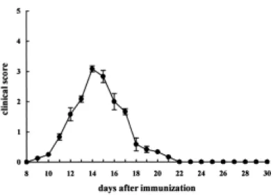
The clinical course of EAN is shown in Figure 1. EAN rats immunized with SP26 peptides developed floppy tails (G1) on days 10-12 post-immunization, and showed progressive hindlimb paralysis (G2 or G3) on days 13-16 post-immunization. All of the rats subsequently recovered on days 24-30 post-immunization (R0) (Figure 1). The clinical and pathological features of EAN in rats, evaluated using the standard morphological analysis, differed somewhat from previous observations (Lee and Shin, 2002), with a longer duration of paralysis at day 24 post-immunization, probably owing to the use of SP26 immunogen.
Figure 1. Clinical course of experimental autoimmune neuritis (EAN) in Lewis rats. EAN was induced by immunization with an emulsion containing SP26 and complete Freund's adjuvant. Each rat was treated with pertussis toxin on the days 0 and 2 after immunization. EAN rats immunized with SP26 peptides developed floppy tails (G1) on days 10-12 post-immunization, and showed progressive hindlimb paralysis (G2 or G3) on days 13-16 post-immunization. All of the rats subsequently recovered on days 24-30 post-immunization. Data are expressed as the mean clinical scores ± S.E.M. (n = 12).
19
-In the normal control rats, no cells were detected infiltrating the sciatic nerves (Figure 2A). Histological examination detected a few inflammatory cells on day 14 post-immunization in the sciatic nerve samples from control rats immunized with CFA only (data not shown). On days 14 and 24, many inflammatory cells infiltrated the sciatic nerves with EAN (Figure 2B). Subsequently, the number declined by day 30 post-immunization. The inflammatory cells in the EAN lesions consisted of R73+(TCR αβ+) T cells (Figure 2C) and ED1+ macrophages (Figure 2D).
Figure 2. Histology of EAN-affected sciatic nerves. There was no histological changes in the rat sciatic nerve (A), while the sciatic nerve of EAN-affected rats (D 14 post-immunization) showed infiltrating inflammatory cells in the parenchyma (B). The inflammatory cells consisted mainly of R73+(TCR αβ+) T cells (C) and ED1+ macrophages (D). A and B, Hematoxylin-eosin staining. C
21
-and D, immunostaining with R73 mAb -and ED1 mAb. A, Normal rat; B-D, EAN-affected rats (day 14 post-immunization). Counterstained with hematoxylin. A, B, and D: paraffin sections. C: frozen section. Scale bars represent: A, B=30μm; C, D=50μm.
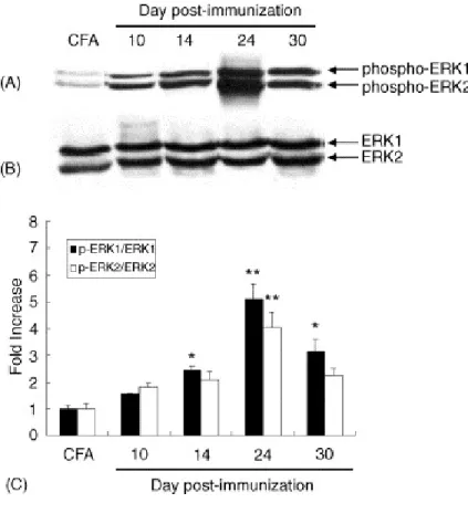
In the Western blot for p-ERK in control rats, two weak bands were detected at 44 and 42 kDa in the sciatic nerves of rats immunized with CFA only. Phosphorylation of ERK1/2 increased significantly with time until day 24 post-immunization, and their phosphorylation declined by day 30 post-immunization (Figure 3A). Moderate total ERK 1/2 (p44/42) was seen in immunoblots of the sciatic nerves of rats with EAN in the matching lane (Figure 3B). Semiquantitative analysis (relative intensity of p-ERK1/2 normalized using the total ERK 1/2) showed a dramatic increase in the phosphorylation of ERK 1/2 on day 24 post-immunization (p<0.05; CFA vs. day 14 post-immunization and day 30 post-immunization, p<0.01; CFA vs. day 24 post-immunization), while the phosphorylation declined slightly by day 30 post-immunization (Figure 3C).
23
-Figure 3. Western blot analysis of phosphorylated ERK1/2 (p-ERK1/2) and total ERK1/2 protein expression in the sciatic nerves of control rats immunized with either complete Freund's adjuvant (CFA) or an emulsion containing SP26 and CFA.
analysis of p-ERK1/2 expression in the sciatic nerves of CFA-immunized controls (CFA) (lane 1), and in EAN 10 (lane 2), 14 (lane 3), 24 (lane 4), and 30 (lane 5) days post-immunization. Both p-ERK1 (p44) and p-ERK2 (p42) produced intense immunoreactivity until day 24 post-immunization in EAN, compared with the control.
(B) A representative photomicrograph of a Western blot of total ERK1/2 expression. A moderate amount of ERK was detected in all samples.
(C) Black bars represent the ratio of p-ERK1/total ERK1 in the densitometric analysis. White bars represent the ratio of p-ERK2/total ERK2. In EAN, the expression of both p-ERK1 and p-ERK2 increased significantly until day 24 post-immunization and declined slightly at day 30 post-immunization. Data are the means ± S.E.M. (n = 3 samples at each time point).
** p<0.01, *p<0.05, compared with the CFA-immunized controls.
25
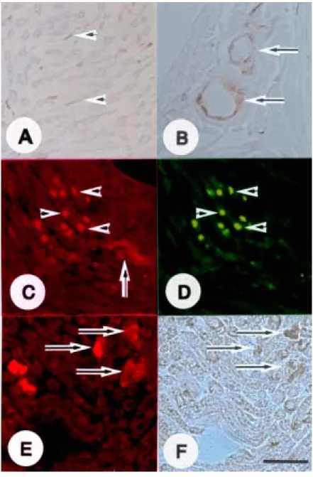
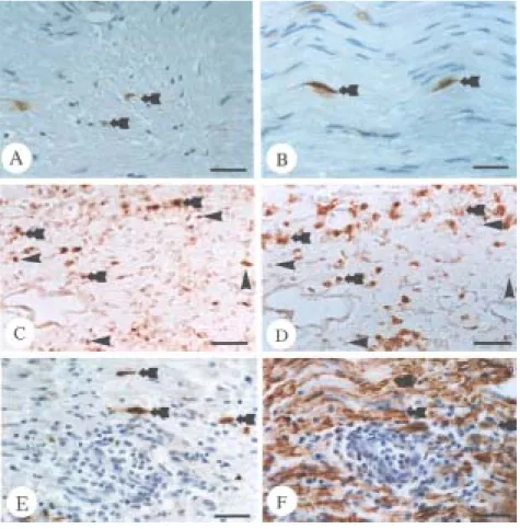
-Immunohistochemical analysis was used to visualize the cell p-ERK phenotype in the sciatic nerves of rats immunized with CFA only (controls) and rats with EAN (days 14 and 24 post-immunization). In controls, p-ERK was seen constitutively in some Schwann (Figure 4A) and vascular endothelial (Figure 4B) cells. On day 14 post-immunization, there was intense p-ERK immunofluorescence (Figure 4C) in ED1-positive macrophages (Figure 4D) subsequently, ERK activation expression (Figure 4E) was enhanced in S100-positive Schwann cells (Figure 4F) on day 24 post-immunization in the sciatic nerves of rats with EAN.
Figure 4. Immunohistochemical staining of p-ERK in the sciatic nerves of CFA-immunized control rats (A and B) and rats with experimental autoimmune neuritis (EAN) (C-F). In
27
-the sciatic nerves of controls (A and B), p-ERK (brown) was expressed constitutively in some Schwann cells (A, arrowheads) and vascular endothelial cells (B, arrows). In the sciatic nerves of rats with EAN (day 14 post-immunization), the p-ERK immunofluorescence was located mainly in some inflammatory cells (C, arrowheads) and vascular endothelial cells (C, arrow). Double immunofluorescence of p-ERK (red) and ED1 (green) showed that some p-ERK positive cells (C, arrowheads) were ED1-immunopositive macrophages (D, arrowheads). Moreover, immunofluorescence of p-ERK (red) (E, arrows) in the sciatic nerve in EAN at day 24 post-immunization was colocalized with Schwann cells that were positive for S100 (brown) (F, arrows). A and B are immunoperoxidase stained for p-ERK. F is immunoperoxidase stained for S100. A, B and F are counterstained with hematoxylin. In sections C and E, goat anti-rabbit IgG-TRITC secondary antibody was used to visualize the p-positive cells (red), and in D, horse anti-mouse IgG-FITC secondary antibody was used to visualizethe ED1-positive cells (green). C and D, and E and F are from the same section, respectively. The sections in C and D were obtained from rats on day 14 post-immunization, and E and F were on day 24 post-immunization. A-F, Scale bars represent 30 µm.
Ⅰ.4. Discussion
This study is the first to show that ERK, one of the MAP kinases, is phosphorylated in host and inflammatory cells in the affected sciatic nerves of animals with EAN. Specifically, Western blot analysis revealed that ERK was activated in the peak and recovery phases during the course of EAN in rats.
In a previous study, we found that the expression of three forms of MAP kinase increased in the spinal cord following encephalomyelitis (Shin et al., 2003). Therefore, if the results of studies of two different models of autoimmune disease (involving the central and peripheral nervous systems, respectively) are considered, it is likely that the phosphorylation of ERK is involved in the activation of host cells (astrocytes and Schwann cells in the central and peripheral nervous systems, respectively) and inflammatory cells (notably macrophages).
ERK regulates gene expression by inducing the phosphorylation of multiple targets, including nuclear transcription factors, such as c-fos, signal transduction proteins, and activators of transcription proteins, depending on the cell phenotype (Shang and Karin, 2001). Phosphorylation of ERK is involved in cell proliferation when cells are exposed to growth
29
-factors and large amounts of growth -factors and cytokines are released either from Schwann cells or inflammatory cells (T cells and macrophages) when these cells infiltrate the sciatic nerves of animals with EAN (Bonvin et al., 2002; Zhu et al., 1998). Moreover, ERK is activated in response to nerve growth factor (NGF), a cell survival and growth factor (Boulton et al., 1991). Furthermore, in the sciatic nerve in EAN following immunization with SP26 peptides, mRNA of an NGF receptor, low-affinity nerve growth factor receptor (p75 NGFR), was present only on days 18 and 23 post-immunization, which corresponded to the maximal severity of the clinical signs, and the onset of clinical recovery, respectively (Conti et al., 1995). It is thought that these factors trigger ERK activation in Schwann cells in cells in a paracrine or autocrine manner.
Ⅰ.5. Conclusion
Considering this, we postulate that a significant increase in the phosphorylation of ERK in rats with EAN is initiated after the infiltration of inflammatory cells, as we showed that ERK is phosphorylated in Schwann cells, vascular endothelial cells, and macrophages during the progression of EAN. Of these cells, both Schwann and vascular endothelial cells showed constitutive phosphorylation of ERK and elevated expression levels after stimulation by inflammatory cells. It is possible that these cells respond to inflammation and propagate a transduction signal via the activation of ERK, irrespective of whether it is beneficial or detrimental.
In summary, we confirmed that the expression of phosphorylated forms of ERK is increased in the sciatic nerve following EAN. This suggests that temporal increases in ERK activity in inflammatory cells and Schwann cells in animals with EAN influence cell signaling, which is crucial for modulating inflammation in the peripheral nervous system.
CHAPTER Ⅱ
Upregulation of osteopontin in
Schwann cells of the sciatic nerves
with experimental autoimmune
neuritis
Ⅱ.1. Introduction
Experimental autoimmune neuritis (EAN) is a T-cell-mediated autoimmune disease of the peripheral nervous system (PNS) that is used as a model of human demyelinating diseases (Hartung and Toyka, 1990; Zhu et al., 1998). The clinical course of EAN is characterized by weight loss, ascending progressive paralysis, and spontaneous recovery. It has been proposed that inflammatory mediators produced in the affected spinal nerve roots and sciatic nerve are involved in the pathogenesis of EAN (Zhu et al., 1998). Several inflammatory mediators, including nitric oxide synthase (NOS) (Conti et al., 2004; Lee and Shin 2002) and pro-inflammatory cytokines (Zhu et al., 1998), have been implicated in the pathogenesis of EAN in the sciatic nerves. Some of these mediators, whether beneficial or detrimental, are known to be closely associated with Schwann cells, the myelinating cells of the PNS.
Osteopontin (OPN) is an Arg-Gly-Asp (RGD)-containing glycoprotein with cytokine-like, chemotactic, and pro-adhesive properties that is constitutively expressed in Schwann cells (Jander et al., 2002). In autoimmune disease models, OPN is a harmful pro-inflammatory cytokine in the autoimmune disease model of experimental autoimmune encephalomyelitis (EAE)
33
-(Chabas et al., 2001). In addition, OPN is abundantly expressed by infiltrating macrophages during wound healing, and has been implicated in tissue repair in the heart (Murry et al., 1994) and in acute renal failure (Fujigaki et al., 2003). In the central nervous system, it was found that OPN is closely associated with reactive astrocytes in EAE lesions (Kim et al., 2004) and in Theiler's virus-induced demyelinating lesions (Shin and Koh, 2004). OPN expression in autoimmune disease of the PNS, however, has not been extensively studied. In the PNS, OPN is constitutively expressed by myelinating Schwann cells in uninjured control nerves; its expression was shown to be reduced following experimental axotomy and in severe axonal polyneuropathies of the human sural nerves, suggesting that OPN expression is regulated in a novel manner by axon-derived signals in Schwann cells (Jander et al., 2002).
Although the previous reports imply that OPN expression changes during the course of mechanical PNS injury, little is known concerning the changes in OPN expression that may occur during autoimmune injury of the PNS, including EAN. Thus the aim of the present study was to elucidate the expression patterns of OPN in the peripheral nerves of rats with EAN, and to investigate the possible involvement of OPN in the pathogenesis of EAN.
Ⅱ.2. Materials and Methods
Ⅱ.2.1. Animals
Female Lewis rats aged 6-8 weeks and weighing 160-200 g were obtained from Harlan Sprague Dawley (Indianapolis, IN) and used in this experiment. Active EAN was induced in Lewis rats, as described previously (Rostami et al., 1990; Shin et al., 2002).
Ⅱ.2.2. EAN induction
Rat was injected in the hind footpads on both sides with an emulsion containing 100 μg of SP26 (Shimadzu, Kyoto, Japan) and complete Freund's adjuvant (CFA; M. tuberculosis H37Ra, 5 mg/ml), and clinically evaluated as previously reported (Lee and Shin, 2002). Each rat was treated with 50 ng of pertussis toxin (Sigma, St. Louis, MO) on days 0 and 2 after immunization. The progress of EAN was divided into four clinical stages: grade 1,
35
-floppy tail; grade 2, ataxia and inability to spread the toes; grade 3, paraplegia; grade 4, tetraplegia or moribund condition.
Ⅱ.2.3. Tissue sampling
On days 14, 24 and 30 post-immunization, during the peak and recovery stages of EAN, respectively, five rats from each group were sacrificed under ether anesthesia and the sciatic nerves (approximately 5cm in length) were removed on both sides. Control rats were immunized with CFA only, and the sciatic nerves were removed on day 14 post-immunization. The nerves were frozen at -70ºC for later protein analysis. Pieces of the sciatic nerves were embedded in paraffin wax after fixation in 4% paraformaldehyde in phosphate-buffered saline (PBS) at pH 7.4.
Ⅱ.2.4. Western blot analysis
Each frozen sciatic nerve was thawed and homogenized in lysis buffer (40mM Tris, 120mM NaCl, 0.1% Noniodet P-40, 2 mM Na3PO4, 1mM phenylmethylsulfonyl fluoride, 10μg/ml aprotinin, 10μg/ml leupeptin) with 20 strokes in a homogenizer. Equal amounts of protein (40 µg/20 µl) were loaded in each lane of an 8% polyacrylamide gel and subjected to denaturing SDS-PAGE. After electrophoresis, the proteins were transferred onto nitrocellulose membranes (Bio-Rad, Hercules, CA). The membranes were incubated with 5% nonfat milk in Tris-buffered saline (TBS; 10 mM Tris-HCl, pH 7.4, and 150 mM NaCl) for 1h to block residual binding sites and then incubated with monoclonal anti-OPN antibody (Akm2A1, sc-21742, IgG1; Santa Cruz Biotechnology, Santa Cruz, CA) for 1h. The blots were washed three times in TBS containing 0.1% Tween-20 and then probed with horseradish peroxidase (HRP)-conjugated anti-mouse IgG (New England Biolabs, Beverly, MA) for 1h. The membrane blots were developed using enhanced chemiluminescence reagents (Amersham, Arlington Heights, IL), according to the manufacturer's instructions. The membranes were reprobed with a monoclonal antibody to beta-actin (Sigma, St. Louis, MO). The
37
-density of each band was measured with a scanning laser densitometer (GS-700; Bio-Rad) and analyzed using Molecular Analyst software (Bio-Rad). The ratios of OPN to beta-actin were compared with a one-way analysis of variance (ANOVA), followed by a Newman-Keuls post hoc test. In all cases, values of p<0.05 were considered statistically significant.
Ⅱ.2.5. Immunohistochemistry
Paraffin tissue sections (5 μm) were deparaffinized and hydrated. The sections were treated with 0.3% hydrogen peroxide in distilled water for 20 min to block endogenous peroxidase activity. After three washes in PBS, the sections were exposed to 10% normal goat serum and then incubated with monoclonal anti-OPN (Akm2A1) or monoclonal anti-macrophage (ED1; Serotec, London, UK) antibodies for 1h at room temperature. Rabbit anti-S100 antiserum (Sigma, St. Louis, MO) was used to label Schwann cells (Mata et al., 1990). After three washes, the sections were sequentially incubated with the appropriate biotinylated secondary antibody and the avidin-biotin peroxidase complex reagents (ABC) Elite kit (Vector,
Burlingame, CA). The peroxidase reaction was developed with diaminobenzidine (Vector) as a substrate. The sections were counterstained with hematoxylin before being mounted. To detect OPN expression in either macrophages or Schwann cells, adjacent sections were immunostained for either OPN or one of the cell-specific markers (ED1 or S100). The co-localization of both antigens in the same cell was discernable.
39
-Ⅱ.3. Results
Ⅱ.3.1.Clinical observation of EAN
EAN was induced in 6-8 week old Lewis rats by immunization with 100 μg of a synthetic peptide obtained from P2 myelin protein (SP26), as shown in a previous study (Conti et al., 2004). EAN symptoms, including weight loss, mild ataxia, and tail and paw weakness appeared by day 10 post-injection (clinical score [mean ± S.E.]: 0.25 ± 0) and peaked by day 14 (clinical score: 3.08 ± 0.1). By day 24 (clinical score: 0), the animals had begun to gain weight and strength. The clinical and pathological features of EAN in rats, evaluated by standard morphological analysis, largely resembled previous observations (Conti et al., 2004; Lee and Shin, 2002), but we noted a more rapid onset of tail weakness (at day 10-post immunization), which was probably induced by the injection of pertussis toxin, as shown in our previous report (Ahn et al., 2001).
Ⅱ.3.2. Western blot analysis of Osteopontin in EAN-affected sciatic nerves
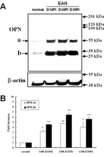
The expression pattern of OPN in the EAN-affected sciatic nerves was examined using Western blot analysis. Figure 1 shows two OPN bands of approximately 60 and 30 kDa, in agreement with a previous study by Leudtke et al. (Luedtke et al., 2002). In rats with EAN, a significant increase in OPN immunoreactivity started at 14 days post-immunization (n = 5) (fold increase [mean ± SE], upper band: 3.16 ± 0.27 [p<0.05], lower band: 4.53 ± 0.04 [p<0.01]), and there was a significant peak at day 24 post-immunization (n = 5) (upper band: 4.73 ± 0.09 [p<0.01], lower band: 6.44 ± 0.36 [p<0.01]) and day 30 post-immunization (n = 5) (upper band: 3.98 ± 0.99 [p<0.05], lower band: 5.61 ± 0.37 [p<0.01]), as compared with normal controls (n = 5), which were arbitrarily set as 1.
To examine the effect of CFA immunization alone, the optical density of OPN expression was compared at 24 days post-immunization. Although CFA immunization alone induces OPN expression at 24 days post-immunization, OPN immunoreactivity was significantly increased in the EAN-affected sciatic nerve (upper band: 4.73 ± 0.09 [p<0.01],
41
-lower band: 6.44 ± 0.36 [p<0.01]) (p<0.05), as compared to control rats immunized with CFA alone (n = 5) (upper band: 3.05 ± 0.15, lower band: 3.95 ± 0.18).
Figure 1. Western blot analysis of OPN in the sciatic nerves from rats. Normal control (normal), EAN-affected (day 14
43
-post-immunization; D14PI), EAN-affected (day 24 post-immunization; D24PI), and EAN-affected (day 30 post-immunization; D30PI).
(A) Representative photographs of Western blot analyses of OPN and beta-actin.
(B) Bar graph: Semiquantitative analysis of OPN immunoreactivity in the sciatic nerves, normalized to beta-actin from the same blot. The normal control (normal) was arbitrarily set as 1. OPN was significantly increased 14 days after inducing EAN. After recovery from EAN-induced hindlimb paralysis, OPN expression continued to increase through days 24 and 30 post-immunization. The data shown are the means ± S.E. obtained from five different experiments. * p<0.05, ** p<0.01 vs. normal controls.
Ⅱ.3.3. Localization of osteopontin in EAN-affected rat sciatic nerves
Although some Schwann cells in the sciatic nerves of normal rats were immunostained for OPN (Figure 2A), the intensity of immunostaining was increased in the Schwann cells of CFA-immunized rats (Figure 2B). At the peak stage of EAN (grade 3, day 14 post-immunization), there was an infiltration of inflammatory cells into the sciatic nerves, but very few of these cells were positive for OPN (Figure 2C). The OPN-positive inflammatory cells were ED1-positive macrophages (Figure 2D); however, the majority of ED1-positive cells in the EAN lesions were OPN-negative. In addition, small round cells in the EAN lesions, apparently identical to T cells, were shown to be negative for OPN by this staining protocol. In the EAN lesions, many OPN-expressing cells (Figure 2E) were also positive for S100, a marker for Schwann cells (Figure 2F).
45
-Figure 2. Immunohistochemical staining for OPN in the sciatic nerve of normal rats (A), CFA-immunized controls at day 14 post-immunization (B), and rats with EAN at day 14 post-immunization (C-F). OPN was expressed weakly in some Schwann cells of the sciatic nerves of normal rats (A, arrows). In the CFA-immunized controls, Schwann
cells (B, arrows) showed increased immunoreactivity, as compared to normal rats. In the EAN lesions, there was cellular infiltration into the sciatic nerve, with some OPN-positive cells (C and E, arrows). Some of these cells were ED1-positive in the adjacent serial section (D, arrows). Arrows indicate cells that expressed both OPN and ED1 (C, D), whereas arrowheads indicate cells that expressed OPN only (C, D). Furthermore, Schwann cells (E, arrows) in sciatic nerves with EAN lesions showed increased OPN expression, as compared to the nerves of CFA-immunized rats. Some of these cells were also S100-positive in the adjacent serial section (F, arrows). Counterstained with hematoxylin. Scale bars represent 30 µm.
47
-Ⅱ.4. Discussion
This is a first quantitative study of OPN expression in the sciatic nerves in a model of autoimmune peripheral nerve disease. The findings using this model, EAN in rats, were generally similar to those obtained using a prototype model of autoimmune disease of the nervous system, experimental autoimmune encephalomyelitis, in our previous study (Kim et al., 2004).
In the PNS, the expression of OPN in Schwann cells is regulated by signals from axons; after a crush injury, Schwann cells adjoining regenerating axons express OPN, but those surrounding transected sciatic nerves do not (Jander et al., 2002). In addition, OPN was not expressed in ED1-positive macrophages in the sciatic nerves after a crush injury. It was shown that Schwann cell expression of OPN was down-regulated following axotomy and reappeared in regenerating, but not permanently transected, nerves (Jander et al., 2002). Therefore, the main difference between axotomy (Jander et al., 2002) and EAN in this study is that the down-regulation of OPN in Schwann cells found in axotomy is not observed in EAN. This may be related to the fact that EAN is a primarily
demyelinating disease without severe axonal damage.
Considering the findings in the two models, OPN appears to be constitutively expressed in Schwann cells and upregulated in response to stimulation, including crush injury and EAN. It is postulated that OPN expression might be induced in macrophages by repeated stimulation (e.g., by pro-inflammatory cytokines), but the macrophages release OPN extracellularly, where it may function to provoke cell migration into the inflammatory lesions.
OPN was shown to be expressed in monocytic lineage/activated macrophages in ischemic brain injury (Wang et al., 1998) and in spinal cord injury (Hashimoto et al., 2003), but in only a small number of macrophages in EAE in our previous study (Kim et al., 2004), and in EAN in the present study. However, OPN was not expressed in sciatic nerves after crush injury (Jander et al., 2002). This discrepancy might be due to differences in the activation status of macrophages, depending on the disease progression. In case of ischemic brain injury (Wang et al., 1998) and cryoinjury in the rat brain, OPN-positive macrophages are abundant in the early stages (37 days) after injury, but become scarce by day 14 after injury, although ED1-positive macrophages remain abundant (unpublished data). We postulated that the majority of the macrophages observed in
49
-the EAE and EAN models, which are OPN-negative, are fully activated macrophages that have migrated from the peripheral immune system and no longer produce OPN. A small number of macrophages (resident microglia) in EAE are positive for OPN, suggesting that they are recently activated (Kim et al., 2004). It is thus consistent with previous studies that the majority of macrophages in EAN lesions are devoid of OPN immunoreactivity; the few macrophages that are positive for OPN are most likely to be macrophages recently activated by other inflammatory cells.
Ⅱ.5. Conclusion
Considering current and previous findings, it is postulated that OPN is upregulated in the sciatic nerves of rats with EAN, and that it might participate in the pro-inflammatory process in the peripheral nervous system during the early stage of EAN. We also postulate that persisting expression of OPN by Schwann cells during the late stages of EAN is related to some process of repair, i.e., remyelination, as shown in a previous study (Selvaraju et al., 2004). Appropriate control of OPN expression in the peripheral nervous system may facilitate the recovery process of diseased nerves, or improve the remyelination capacity of Schwann cells.
51
-자기면역성신경염에서 extracellular
signal-regulated kinase와 Osteopontin의 발현
(지도교수 : 신 태 균) 안 미 정 제주대학교 수의학과 자기면역성신경염은 T세포 매개성 말초신경계 질환으로 사람 의 길랑-바레증후군의 동물모델로 이용되고 있다. 이러한 자기면역 성신경염은 말초신경 조직에 반응하는 림프구에 의해 야기되는데 뒷다리의 마비를 주 증상으로 한다. 신경염의 조직학적 소견은 림 프구의 침윤, 큰포식세포의 증가, 신경을 에워싸고 있는 수초 변성 이 특징이며, 침윤된 염증세포로부터 분비되는 사이토카인, 반응성 질소화합물 등 염증 유도성 물질이 이러한 병변을 진행시키며, 시 간의 경과에 따라 염증세포의 세포의 자연사와 슈반세포의 활성에 의해 질병이 자연회복 되는 특징이 있다.
ERK (extracellular signal-regulated kinase)는 mitogen -activated protein 키나제 족의 하나로 ERK1과 ERK2로 구분된다.
많은 세포 표면의 수용체는 신호전달을 위해 ERK1과 ERK2뿐만 아니라 많은 신호전달체계를 자극할 수 있으며, ERK 경로는 성장 인자나 산화적 스트레스에 반응하여 활성화되며, 인산화된 ERK는 또한 세포생존, 증식, 분화, 운동과 같은 세포활성 등에 관여한다. 오스테오폰틴 (Osteopontin)은 아르기닌-글라이신-아스파르틱 산이 포함된 사이토카인과 유사한 접착분자인 당단백질이다. 이러 한 오스테오폰틴는 슈반세포에서 상존적으로 발현하며, 자기면역성 모델에서의 오스테오폰틴의 발현은 해로운 전-염증성 사이토카인으 로 작용한다고 연구되었다. 그러나 이러한 물질들이 자기면역성신 경염에서 어떠한 역할을 하는지에 대한 연구는 알려진 바 없다. 이 연구에서는 자기면역성 신경염의 진행과정에서 ERK와 오 스테오폰틴이 어떠한 형태로 관여되는지를 조사하였다. 자기면역성 신경염을 유도하기 위해 SP26 펩티드와 CFA (complete Freund adjuvant)를 혼합한 항원으로 면역하였으며, 매일 체중의 변화와 마 비정도를 검사하였다. 말초신경 조직 내 ERK와 오스테오폰틴의 발 현을 비교하기 위해 면역 후 10일, 14일, 24일, 그리고 30일에 좌골 신경을 채취하여 웨스턴 블럿트와 조직검사에 이용하였다. 자기면역성신경염의 임상증상은 면역 후 10-12일째 꼬리의 마비를 나타내기 시작하여 13-16일째에 뒷다리가 마비되었다가 점 차 회복하여 24-30일째에 정상적으로 회복하였다. 웨스턴 블럿트 결과 인산화된 ERK의 발현은 면역 후 14일째 부터 CFA만을 면역한 대조군에 비해 유의성 있는 증가를 나타내었 고, 24일째까지 증가하다가 30일째 감소하는 경향을 나타내었고, 오
53 -스테오폰틴은 면역 후 14일째부터 유의성 있는 증가를 나타내었다. 면역염색결과 인산화 된 ERK는 CFA만을 면역한 대조군의 좌골신경에서는 슈반세포와 혈관내피세포에서 약하게 발현하였으 며, 신경염을 유도한 좌골신경에서는 면역 후 14일째와 24일째에 주로 큰포식세포에서 강하게 발현하였다. 또한 면역 후 24일째와 30일째에는 슈반세포에서 증가된 발현이 관찰되었다. 오스테오폰틴 의 발현은 대조군의 좌골신경에서는 슈반세포에서 발현하였고, 신 경염을 유도한 좌골신경에서는 면역 후, 14일째에 슈반세포에서 과 립형태로 양성 반응을 나타내었다. 이 때 염증세포에서는 오스테오 폰틴의 발현이 거의 나타내지 않았으며, 마비가 회복된 24일째에서 오스테오폰틴은 슈반세포에서 발현이 증가되었다. 결론적으로, 자기면역성신경염에서 인산화된 ERK의 발현은 염증세포와 슈반세포의 분화와 생존에 중요한 역할을 하며, 특히 ERK의 활성은 자기면역성신경염의 회복단계에서 슈반세포의 생존 에 관여할 수 있을 것으로 생각된다. 또한 슈반세포에서 오스테오 폰틴의 발현은 면역자극에 의해 쉽게 유도될 수 있고, 자기면역성 신경염에서 염증반응에 의해 더욱 증가된 발현을 나타낸다. 또한 마비가 회복된 후, 오스테오폰틴의 계속 증가되는 발현은 일시적인 염증손상 후의 기능적 회복을 의미할 수 있다고 생각된다. 주요어 : 수초탈락, 슈반세포, 자기면역성신경염, 좌골신경,
References
Ahn, M., Kang, J., Lee, Y., Riu, K., Kim, Y., Jee, Y., Matsumoto, Y., Shin, T., 2001. Pertussis toxin-induced hyperacute autoimmune encephalomyelitis in Lewis rats is correlated with increased expression of inducible nitric oxide synthase and tumor necrosis factor alpha. Neurosci. Lett. 308, 41-44.
Aikawa, R., Komuro, I., Yamazaki, T., Zou, Y., Kudoh, S., Tanaka, M., Shiojima, I., Hiroi, Y., Yazaki, Y., 1997. Oxidative stress activates extracellular signal-regulated kinases through Src and Ras in cultured cardiac myocytes of neonatal rats. J. Clin. Invest. 100, 1813-1821.
Bonvin, C., Guillon, A., van Bemmelen, M.X., Gerwins, P., Johnson, G.L., Widmann, C., 2002. Role of the amino-terminal domains of MEKKs in the activation of NF kappa B and MAPK pathways and in the regulation of cell proliferation and apoptosis. Cell Signal. 14, 123-131.
55
-Boulton, T.G., Nye, S.H., Robbins, D.J., Ip, N.Y., Radziejewska, E., Morgenbesser, S.D., DePinho, R.A., Panayotatos, N., Cobb, M.H., Yancopoulos, G.D., 1991. ERKs: a family of protein-serine/threonine kinases that are activated and tyrosine phosphorylated in response to insulin and NGF. Cell 65, 663-675.
Chabas, D., Baranzini, S.E., Mitchell, D., Bernard, C.C., Rittling, S.R., Denhardt, D.T., Sobel, R.A., Lock, C., Karpuj, M., Pedotti, R., Heller, R., Oksenberg, J.R., Steinman, L., 2001. The influence of the proinflammatory cytokine, osteopontin, on autoimmune demyelinating disease. Science 294, 1731-1735.
Chang, L., Karin, M.K., 2001. Mammalian MAP kinase signaling cascades. Nature 410, 37-40.
Conti, G., Baron, P.L., Scarpini, E., Vedeler, C., Rostami, A., Pleasure, D., Scarlato, G., 1995. Low-affinity nerve growth factor receptor expression in sciatic nerve during P2-peptide induced experimental allergic neuritis. Neurosci. Lett. 199, 135-138.
Conti, G., Rostami, A., Scarpini, E., Baron, P., Galimberti, D., Bresolin, N., Contri, M., Palumbo, C., De Pol, A., 2004. Inducible nitric oxide synthase (iNOS) in immune-mediated demyelination and Wallerian degeneration of the rat peripheral nervous system. Exp. Neurol. 187, 350-358.
Fujigaki, Y., Sun, D.F., Goto, T., Hishida, A., 2003. Temporary changes in macrophages and MHC class-II molecule-expressing cells in the tubulointerstitium in response to uranyl acetate-induced acute renal failure in rats. Virchows Arch. 443, 206-216.
Gold, R., Hartung, H.P., Toyka, K.V., 2000. Animal models for autoimmune demyelinating disorders of the nervous system. Mol. Med. Today 6, 88-91.
Gregorian, S.K., Clark, L., Heber-Katz, E., Amento, E.P., Rostami, A., 1993. Induction of peripheral tolerance with peptide-specific anergy in experimental autoimmune neuritis. Cell Immunol. 150(2), 298-310.
57
-Gutkind, J.G., 1998. The pathway connecting G protein-coupled receptors to the nucleus through divergent mitogen-activated protein kinase cascades. J. Biol. Chem. 273, 1839-1842.
Hartung, H.P., Toyka, K.V., 1990. T-cell and macrophage activation in experimentally induced autoimmune neuritis and Guillain-Barre syndrome. Ann. Neurol. 27, S57-S63.
Hartung, H.P., Willison, H., Jung, S., Pette, M., Toyka, K.V., Giegerich, G., 1996. Autoimmune response in peripheral nerve. Springer Semin Immunopathol. 18, 97-123.
Hashimoto, M., Koda, M., Ino, H., Murakami, M., Yamazaki, M., Moriya, H., 2003. Upregulation of osteopontin expression in rat spinal cord microglia after traumatic injury. J. Neurotrauma 20, 287-296.
Hughes, R.A., Atkinson, P.F., Gray, I.A., Taylor, W.A., 1987. Major histocompatibility antigens and lymphocyte subsets during experimental allergic neuritis in the Lewis rat. J. Neurol. 234, 390-395.
Jander, S., Bussini, S., Neuen-Jacob, E., Bosse, F., Menge, T., Muller, H.W., Stoll, G., 2002. Osteopontin: a novel axon-regulated Schwann cell gene. J. Neurosci. Res. 67, 156-166.
Jung, S., Huitinga, I., Schmidt, B., Zielasek, J., Dijkstra, C.D., Toyka, K.V., Hartung, H.P., 1993. Selective elimination of macrophages by dichlormethylene diphosphonate-containing liposomes suppresses experimental autoimmune neuritis. J. Neurol. Sci. 119(2), 195-202.
Kim, M.D., Cho, H.J., Shin, T., 2004. Expression of osteopontin and its ligand, CD44, in the spinal cords of Lewis rats with experimental autoimmune encephalomyelitis. J. Neuroimmunol. 151, 80-86.
Lee, Y., Shin, T., 2002. Expression of constitutive endothelial and inducible nitric oxide synthase in the sciatic nerve of Lewis rats with experimental autoimmune neuritis. J. Neuroimmunol. 126, 78-85.
59
-Lewis, T.S., Shapiro, P.S., Ahn, N.G., 1998. Signal transduction through MAP kinase cascades. Adv. Cancer Res. 74, 149-139.
Luedtke, C.C., McKee, M.D., Cyr, D.G., Gregory, M., Kaartinen, M.T.. Mui, J., Hermo, L., 2002. Osteopontin expression and regulation in the testis, efferent ducts, and epididymis of rats during postnatal development through to adulthood. Biol. Reprod. 66, 1437-1448.
Mata, M., Alessi, D., Fink, D.J., 1990. S100 is preferentially distributed in myelin-forming Schwann cells. J. Neurocytol. 19, 432-442.
Matsumoto, Y., Kim, G., Tanuma, N., 2000. Characterization of T cell receptor associated with the development of P2 peptide-induced autoimmune neuritis. J. Neuroimmunol. 102, 67-72.
Moses, S., Franzen, A., Lovdahl, C., Hultgardh-Nilsson, A., 2001. Injury-induced osteopontin gene expression in rat arterial smooth muscle cells is dependent on mitogen-activated protein kinases ERK1/ERK2. Arch. Biochem. Biophys. 396,
133-137.
Murry, C.E., Giachelli, C.M., Schwartz, S.M., Vracko, R., 1994. Macrophages express osteopontin during repair of myocardial necrosis. Am. J. Pathol. 145, 1450-1462.
Noguchi, K., Obata, K, 2004. MAPK activation in nociceptive neurons and pain hypersensitivity. Life Sci. 74, 2643-2653.
Olee, T., Powers, J.M., Brostoff, S.W., 1988. A T cell epitope for experimental allergic neuritis. J. Neuroimmunol. 19(1-2), 167-173.
Olee, T., Powell, H.C., Brostoff, S.W., 1990. New minimum length requirement for a T cell epitope for experimental allergic neuritis. J. Neuroimmunol. 27(2-3), 187-190.
Olsson, T., Holmdahl, R., Klareskog, L., Forsum, U., Kristensson, K., 1984. Dynamics of Ia-expressing cells and T lymphocytes of different subsets during experimental allergic neuritis in Lewis rats. J. Neurol. Sci. 66(2-3), 141-149.
61
-Ota, K., Irie, H., Takahashi, K., 1987. T cell subsets and Ia-positive cells in the sciatic nerve during the course of experimental allergic neuritis. J. Neuroimmunol. 13(3), 283-292.
Robinson, M.J., Stippec, S.A., Goldsmith, E., Cobb, M.A., White, M.H., 1998. A constitutively active and nuclear form of the MAP kinase ERK2 is sufficient for neurite outgrowth and cell transformation. Curr. Biol. 8, 1141-1150.
Rosen, J.L., Brown, M.J., Rostami, A., 1992. Evolution of the cellular response in P2-induced experimental allergic neuritis. Pathobiology 60(2), 108-112.
Rostami, A., Gregorian, S.K., Brown, M.J., Pleasure, D.E., 1990. Induction of severe experimental autoimmune neuritis with a synthetic peptide corresponding to the 53-78 amino acid sequence of the myelin P2 protein. J. Neuroimmunol. 30, 145-151.
Selvaraju, R., Bernasconi, L., Losberger, C., Graber, P., Kadi, L., Avellana-Adalid, V., Picard-Riera, N., Van Evercooren, A.B., Cirillo, R. Kosco-Vilbois, M., Feger, G., Papoian, R.,
Boschert, U., 2004. Osteopontin is upregulated during in vivo demyelination and remyelination and enhances myelin formation in vitro. Mol. Cell Neurosci. 25, 707-721.
Shin, T., Ahn, M., Jung, K., Heo, S., Kim, D., Jee, Y., Lim, Y.K., Yeo,E.J., 2003. Activation of mitogen-activated protein kinases in experimental autoimmune encephalomyelitis. J. Neuroimmunol. 140, 118-125.
Shin, T., Koh, C.S., 2004. Immunohistochemical detection of osteopontin in the spinal cords of mice with Theiler's murine encephalomyelitis virus-induced demyelinating disease. Neurosci. Lett. 356, 72-74.
Shin, T., Min, D.S., Ahn, M., Son, W., Matsumoto, Y., 2002. Increased expression of phospholipase D1 in the sciatic nerve of rats with experimental autoimmune neuritis. Immunol. Invest. 31, 169-176.
Wang, X., Louden, C., Yue, T.L., Ellison, J.A., Barone, F.C., Solleveld, H.A., Feuerstein, G.Z., 1998. Delayed expression of osteopontin after focal stroke in the rat. J. Neurosci. 18, 2075-2083.
63
-You, J., Reilly, G.C., Zhen, X., Yellowley, C.E., Chen, Q., Donahue, H.J., Jacobs CR., 2001. Osteopontin gene regulation by oscillatory fluid flow via intracellular calcium mobilization and activation of mitogen-activated protein kinase in MC3T3-E1 osteoblasts. J. Biol. Chem. 276, 13365-13371.
Zhu, J., Mix, E., Link, H., 1998. Cytokine production and the pathogenesis of experimentally induced autoimmune neuritis and Guillain-Barré syndrome. J. Neuroimmunol. 84, 40-52.
Zhu, J., Link, H., Mix, E., Olsson, T., Huang, W.X., 1994. Th1-like cell responses to peripheral nerve myelin components over the course of experimental allergic neuritis in Lewis rats. Acta Neurol. Scand. 90, 19-25.
Zhu, J., Link, H., Weerth, S., Linington, C., Mix, E., Qioa, J., 1994. The B cells repertoire in experimental allergic neuritis involved multiple myeli proteins and GM1. J. Neurol. Sci. 125, 132-137.
CURRICULUM VITAE
Name Meejung Ahn, D.V.M
Date of Birth July 03, 1977
Birthplace Jeju, Korea (South) Citizenship Korean
Address
#1165-1 Ara-2-dong, Jeju 690-122, Republic of Korea, Tel.: +82-64-754-3363;
Fax.:+82-64-756-3354
E-mail: [email protected], EDUCATION
2004.3 - Present
Student for Doctorate, Dept. of Veterinary Medicine, Graduate School, Cheju National University
2000.3 - 2002.2 M.S., Dept. of Veterinary Medicine, Graduate School, Cheju National University
1996.3 - 2000.2 D.V.M., Dept. of Veterinary Medicine, Cheju National University
1993.3 - 1996.2 Shin Sung girls high School, Jeju, Korea CAREER
2003.3 - 2005.8 Teaching Assistant, Dept. of Veterinary Medicine, Cheju National University
2001.3 - 2002.2
Research Assistant, Research Instrument Center, Cheju National University, Jeju, Kore
65
-<PUBLICATION>
1) Kim H, Moon C, Ahn M, Lee Y, Kim H, Kim S, Ha T, Jee Y, Shin T. Expression of nitric oxide synthase isoforms in the porcine ovary during follicular development. J. Vet. Sci. 2005 Jun;6(2):97-101.
2) Shin T, Kim H, Jin JK, Moon C, Ahn M, Tanuma N, Matsumoto Y. Expression of caveolin-1, -2, and -3 in the spinal cords of Lewis rats with experimental autoimmune encephalomyelitis. J. Neuroimmunol. 2005 Aug;165(1-2):11-20. 3) Shin T, Ahn M, Kim H, Moon C, Kang TY, Lee JM, Sim
KB, Hyun JW. Temporal expression of osteopontin and CD44 in rat brains with experimental cryolesions. Brain Res. 2005 Apr 11; 1041(1): 95-101.
4) Moon C, Ahn M, Kim H, Lee Y, Koh CS, Matsumoto Y, Shin T. Activation of p38 mitogen-activated protein kinase in the early and peak phases of autoimmune neuritis in rat sciatic nerves. Brain Res. 2005 Apr 8; 1040(1-2): 208-213.
5) Moon C, Ahn M, Yasuzumi F, Shin T. Increased expression of both constitutive and inducible forms of nitric oxide synthase in the delayed phase of acute experimental testicular torsion. J. Vet. Med. Sci. 2005 April; 67(4):453-456.
6) Moon C, Lee J, Ahn M, Shin T. Involvement of Disabled-2 protein in the central nervous system inflammation following experimental cryoinjury of rat brains. Neurosci. Lett. 2005 Apr 18; 378(2): 88-91.
7) Kang J, Ahn M, Moon C, Min DS, Matsumoto Y, Shin T. Phospholipase D1 is up-regulated in the retinas of Lewis rats with experimental autoimmune uveoretinitis. Immunol. Invest. 2005 Feb; 1(1-2): 27-34.
8) Jee YH, Jeong WI, Kim TH, Hwang IS, Ahn MJ, Joo HG, Hong SH, Jeong KS. p53 and cell-cycle-regulated protein expression in small intestinal cells after fast-neutron irradiation in mice. Mol Cell Biochem. 2005 Feb; 270(1-2): 21-28.
9) Moon C, Yasuzumi F, Okura N, Kim H, Ahn M, Shin T. Enhanced expression of tyrosine kinase receptor A in germ cells of the rat testis with acute experimental testicular torsion. Urol. Int. 2005 Jan; 74(1): 79-85.
10)Moon C, Ahn M, Wie M-B, Kim H-M, Koh C-S, Hong S-C, Kim M-D, Tanuma N, Matsumoto Y, Shin T. Phenidone, a dual inhibitor of cyclooxygenases and lipoxygenases, ameliorates rat paralysis in experimental autoimmune encephalomyelitis by suppressing its target enzymes. Brain Res. 2005 Jan; 1035: 206-210.
11) Ahn M, Lee Y, Sim KB, Min do S, Matsumoto Y, Wie MB, Shin YG, Shin T. Increased expression of phospholipase D in the heart with experimental autoimmune myocarditis in Lewis rats. Immunol Invest. 2004 Feb; 33(1): 95-105.
12)Moon C, Ahn M, Kim S, Jin J-K, Sim K-B, Kim H-M, Lee M-Y, Shin T. Temporal patterns of the embryonic intermediate filaments nestin and vimentin expression in the
67
-cerebral cortexof adult rats after cryoinjury. Brain Res. 2004 Dec 3; 1028:238-242.
13)Ahn M, Moon C, Lee Y, Koh CS, Kohyama K, Tanuma N, Matsumoto Y, Kim H-M, Kim S-R, Shin T. Activation of extracellular signal-related kinases in the sciatic nerves of rats with experimental autoimmune neuritis. Neurosci. Lett. 2004 Nov 30; 372(1-2), 57-61.
14)Moon C, Ahn M, Kim S, Jin JK, Sim KB, Kim HM, Lee MY, Shin T. Temporal patterns of the embryonic intermediate filaments nestin and vimentin expression in the cerebral cortex of adult rats after cryoinjury. Brain Res. 2004 Dec 3;1028(2):238-242.
15)Ahn M, Lee Y, Moon C, Jin J-K, Matsumoto Y, Koh C-S, Kim H-M, Shin T. Upregulation of osteopontin in Schwann cells of the sciatic nerves of Lewis rats with experimental autoimmune neuritis. Neurosci. Lett. 2004 Nov 30; 372(1-2), 137-141.
16)Moon C, Heo S, Ahn M, Kim H, Shin M, Sim K-M, Kim HM, Shin T. Immunohistochemical study of osteopontin in the spinal cords of rats with clip compression injury. J. Vet. Med. Sci. 2004 Oct; 66(10): 1307-1310.
17)Kim H, Joo H-G, Moon C, Ahn M, Jee Y, Lim Y-K, Koh C-S, Shin T. Expression of galectin-3 in the spinal cords of Lewis rats and NOD mice with experimental autoimmune encephalomyelitis. Korean J. Vet. Res. 2004 Sep; 44(3): 349-355.
18)Ahn M, Lee Y, Sim KB, Min do S, Matsumoto Y, Wie MB, Shin YG, Shin T. Increased expression of phospholipase D in the heart with experimental autoimmune myocarditis in Lewis rats. Immunol Invest. 2004 Feb;33(1):95-105.
19)Moon C, Ahn M, Jee Y, Heo S, Kim S, Kim H, Sim KB, Koh CS, Shin YG, Shin T. Sodium salicylate-induced amelioration of experimental autoimmune encephalomyelitis in Lewis rats is associated with the suppression of inducible nitric oxide synthase and cyclooxygenases. Neurosci Lett. 2004 Feb 12;356(2):123-6.
20)Kim DH, Heo SD, Ahn MJ, Sim KB, Shin TK. Activation of embryonic intermediate filaments contributes to glial scar formation after spinal cord injury in rats.J Vet Sci. 2003 Aug;4(2):109-112.
21)Shin T, Ahn M, Jung K, Heo S, Kim D, Jee Y, Lim YK, Yeo EJ. Activation of mitogen-activated proteinkinases in experimental autoimmune encephalomyelitis. J Neuroimmunol. 2003 Jul; 140(1-2): 118-25.
22)Jung K, Min do S, Sim KB, Ahn M, Kim H, Cheong J, Shin T Upregulation of phospholipase D1 in the spinal cords of rats with clip compression injury. Neurosci. Lett. 2003 Jan 16; 336(2): 126-30.
23)Ha TY, Ahn MJ, Lee YD, Yang JH, Kim HS, Shin TK. Histochemical detection of glycoconjugates in the male reproductive system of the horse. J. Vet. Sci. 2003 Apr;4(1):21-8.
69
-24)Jung KS, Ahn MJ, Lee YD, Go GM, Shin TK. Histochemistry of six lectins in the tissues of the flat fish Paralichthys olivaceus. J. Vet. Sci. 2002 Dec;3(4):293-301.
25)Shin T, Min DS, Ahn M, Son W, Matsumoto Y Increased expression of phospholipase D1 in the sciatic nerve of rats with experimental autoimmune neuritis. Immunol. Invest. 2002 Aug-Nov; 31(3-4): 169-76.
26)Moon C, Terashima S, Ahn M, Kang J, Shin T. Immunohistochemical analysis of neuronal nitric oxide synthase in the trigeminal ganglia of the crotaline snake Trimeresurus flavoviridis. Neurosci. Lett. 2002 Feb 8; 319(1): 21-24.
27)Ahn M, Min D, Kang J, Cheong K, and Shin T. Increased expression of phospholipase D1 in the spinal cords of rats with experimental autoimmune encephalomyelitis. Neurosci. Lett. 2001 Dec 306(2): 95-98.
28)Kang J, Ahn M, Kim Y-S, Moon C, Lee Y, Wie M, Lee Y, Shin T. Melatonin ameliorates autoimmune encephalomyelitis through suppression of intercellular adhesion molecule-1. J. Vet. Sci. 2001 Aug; 2(2): 85-89.
29)Ahn M, Kang J, Lee Y, Riu K, Kim Y, Jee Y, Matsumoto Y and Shin T. Pertussis toxin -induced hyperacute EAE is correlated with theexpression of iNOS and TNF-a. Neurosci. Lett. 2001 Jul; 308(1): 41-44.
30)Shin T, Kang B, Tanuma N, Matsumoto Y, Wie M, Ahn M and Kang J. Intrathecal administration of endothelin-1
receptor antagonist ameliorates autoimmune encephalomyelitis in Lewis rats. NeuroReport 2001 May 12(7): 1465-1468.
31)Shin T, Weinstock D, Castro MD, Acland H, Walter M, Kim H, Ahn M, Purchase HG. Neuronal constitutive and inducible nitric oxide synthase expression in the brain of Listeria monocytogenes-infected cattle. Acta. Vet. Brno. 2001 March 70:43-47.
32)Shin T, Moon C, Ahn M, Wie M. Immunohistochemical study of CPP32 (Caspase-3) in the spinal cords of rats with experimental autoimmune encephalomyelitis. Korean J. Vet. Res. 2000 Sep; 40(3):431-437.
33)Moon C, Kim S, Ahn M, Lee S, Park S, Jeong K, Yoon D Y, Choe Y K, Shin T. Effects of Opuntia ficus-indica extract on immune cell activation. Kor. J. Life. Sci. 2000 Aug;10(4):362-364.
71 -감사의 글 몰랐습니다 나도 모르게 내 안에 꽃씨 하나 심었던가 봅니다 딱히 마음 모아 북돋아 준 기억조차 없는데 딱히 마음 쏟아 물 한번 준 기억조차 없는데 어느새 쑥쑥 자랐던가 봅니다 글쎄 은은히 향 풍기며 목을 끌어안지 뭐예요 그저 미안할 뿐입니다 그저 고마울 뿐입니다 그저 사랑스러울 뿐입니다 - 양숙의 시집《당신 가슴에》에 실린 시 <몰랐습니다>(전문)에서 -이제 인생의 또 다른 한발을 내딛었습니다. 많은 가르침과 사랑주시고, 격려해주시고, 지켜봐주신 모든 분들께 진심으로 감사드립니다.