저작자표시-동일조건변경허락 2.0 대한민국 이용자는 아래의 조건을 따르는 경우에 한하여 자유롭게 l 이 저작물을 복제, 배포, 전송, 전시, 공연 및 방송할 수 있습니다. l 이차적 저작물을 작성할 수 있습니다. l 이 저작물을 영리 목적으로 이용할 수 있습니다. 다음과 같은 조건을 따라야 합니다: l 귀하는, 이 저작물의 재이용이나 배포의 경우, 이 저작물에 적용된 이용허락조건 을 명확하게 나타내어야 합니다. l 저작권자로부터 별도의 허가를 받으면 이러한 조건들은 적용되지 않습니다. 저작권법에 따른 이용자의 권리는 위의 내용에 의하여 영향을 받지 않습니다. 이것은 이용허락규약(Legal Code)을 이해하기 쉽게 요약한 것입니다. Disclaimer 저작자표시. 귀하는 원저작자를 표시하여야 합니다. 동일조건변경허락. 귀하가 이 저작물을 개작, 변형 또는 가공했을 경우 에는, 이 저작물과 동일한 이용허락조건하에서만 배포할 수 있습니다.
A Thesis
for the Degree of Master of Science in Medicine
Cytoprotective Effects of Americanin B against
Oxidative Stress in Human Keratinocytes
Jian Zheng
Department of Medicine
Graduate School
Jeju National University
인간 피부세포에서 산화적 스트레스에 대한
Americanin B의 보호효과
지도교수 현진원
정 건
이 논문을 의학 석사학위 논문으로 제출함
2014년 8월
정건의 의학 석사학위 논문을 인준함
심사위원장
위 원
위 원
제주대학교 대학원
2014년 8월
Cytoprotective Effects of Americanin B against
Oxidative Stress in Human Keratinocytes
Jian Zheng
(Supervised by Professor Jin-Won Hyun)
A thesis submitted in partial fulfillment of the requirement for the
degree of Master of Science in Medicine
2014. 08.
This thesis has been examined and approved
.
.
Department of Medicine
GRADUATE SCHOOL
JEJU NATIONAL UNIVERSITY
Abstract
The aim of this study was to evaluate the cytoprotective effects of americanin B, a lignan
compound, against hydrogen peroxide (H2O2)-induced cell damage. Americanin B decreased
the level of DPPH radicals, superoxide anions, hydroxyl radicals and intracellular reactive
oxygen species. Americanin B also attenuated DNA damage induced by H2O2 treatment, as
shown by the inhibition of formation of comet tails indicative of DNA strand breakage, and
prevented the oxidation of protein and peroxidation of lipid, as determined by protein
carbonyls and 8-isoprostane. Furthermore, americanin B protected against H2O2-induced
apoptotic cell death, as determined by a reduction in the numbers of apoptotic bodies stained
with Hoechst 33342. These findings suggest that americanin B protects cells against oxidative
II
Contents Abstract... Ӏ Contents... ӀӀ List of figures... ӀV 1. Introduction... 12. Materials and Methods... 3
2-1. Reagents
2-2. Cell culture
2-3. Cell viability assay
2-4. DPPH radical scavenging activity
2-5. Detection of the superoxide anions
2-6. Detection of hydroxyl radicals
2-7. Detection of intracellular ROS
2-8. Single-cell gel electrophoresis (comet assay)
2-9. Protein carbonyl formation
2-10. Lipid peroxidation assay
2-11. Nuclear staining with Hoechst 33342
2-12. Statistical analysis
3. Results... 8
3-1.
Americanin B attenuates ROS generation3-2.
Americanin B protects cell components against H2O2-inducedoxidative damage to lipids, proteins and DNA
4. Dissussion... 19
5. Reference... 22
6. Abstract in korean... 30
IV
List of figures
Figure 1. Americanin B attenuates ROS generation... 9 Figure 2. Americanin B protects against H2O2-induced oxidative lipid, protein, and
DNA damage... 14
1. Introduction
Reactive oxygen species (ROS), are an unavoidable consequence of aerobic metabolism,
which generates free radicals such as superoxide anions (O2-) and hydroxyl radicals (·OH), as
well as non-radical molecules like hydrogen peroxide (H2O2) and singlet oxygen (1O2)
(Sharma et al. 2012). At high concentrations, ROS are extremely harmful to living organisms.
Although possessing endogenous defense mechanisms responsible for reduction of ROS in
organisms, lack of the capacity of cellular antioxidant systems or accumulation in ROS
production can give rise oxidative damage to cellular components, potentially resulting in
initiation of several pathologic processes (Bandyopadhyay et al. 1999). When cellular
antioxidant capacity, which prevents oxidative injury, can’t neutralize ROS generation, cells expose to condition termed “oxidative stress” (Tezel 2006). The accumulation of ROS in
cytoplasm or organelle can disrupt internal balance of cells by causing peroxidation of lipids
(Dmitriev and Titov 2010), oxidative modification of proteins (Lee and Yun, 2012), breaking
the structure and function of nucleic acids (Mena et al. 2009), inhibition of antioxidant
enzymes (Valko et al. 2007), activation of apoptotic pathways, and eventually cell dysfunction
(Sinha et al. 2013).
In this study, the human keratinocyte (HaCaT) cell was chosen as cell model to examine the
effect of oxidative stress induced by ROS. The skin is the largest organ of the body and has
three main layers, the epidermis, the dermis and the hypodermis. The HaCaT cells are original
from the most outer layer of skin, the epidermis, which serves as the physical and chemical
barrier between the interior body and exterior environment (Proksch et al. 2008). In order to
retard oxidative stress, cells have to precisely and dynamically keep the equilibrium between
increase and reduction of ROS. H2O2, an inducer of oxidative stress, is capable of induction of
cell injury both in vitro and in vivo (Hsu et al. 2013; Kim et al. 2012; Terashvili et al. 2012).
Exogenous abruptly H2O2 can exacerbate oxidative stress beyond the counteraction capacity
2
mitochondrial dysfunction (Maroto and Perez-Polo 1997). On the other hand, UVB exposure
also is a common inducer of oxidative stress, which can induce DNA damage by forming
pyrimidine dimers directly (Bohr et al. 1985). In addition, the cytoprotective enzymes in cells
protect critical macromolecules such as DNA, proteins, and lipids from ROS-mediated
damage, and thus function as the line of defense against oxidative stress (Nguyen et al. 2013).
Plants are rich in flavonoids, stilbenes, and lignans, among which lignans are well studied
because of their beneficial biological activity (Dixon 2004). Current work has gradually
concentrated on lignans, which show antioxidants (Lee et al. 2009; Mei et al. 2009; Wang et
al. 2005; Harper et al. 1999) and anti-inflammatory properties (Lee et al. 2012). Moreover, the
pharmacological activity of lignans exerts beneficial health effects including inhibition of
cyclooxygenase-2 (Schühly et al. 2009) as well as anti-neuroinflammatory (Kim et al. 2010),
and antioxidant activities (Haraguchi et al. 1997; Lee et al. 2004). The skeletal structure of
lignan is a group of dimeric phenylpropanoids, where two C6-C3 units are connected by its
central C-8 carbon and the link of two C6-C3 units is taken place of carbon-carbon bond or
ether oxygen atom in the absence of the C-8 bond, which is named after neolignan (Moss
2000). Lignan has many derivatives by adding different functional groups based on the
primary framework and one of these derivatives, bearing electron-donating group or
converting to electron-donating group of lignan, such as phenolic hydroxyl group, α,
β-unsaturated carbonyl group or alkenyl by specific metabolites and biosynthesized ways
(Cunha et al. 2012), can show antioxidant effects (Chen et al. 2002; Farombi et al. 2008).
Americanin B (C27H24O9) belonging to lignan possesses phenolic hydroxyl group and
unsaturated carbonyl group (Yu et al. 2001) which are supposed to excert antioxidant activity.
This study investigated the ability of americanin B to protect human keratinocytes against
2. Materials and Methods
2-1. ReagentsN-acetyl cysteine (NAC), 5,5-dimethyl-1-pyrroline-N-oxide (DMPO),
2',7'-dichlorodihydrofluorescein diacetate (DCF-DA), 1,1-diphenyl-2-picrylhydrazyl (DPPH),
(3-(4,5-dimethylthiazol-2-yl)-2,5-diphenyltetrazolium) bromide (MTT), and Hoechst 33342 dye
were purchased from Sigma Chemical Company (St. Louis, MO, USA).
Diphenyl-1-pyrenylphosphine (DPPP) was purchased from Molecular Probes (Eugene, OR, USA).
Americanin B was provided by Professor Sam Sik Kang (Seoul National University, Seoul,
Korea). All other chemicals and reagents were of analytical grade.
2-2. Cell culture
The human keratinocyte cell line HaCaT was supplied by the Amore Pacific Company
(Gyeonggi-do, Korea) and maintained at 37C in an incubator supplemented with an atmosphere containing humidified 5% CO2 plus95% air. These cells were grown in RPMI
1640 medium which contains 10% fetal calf serum, streptomycin (100 µg/ml), and penicillin
(100 units/ml).
2-3. Cell viability assay
The effect of americanin B on the viability of HaCaT cells was applied by the MTT assay.
Cells were seeded on a 96-well plate at a density of 1 × 10⁵ cells/ml, cultured for 16 h, and
then treated with 1, 5, 10, or 20 μM of americanin B. After incubation for a further 16 h, MTT
stock solution (50 µl, 2 mg/ml) was added to each well to yield a total reaction volume of 250
µl. Four hours later, the supernatants were sucked, and application of the dimethylsulfoxide
(DMSO) dissolved formazan crystals in each well, and the absorbance of solution was read at
4
2-4. DPPH radical scavenging activity
Americanin B (1, 5, 10, or 20 μM) or NAC (1 mM) was added to a solution of DPPH (0.1
mM) dissolved in methanol. These mixtures were shaken vigorously and reacted at room
temperature for 3 h. After 3 h, the amount of residual DPPH was measured at 520 nm using a
spectrophotometer (Brand-Williams et al. 1995). The DPPH inhibition (%) was calculated as
follows: % inhibition of DPPH = (absorbance of control - absorbance of americanin B or
NAC)/absorbance of control × 100.
2-5. Detection of the superoxide anions
The xanthine/xanthine oxidase system produces superoxide anions, which reacted with a
nitrone spin trap, DMPO and formed the reaction product the DMPO/·OOH adducts. The
DMPO/·OOH adducts were detected by using a JES-FA electron spin resonance (ESR)
spectrometer (JEOL, Tokyo, Japan) (Ueno et al. 1984; Kohno et al. 1994). ESR signaling was
read 2.5 min after 20 µl of xanthine oxidase (0.25 U/ml) was mixed with 20 µl each of xanthine
(10 mM), DMPO (3 M), and americanin B (10 μM). The ESR spectrometer parameters were
set as follows: magnetic field = 336.8 mT, power = 1.00 mW, frequency = 9.4380 GHz,
modulation amplitude = 0.2 mT, gain = 500, scan time = 0.5 min, scan width = 10 mT, time
constant = 0.03 sec, and temperature = 25C.
2-6. Detection of hydroxyl radicals
The Fenton reaction (H2O2 + FeSO4)system produces hydroxyl radicals, which reacted with
a nitrone spin trap, DMPO and formed the reaction product the DMPO/·OH adducts (Li et al.
2003, 2004). The ESR spectrum was recorded 2.5 min after phosphate buffer solution (pH 7.4)
was reacted with 20 µl each of 0.3 M DMPO, 10 mM FeSO4, 10 mM H2O2, and 10 μM
americanin B. The ESR spectrometer parameters were set as follows: magnetic field = 336.8
scan time = 0.5 min, scan width = 10 mT, time constant = 0.03 sec, and temperature = 25C.
2-7. Detection of intracellular ROS
After seeding in 96-well plate at a density of 1 × 10⁵ cells/ml, cells were incubated in
incubator for 16 h. Later, they were treated with americanin B and the compound NAC (2
mM), as an antioxidant, was performed for a positive control. After incubation of samples for
1 h at 37°C, H2O2 (1 mM) was added to these wells, and the plates were again incubated for
30 min at 37°C. Thirty minutes later, DCF-DA solution (25 µM) was added and waited for ten
minutes in dark, and then the fluorescence of 2',7'-dichlorofluorescein (DCF) was detected and
recorded with PerkinElmer LS-5B spectrofluorometer (PerkinElmer, Waltham, MA)
(Rosenkranz et al. 1992). For imaging analysis of the generation of intracellular ROS, cells
were seeded on a 4-well chamber slide at a density of 2 × 10⁵ cells/ml. After plating for 16 h,
these cells were exposed to 10 µM americanin B. After 1 h, 1 mM H2O2 was added to the plate.
After 2 h, 100 µM of DCF-DA was added to each well, and the cells were incubated for an
additional 30 min at 37C. After washing with PBS, the stained cells were mounted on the chamber slide with mounting medium (DAKO). The images were obtained on a confocal
microscope using the Laser Scanning Microscope 5 PASCAL software (Carl Zeiss).
2-8. Single-cell gel electrophoresis (comet assay)
The breakdown of DNA strand is assessed by the comet assay (Rajagopalan et al. 2003;
Singh et al. 2000). The cell pellet was suspended with 0.5% low-melting agarose (LMA, 75
µl) at 39C, and then it was transfered to a microscopic slide pre-coated with 200 µl of 1 % normal melting agarose. After solidification of the agarose, the slide was covered with another
75 µl of 0.5% LMA and then immersed in a lysis solution (2.5 M NaCl, 100 mM Na-EDTA,
10 mM Tris, 1% Triton X-100, 10% DMSO, pH 10) for 1 h at 4C. The slides were subsequently placed in a gel electrophoresis apparatus containing 300 mM NaOH and 10 mM
6
Na-EDTA (pH 13) for 40 min to allow for DNA unwinding and the expression of alkali-labile
damage. An electrical field was then applied (300 mA, 25 V) for 20 min at 4C to draw the negatively charged DNA towards the anode. The slides were washed three times for 5 min at
4C in a neutralizing buffer (0.4 M Tris, pH 7.5), stained with ethidium bromide, and observed under a fluorescence microscope and image analyzer (Kinetic Imaging, Komet 5.5, UK). The
percentage of the total fluorescence in the comet tails and the tail lengths of 50 cells per slide
were recorded.
2-9. Protein carbonyl formation
Cells were treated with 10 μM of americanin B, followed 1 mM of H2O2 was added to the
plate 1 h later, and the mixture was incubated for 12 h. The amount of carbonyl formation in
protein was determined using an OxiselectTM protein carbonyl enzyme-linked immunosorbent
assay kit purchased from Cell Biolabs (San Diego, CA).
2-10. Lipid peroxidation assay
8-Isoprostane, an indicator of lipid peroxidation, is formed in medium secreted from cells
and measured by a commercial enzyme immune assay (Cayman Chemical, Ann Arbor, MI,
USA) (Beauchamp et al. 2002). Lipid peroxidation was also detected by the image analysis
after staining of DPPP (Okimoto et al. 2000). Cells were treated with 10 µM americanin B for
1 h, followed by exposure to 1 mM H2O2. Five hours later, 5 mM DPPP was added and
incubated for 30 min in the dark. Images of DPPP fluorescence were saved with a Zeiss
Axiovert 200 inverted microscope at an excitation wavelength of 351 nm and an emission
wavelength of 380 nm.
2-11. Nuclear staining with Hoechst 33342
Hoechst 33342, the DNA-specific fluorescent dye, can interact with DNA and mark the
μM) or NAC (2 mM), and H2O2 (1 mM) was added to the plate 1 h later. After additional 12 h
incubation at 37C, cells were incubated with Hoechst 33342 (1.5 µl of a 10 mg/mlstock) for 10 min at 37C. The bright color of being dyed cells (apoptotic cells) were visualized under a fluorescence microscope equipped with a CoolSNAP-Pro color digital camera.
2-12. Statistical analysis
All measurements were performed in triplicate, and all values are expressed as means ±
standard error. The results were subjected to an analysis of variance using Tukey’s test to analyze differences between means. In each case, a p-value < 0.05 was considered statistically
8
3. Results
3-1. Americanin B attenuates ROS generation.
MTT assay revealed that americanin B itself was not cytotoxic towards HaCaT cells at any
concentration used, up to 20 µM (Fig. 1A). Americanin B scavenged the DPPH radical in a
concentration-dependent manner__ 4% of radicals at 1 µM, 16% at 5 µM, 36% at 10 µM, and
58% at 20 µM. By comparison, the well-known ROS scavenger NAC scavenged 90 % of
radicals at a concentration of 2 mM (Fig. 1B). Next, ESR spectrometry was used to investigate
the ability of americanin B at 10 µM to eliminate the superoxide anions and hydroxyl radicals.
The ESR outcomes indicated that the xanthine/xanthine oxidase system distinctly enhanced
the value of superoxide anion signal from 957 in the control to 3637; however, americanin B
treatment reduced the superoxide anion signal to 2834 (Fig. 1C). In the same way, americanin
B treatment attenuated the production of the hydroxyl radicals from 3011 in the Fenton
reaction (H2O2 + FeSO4) to 2231 (Fig. 1D).
Next, the ROS-removing activity of americanin B in cells treated with H2O2 was explored
by probe the intensity of fluorescent dye DCF-DA. The fluorescence spectrometric data
revealed that the reduction of intracellular ROS of americanin B was 7% at 1 µM, 31% at 5
µM, 36% at 10 µM, and 45% at 20 µM, compared with 70% for 2 mM NAC (Fig. 1E).
Furthermore, confocal microscopy revealed that americanin B at 10 µM ameliorated the
increase of intensity caused by H2O2 treatment (Fig. 1F), indicating that americanin B
Based on the results of these experiments, the study chose a dose of 10 µM americanin B for
12
Figure 1. Americanin B attenuates ROS generation. (A) HaCaT cells were treated with 0,
1, 5, 10, or 20 µM americanin B, or with 2 mM NAC. After 16 h, cell viability was determined
by the MTT assay. (B) Levels of the DPPH radicals scavenged by various concentrations of
americanin B were measured spectrophotometrically at 520 nm. *p<0.05, significantly
different from control. (C) Superoxide anions generated by the xanthine/xanthine oxidase
system were reacted with DMPO, and the resulting DMPO/·OOH adducts are detected by ESR
spectrometry. The results are expressed as representative peak data and histogram. Control:
PBS + DMPO; americanin B: PBS + americanin B + DMPO; superoxide anions: PBS +
xanthine + xanthine oxidase + DMPO; americanin B + superoxide anions: americanin B +
xanthine + xanthine oxidase + DMPO. **p<0.001, significantly different from control;
#p<0.001, significantly different from superoxide anions. (D) The hydroxyl radicals generated
by the Fenton reaction (H2O2 + FeSO4) were reacted with DMPO, and the resulting
DMPO/·OH adducts were detected by ESR spectrometry. Results are expressed as
representative peak data and histogram. Control: PBS + DMPO; americanin B: PBS +
americanin B + DMPO; hydroxyl radicals: PBS + FeSO4 + H2O2 + DMPO; americanin B +
hydroxyl radicals: americanin B + FeSO4 + H2O2 + DMPO. **p<0.001, significantly different
from control; #p<0.001, significantly different from hydroxyl radicals. (E) HaCaT cells were
treated with 0, 1, 5, 10, or 20 µM americanin B, or with 2 mM NAC. One hour later, 1 mM
H2O2 was added to the plate. After an additional 30 min, cells were stained with DCF-DA, and
intracellular ROS were detected using a spectrofluorometer. **p<0.001, significantly different
from control. (F) Representative confocal images illustrated that H2O2 treatment increased the
red fluorescence intensity of DCF (produced by ROS) compared to the control, and americanin
B treatment of H2O2-treated cells reduced the fluorescence intensity. **p<0.001, significantly
different from control; #p<0.001, significantly different from H
3-2. Americanin B protects cell components against H2O2-induced oxidative damage to
lipids, proteins, and DNA.
The studies investigated whether americanin B was capable of inhibition damage to
macromolecules in H2O2-treated cells; it firstly monitored H2O2-induced DNA damage by
using the alkaline comet assay. The length of comet tails in microscopic images and the
percentage of cellular fluorescence were shown in Fig. 2A. After treatment of cells with H2O2,
comet tail length was distinctly elongated, as well as the ratio of injury DNA strand outside of
nucleus. Nevertheless, treatment of H2O2-treated cells with americanin B clearly recovered the
portion of injury DNA from 47% to 23%. Second, protein carbonylation was measured, which
is reaction taken place during the process of oxidation in protein to form carbonyl groups
(Pirinccioglu et al. 2010). Management of H2O2 obviously increased the content of carbonyl
moieties, whereas pre-treatment with americanin B in H2O2-treated cells notably suppressed
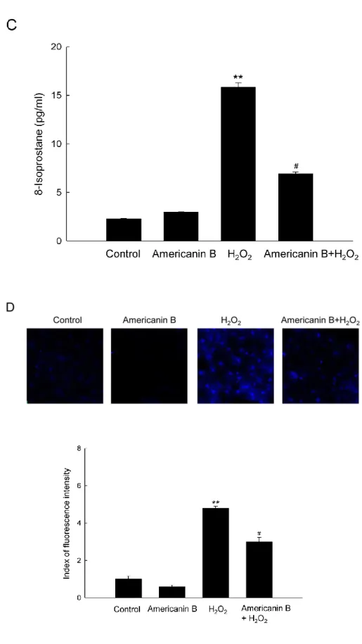
formation of protein carbonyls (Fig. 2B). Finally, the degree of 8-isoprostane was examined,
a hallmarker of lipid peroxidation, which is released from the oxidative cells into the culture
medium. As shown in Fig. 2C, cells treated with H2O2 secreted higher levels of 8-isoprostane
than untreated cells, but pre-treatment with americanin B in H2O2-treated cells significantly
reduced the 8-isoprostane level. In addition, lipid peroxidation was also verified by fluorescent
product DPPP oxide produced from DPPP (Okimoto et al. 2000). The intensity of DPPP oxide
was dramatically increased in H2O2-treated cells relative to control. Pre-treatment with
americanin B in H2O2-treated cells led to a reduction in fluorescence intensity (Fig. 2D). Taken
together, these results presented in Fig. 2 confirm that americanin B significantly protects
Figure 2. Americanin B protects against H2O2-induced oxidative lipid, protein, and DNA
16
H2O2. (A) The comet assay was performed to assess DNA damage. Representative images and
percentage of cellular fluorescence within comet tails are shown. (B) Protein oxidation was
assayed by measuring the amount of carbonyl formation. Lipid peroxidation was assayed by
(C) measuring 8-isoprostane levels in the conditioned medium and (D) detecting lipid
hydroperoxide by fluorescence microscopy after the DPPP reaction. **p<0.001, significantly
different from control; #p<0.001, significantly different from H
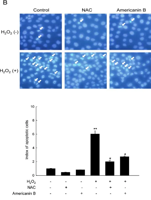
3-3. Americanin B reduces apoptosis induced by H2O2.
To elucidate the cytoprotective effect of americanin B against H2O2-induced apoptosis,the
research examined the viability of HaCaT cells exposed to 1 mM H2O2, either pre-treated or
not with 10 µM americanin B. In H2O2-treated cells, cell viability was reduced to 64% relative
to the control group, but pre-treatment with americanin B recovered the viability to 83% (Fig.
3A). In parallel, cells were stained with nuclei with Hoechst 33342, and then assessed the cells
by microscopy. In control groups or americanin B-treated cells, normal nuclei could be
visualized, whereas significant nuclear condensation was found in H2O2-treated cells
(apoptotic index = 6). However, when these cells were pre-treated with americanin B or NAC,
the number of nuclear fragmentation was declined (americanin B: apoptotic index = 2.7; NAC:
apoptotic index = 2) (Fig. 3B). These results indicate that americanin B protects cells against
18
Figure 3. Americanin B reduces apoptosis induced by H2O2. (A) Cells were treated with
10 µM americanin B and exposed to H2O2 1 h later. After incubation for a further 24 h, cell
viability was determined by the MTT assay and is expressed as a percentage of the control.
*p<0.05, significantly different from control; p<0.05, significantly different from H2O2
-treated cells. (B) Cells were stained with Hoechst 33342 dye and observed by fluorescence
microscopy; apoptotic bodies were quantitated. **p<0.001, significantly different from
control; #p<0.001, significantly different from H
4. Discussion
Previous researches show that plenty of natural compounds exert pharmaceutical activity
such as anticancer, antioxidant, or anti-inflammatory activities because of containing active
pharmaceutical ingredient phenolic groups(Chung et al. 1998; Cassidy et al. 2000; Tapiero et
al. 2002). Moreover, the antioxidant activities of many natural compounds are related to their
phenolic structures (Rice-Evans et al. 1996; Heim et al. 2002). Several lines of evidences
suggest that the protective mechanism is based on formation of phenoxyl radical by upon
donating a hydrogen atom from phenolic hydroxyl groups. The resultant radical can effectively
act as antioxidant to quench reactive oxygen and nitrogen species by giving radicals to them
and forming the stable state (Valentão et al. 2002, 2003; Heim et al. 2002; Payá et al. 1992;
Choi et al. 2002), and interdict the process of lipid peroxidation (Cheng et al. 2003; Foti et al.
2001) by inhibition of the cycle of radicals. The number and position of aromatic hydroxyl
groups strongly affect the property of phenolic antioxidants (Cai et al. 2006; Lien et al. 1998;
Tyrakowska et al. 1999). The main requirement for effective radical quenching in aspect of
structure is the bearing an ortho-dihydroxy group (catechol structure), which confers intrinsic
antioxidant properties by providing an easily donated electron (Kang et al. 2012).
Recent studies have shown that the presence of a catechol group is associated with a high
capacity to protect DNA against oxidative damage (Melidou et al. 2005; Noroozi et al. 1998).
The antioxidant capacity of phenolic compounds has also been attributed to their ability to
chelate metal ions involved in the production of free radicals (Yang et al. 2001). Furthermore,
a great many studies have certified that an unsaturated group within its structure (e.g.,
carbon-carbon double bond, carbon-carbon-oxygen double bond, or conjugated system) can enhance the
antioxidant capacity of a compound. Compounds bearing an electrophilic α, β-unsaturated
carbonyl group can interact with nucleophiles, including ROS such as the superoxide anions,
hydroxyl radicals, H2O2, and singlet oxygen (Farombi et al. 2008).
20
group in its structure (Yu et al. 2001), and it is likely that these groups provide it with its
intrinsic antioxidant properties. Based on its structural properties, americanin B appears to
exert its antioxidant effects by directly quenching ROS. When recognizing ROS, the catechol
moieties of americnanin B become rich-electronic groups by releasing one hydrogen atom and
form large conjugated groups with the help of benzene ring, providing some free radicals to
unstable molecules because of lacking of electron in electron shell. In addition, α,
β-unsaturated carbonyl group itself is a conjugated group and forms covalent bonds with ROS
molecules via sharing one or more electron pairs to stable structures in outer electrons. ROS
disable the ability to attack cells or organism when they become steady state through been
given or sharing electrons from americanin B. Consistent with this assumption, the
investigation showed that americanin B decreased generation of free radicals such as DPPH
radicals, superoxide anions and hydroxyl radicals as well as intracellular ROS.
In H2O2-treated cells, americanin B markedly decreased the tail length and the proportion
of damaged DNA. H2O2 promoted the modification of protein-bound carbonyl groups and
disrupted cell function; americanin B blocked the formation of protein carbonyl following
H2O2 treatment. Similarly, americanin B also protected membrane lipids from H2O2-induced
peroxidation, as shown by the DPPP data and levels of 8-isoprostane. These protective effects
of americanin B against DNA, lipid, and protein damage ultimately help to protect cells against
oxidative stress-induced cell death. Cells exposed to H2O2 exhibited the distinct nuclear
fragmentation of apoptosis, but americanin B suppressed these manifestations of apoptosis and
increased cell viability.
In this study, these data showed that americanin B is an antioxidant that protects cells against
the oxidative damage caused by H2O2 via scavenging ROS and inhibiting apoptosis. In future
experiments, the subsequent investigation will explore the mechanisms by which americanin
B blocks ROS generation and prevents apoptosis.
and contents is from “Americanin B protects cultured human keratinocytes against
oxidative stress by exerting antioxidant effects”, which has been published in the journal
of “In Vitro Cellular & Developmental Biology - Animal” with “DOI
22
5. References
Bandyopadhyay U.; Das D.; Banerjee R. K. Reactive oxygen species: oxidative damage and
pathogenesis. Curr. Sci. 77: 658-666; 1999.
Beauchamp M. C.; Letendre E.; Renier G. Macrophage lipoprotein lipase expression is
increased in patients with heterozygous familial hypercholesterolemia. J. Lipid Res. 43:
215-222; 2002.
Brand-Williams W.; Cuvelier M. E.; Berset C. L. W. T. Use of a free radical method to evaluate
antioxidant activity. Lebensm. Wiss. Technol. 28: 25-30; 1995.
Bohr V. A.; Smith C. A.; Okumoto D. S.; Hanawalt P. C. DNA repair in an active gene: removal
of pyrimidine dimers from the DHFR gene of CHO cells is much more efficient than in the
genome overall. Cell 40: 359-369; 1985.
Cai Y. Z.; Sun M.; Xing J.; Luo Q.; Corke H. Structure-radical scavenging activity
relationships of phenolic compounds from traditional Chinese medicinal plants. Life Sci.
78: 2872-2888; 2006.
Carmichael J.; DeGraff W. G.; Gazdar A. F.; Minna J. D.; Mitchell J. B. Evaluation of a
tetrazolium-based semiautomated colorimetric assay: assessment of chemosensitivity
testing. Cancer Res. 47: 936-942; 1987.
Cassidy A.; Hanley B.; Lamuela-Raventos R. M. Isoflavones, lignans and stilbenes-origins,
metabolism and potential importance to human health. J. Sci. Food Agric. 80: 1044-1062;
Chen J. W.; Zhu Z. Q.; Hu T. X.; Zhu D. Y. Structure-activity relationship of natural flavonoids
in hydroxyl radical-scavenging effects. Acta Pharmacol. Sin. 23: 667-672; 2002.
Cheng Z.; Ren J.; Li Y.; Chang W.; Chen Z. Establishment of a quantitative structure-activity
relationship model for evaluating and predicting the protective potentials of phenolic
antioxidants on lipid peroxidation. J. Pharm. Sci. 92: 475-484; 2003.
Choi H. R.; Choi J. S.; Han Y. N.; Bae S. J.; Chung H. Y. Peroxynitrite scavenging activity of
herb extracts. Phytother. Res. 16: 364-367; 2002.
Chung K. T.; Wong T. Y.; Huang Y. W.; Lin Y. Tannins and human health: a review. Crit.
Rev. Food Sci. Nutr. 38: 421-464; 1998.
Cunha W. R.; e Silva M. L. A.; Sola R. C.; Veneziani S. R. A.; Bastos J. K. Lignans: chemical
and biological properties. In: Venketeshwer R (ed). Phytochemicals-a global perspective of
their role in nutrition and health. In Tech, Rijeka, Croatia, 213-234; 2012.
Dixon R. A. Phytoestrogens. Annu. Rev. Plant Biol. 55: 225-261; 2004.
Dmitriev L. F.; Titov V. N. Lipid peroxidation in relation to ageing and the role of endogenous
aldehydes in diabetes and other age-related diseases. Ageing Res. Rev. 9: 200-210; 2010.
Farombi E. O.; Shrotriya S.; Na H. K.; Kim S. H.; Surh Y. J. Curcumin attenuates
dimethylnitrosamine-induced liver injury in rats through Nrf2-mediated induction of heme
oxygenase-1. Food Chem. Toxicol. 46: 1279-1287; 2008.
Foti M.; Ruberto G. Kinetic solvent effects on phenolic antioxidants determined by
spectroscopic measurements. J. Agric. Food Chem. 49: 342-348; 2001.
24
from Magnolia obovata. J. Pharm. Pharmacol. 49: 209-212; 1997.
Harper A.; Kerr D. J.; Gescher A.; Chipman J. K. Antioxidant effects of isoflavonoids and
lignans, and protection against DNA oxidation. Free Radic. Res. 31: 149-160; 1999.
Heim K. E.; Tagliaferro A. R.; Bobilya D. J. Flavonoid antioxidants: chemistry, metabolism
and structure-activity relationships. J. Nutr. Biochem. 13: 572-584; 2002.
Hsu J. Y.; Chu J. J.; Chou M. C.; Chen Y. W. Dioscorin pre-treatment protects A549 human
airway eithelial cells from hydrogen peroxide-induced oxidative stress. Inflammation 2013:
1-7; 2013.
Kang K. A.; Zhang R.; Piao M. J.; Chae S. W.; Kim H. S.; Park J. H.; Jung K. S.; Hyun J. W.
Baicalein inhibits oxidative stress-induced cellular damage via antioxidant effects. Toxicol.
Ind. Health 28: 412-421; 2012.
Kim H. S.; Lee K.; Kang K. A.; Lee N. H.; Hyun J. W. Phloroglucinol exerts protective effects
against oxidative stress-induced cell damage in SH-SY5Y cells. J. Pharmacol. Sci.119:
186-192; 2012.
Kim K. H.; Choi J. W.; Ha S. K.; Kim S. Y.; Lee K. R. Neolignans from Piper kadsura and
their anti-neuroinflammatory activity. Bioorg. Med. Chem. Lett. 20: 409-412; 2010.
Kohno M.; Mizuta Y.; Kusai M.; Masumizu T.; Makino K. Measurements of superoxide anion
radical and superoxide anion scavenging activity by electron spin resonancespectroscopy
coupled with DMPO spin trapping. Bull. Chem. Soc. Jpn. 67: 1085-1090; 1994.
Lee D. Y.; Lee D. G.; Cho J. G.; Bang M. H.; Lyu H. N.; Lee Y. H.; Kim S. Y.; Baek N. I.
effects. Arch. Pharm. Res. 32: 1345-1349; 2009.
Lee D. Y.; Seo K. H.; Jeong R. H.; Lee S. M.; Kim G. S.; Noh H. J.; Kim G. W.; Kim J. Y.;
Baek N. I. Anti-inflammatory lignans from the fuits of Acanthopanax sessiliflorus.
Molecules 18: 41-49; 2012.
Lee K. M.; Yun C. H. Potential in vitro protective effect of quercetin, catechin, caffeic acid
and phytic acid against ethanol-induced oxidative stress in SK-Hep-1 cells. Biomol.
Ther. 20: 492-498; 2012.
Lee W. S.; Baek Y. I.; Kim J. R.; Cho K. H.; Sok D. E.; Jeong T. S. Antioxidant activities of a
new lignan and a neolignan from Saururus chinensis. Bioorg. Med. Chem. Lett. 14:
5623-5628; 2004.
Li L.; Abe Y.; Kanagawa K.; Usui N.; Imai K.; Mashino T.; Mochizuki M.; Miyata N.
Distinguishing the 5,5-dimethyl-1-pyrroline N-oxide (DMPO)-OH radical quenching
effect from the hydroxyl radical scavenging effect in the ESR spin-trapping method. Anal.
Chim. Acta 512: 121-124; 2004.
Li L.; Abe Y.; Mashino T.; Mochizuki M.; Miyata N. Signal enhancement in ESR spin-trapping
for hydroxyl radicals. Anal. Sci. 19: 1083-1084; 2003.
Lien E. J.; Ren S.; Bui H. H.; Wang R. Quantitative structure-activity relationship analysis of
phenolic antioxidants. Free Radic. Biol. Med. 26: 285-294; 1998.
Maroto R.; Perez-Polo J. R. BCL-2-related protein expression in apoptosis: oxidative stress
versus serum deprivation in PC12 cells. J. Neurochem. 69: 514-523; 1997.
26
the fruits of Broussonetia papyrifera. J. Nat. Prod. 72: 621-625; 2009.
Melidou M.; Riganakos K.; Galaris D. Protection against nuclear DNA damage offered by
flavonoids in cells exposed to hydrogen peroxide: the role of iron chelation. Free Radic.
Biol. Med. 39: 1591-1600; 2005.
Mena S.; Ortega A.; Estrela J. M. Oxidative stress in environmental-induced carcinogenesis.
Mutat. Res. 674: 36-44; 2009.
Moss G. P. Nomenclature of lignans and neolignans (IUPAC Recommendations 2000). Pure
Appl. Chem. 72: 1493-1523; 2000.
Nguyen C. N.; Kim H. E.; Lee S. G. Caffeoylserotonin protects human keratinocyte HaCaT
cells against H2O2-induced oxidative stress and apoptosis through upregulation of HO-1
expression via activation of the PI3K/Akt/Nrf2 pathway. Phytother. Res. 17: 1810-1818;
2013.
Noroozi M.; Angerson W. J.; Lean M. E. Effects of flavonoids and vitamin C on oxidative
DNA damage to human lymphocytes. Am. J. Clin. Nutr. 67: 1210-1218; 1998.
Okimoto Y.; Watanabe A.; Niki E.; Yamashita T.; Noguchi N. A novel fluorescent probe
diphenyl-1-pyrenylphosphine to follow lipid peroxidation in cell membranes. FEBS Lett.
474: 137-140; 2000.
Payá M.; Halliwell B.; Hoult J. R. S. Interactions of a series of coumarins with reactive oxygen
species: Scavenging of superoxide, hypochlorous acid and hydroxyl radicals. Biochem.
Pharmacol. 44: 205-214; 1992.
and protein carbonyl (PCO) levels as biomarkers of oxidative stress in subjects with
familial hypercholesterolemia. Clin. Biochem. 43: 1220-1224; 2010.
Proksch E.; Brandner J. M.; Jensen J. M. The skin: an indispensable barrier. Exp. Dermatol. 17:
1063-1072; 2008.
Rajagopalan R.; Ranjan S.; Nair C. K. Effect of vinblastine sulfate on
gamma-radiation-induced DNA single-strand breaks in murine tissues. Mutat. Res. 536: 15-25; 2003.
Rice-Evans C. A.; Miller N. J.; Paganga G. Structure-antioxidant activity relationships of
flavonoids and phenolic acids. Free Radic. Biol. Med. 20: 933-956; 1996.
Rosenkranz A. R.; Schmaldienst S.; Stuhlmeier K. M.; Chen W.; Knapp W.; Zlabinger G. J. A
microplate assay for the detection of oxidative products using 2′,7′-dichlorofluorescin-diacetate. J. Immunol. Methods 156: 39-45; 1992.
Schühly W.; Khan S. I.; Fischer N. H. Neolignans from North American Magnolia species
with cyclooxygenase 2 inhibitory activity. Inflammopharmacology 17: 106-110; 2009.
Singh N. P. Microgels for estimation of DNA strand breaks, DNA protein crosslinks and
apoptosis. Mutat. Res. 455: 111-127; 2000.
Sinha K.; Das J.; Pal P. B.; Sil P. C. Oxidative stress: the mitochondria-dependent and
mitochondria-independent pathways of apoptosis. Arch. Toxicol. 87: 1157-1180; 2013.
Tapiero H.; Tew K. D.; Ba N.; Mathé G. Polyphenols: do they play a role in the prevention of
human pathologies? Biomed. Pharmacother. 56: 200-207; 2002.
Terashvili M.; Sarkar P.; Nostrand M. V.; Falck J. R.; Harder D. R. The protective effect of
28
injury in astrocyte-dopaminergic neuronal cell line co-culture. Neuroscience 223: 68-76;
2012.
Tezel G. Oxidative stress in glaucomatous neurodegeneration: mechanisms and consequences.
Prog. Retin. Eye Res. 25: 490-513; 2006.
Tyrakowska B.; Soffers A. E. M. F.; Szymusiak H.; Boeren S.; Boersma M. G.; Lemanska K.;
Vervoort J.; Rietjens I. M. C. M. TEAC antioxidant activity of 4-hydroxybenzoates.
Free Radic. Biol. Med. 27: 1427-1436; 1999.
Ueno I.; Kohno M.; Yoshihira K.; Hirono I. Quantitative determination of the superoxide
radicals in the xanthine oxidase reaction by measurement of the electron spin resonance
signal of the superoxide radical spin adduct of 5,5-dimethyl-1-pyrroline-1-oxide. J.
Pharmacobio-dyn. 7: 563-569; 1984.
Valentão P.; Fernandes E.; Carvalho F.; Andrade P. B.; Seabra R. M.; Bastos M. L.
Antioxidative properties of cardoon (Cynara cardunculus L.) infusion against superoxide
radical, hydroxyl radical and hypochlorous acid. J. Agric. Food Chem. 50: 4989-4993;
2002.
Valentão P.; Fernandes E.; Carvalho F.; Andrade P. B.; Seabra R. M.; Bastos M. L. Hydroxyl
radical and hypochlorous acid scavenging activity of small centaury (Centaurium
erythraea) infusion. A comparative study with green tea (Camellia sinensis). Phytomedicine 10: 517-522; 2003.
Valko M.; Leibfritz D.; Moncol J.; Cronin M. T.; Mazur M.; Telser J. Free radicals and
Biol. 39: 44-84; 2007.
Wang L. Y.; Unehara N.; Kitanaka S. Lignans from the roots of Wikstroemia indica and their
DPPH radical scavenging and nitric oxide inhibitory activities. Chem. Pharm. Bull. 53:
1348-1351; 2005.
Yang C. S.; Landau J. M.; Huang M. T.; Newmark H. L. Inhibition of carcinogenesis by dietary
polyphenolic compounds. Annu. Rev. Nutr. 21: 381-406; 2001.
Yu J. G.; Li T. M.; Sun L.; Luo X. Z.; Ding W.; Li D. Y. Studies on chemical constituents of
the seeds from Atrabotrys hexapetalus (Annonaceae). Acta Pharmaceutica Sin. 36:
30
6. Abstract in Korean
이 연구는 리그난계 화합물인 americanin B 의 hydrogen peroxide (H2O2)로 유도된
세포 손상에 대한 세포 보호 효과를 평가하였다. Americanin B 는 DPPH radicals, superoxide anions, hydroxyl radicals 의 레벨을 감소시킬 뿐만 아니라 세포 내 ROS 의 레벨도 감소시킨다. Americanin B 는 또한 DNA 가닥 파괴를 나타내는 comet tails 형성의 억제에 의해 나타낸 바와 같이, H2O2 처리에 의해 유도된 DNA 손상을
감소시키며, protein carbonyls 및 8–isoprostane 의 측정을 통해 단백질의 oxidation 과 지질의 peroxidation 을 보호한다는 것을 나타내었다. 또한, americanin B 는 H2O2에
유도된 apoptotic cell death 에 대한 보호를 하며, 이것은 Hoechst 33342 로 염색된 apoptotic bodies 의 숫자의 감소에 의해 측정되었다. 이러한 연구 결과는 americanin
B 가 antioxidant effects 와 apoptosis 억제를 발휘함으로써 oxidative damage 에 대한 세포를 보호하는 것을 제안한다.
7. Acknowledgements
First and foremost, I would like to express my sincere gratitude to my dedicated supervisor
Prof. Jin-Won Hyun to offer me the opportunity to commence my master degree in Jeju
National University. Her modest, encouragement and guidance have provided a good basis for
me during all the time of my two-year study in South Korean.
I owe my sincere appreciation to Prof. Eun-Sook Yoo and Bae-Beok Park, school of
medicine in Jeju National University, who gave me the instructions and suggestions to
complete my thesis. And all professors who have guided me in school of medicine are also
greatly appreciated.
I am particularly grateful to my lab teacher Mei-Jing Piao for her profound knowledge, rich
research experience and meticulous guidance. Senior brother Ki-Cheon Kim and senior sister
Areum-Daseul Kim are deeply appreciated for their valuable ideas, suggestions, patience and
kindness on my learning and life. I would like to express my heartfelt gratitude to my classmate
and friend Cheng-Wen Yao, who supplies selfless assistance on experiments and thesis writing,
excellent knowledge and ideas, nice moment over the two years. Thanks are also due to my
labmate Jin-Won Cha for her great encouragement and suggestions not only on my research
but also convenience to my personal life. I am also thankful to Xia Han, Susara M.H. and
Min-Chang Oh because of their kind help and improvements.
My special thanks go to my girlfriend Chun-Ling Wang, who helped me by various ways to
finish my studies in Korea. I would also like to express my special express gratitude to Hao
Li-32
Jie Min, Ye Tian, Zhen-Yu Zhong, Wen-Hao Shi, Zhou-Zhou Xu, Shuo He, Ke-Ru Chen,
Zhang-Ming He-Lian, Sheng Piao and other friends for share of joy, laughter and happy time.
Last but not least, I would like to thank my family for their support all the way from the
very beginning of my study. I am thankful to all my family members for their thoughtfulness