ABSTRACT
Invasive fungal infection (IFI) is a serious threat to pediatric patients with cancer given high morbidity and mortality. We present an 18-year-old male with precursor T-cell lymphoblastic leukemia who developed Pancoast syndrome, presented with paresthesia and numbness in the right shoulder and arm during a neutropenic fever period. He was diagnosed with pneumonia in the right upper lung field. He was later found to have an invasive pulmonary fungal infection caused by multiple fungi species, including Rhizomucor, confirmed by histology and polymerase chain reaction (PCR) (proven infection), Penicillium decumbens diagnosed by PCR, and Aspergillus suspected from galactomannan assay (probable infection). Unfortunately, the patient's condition further worsened owing to the aggravation of leukemia, chemotherapy-induced neutropenia, and bacterial coinfection, leading to multiorgan failure and death. Here, we report a case of IFI caused by multiple fungal species that presented as Pancoast syndrome.
Keywords: Pancoast syndrome; Lung diseases, fungal; Coinfection
INTRODUCTION
Fungal infections by Rhizopus, Mucor, and Rhizomucor species account for up to 75% of mucormycosis cases encountered in patients with hematologic malignancy.1) In addition, Aspergillus species are an important cause of life threatening infection in
immunocompromised patients. This at-risk population includes allogeneic hematopoietic stem cell transplant recipients, solid organ transplant recipients, and patients using corticosteroids; this group is predisposed to invasive fungal infections (IFIs).2) High rates of mortality and morbidity have been reported among patients with IFI caused by multiple
Case Report
Received: Jun 23, 2020 Revised: Oct 31, 2020 Accepted: Oct 31, 2020 Correspondence to Yae-Jean KimDepartment of Pediatrics, Samsung Medical Center, Sungkyunkwan University, School of Medicine, 81 Irwon-ro, Gangnam-gu, Seoul 06351, the Republic of Korea.
E-mail: [email protected] Copyright © 2021 The Korean Society of Pediatric Infectious Diseases
This is an Open Access article distributed under the terms of the Creative Commons Attribution Non-Commercial License (https:// creativecommons.org/licenses/by-nc/4.0/) which permits unrestricted non-commercial use, distribution, and reproduction in any medium, provided the original work is properly cited. ORCID iDs Hyungsuk Jin https://orcid.org/0000-0001-7857-3886 Dongsub Kim https://orcid.org/0000-0002-9836-6769 Joon-sik Choi https://orcid.org/0000-0002-5587-2960 Hee Jae Huh
https://orcid.org/0000-0001-8999-7561 Nam Yong Lee
https://orcid.org/0000-0003-3688-0145 Joungho Han
https://orcid.org/0000-0003-4424-7008 Hee Won Cho
https://orcid.org/0000-0002-0440-645X Youngeun Ma
https://orcid.org/0000-0002-5862-6319 Tae Yeon Jeon
https://orcid.org/0000-0002-7796-1307
Hyungsuk Jin ,1 Dongsub Kim ,1,2 Joon-sik Choi ,1,3 Hee Jae Huh ,4 Nam Yong Lee ,4 Joungho Han ,5 Hee Won Cho ,1 Youngeun Ma ,1,6 Tae Yeon Jeon ,7 So-Young Yoo ,7 Keon Hee Yoo ,1 Hong Hoe Koo ,1 Yae-Jean Kim 1
1 Department of Pediatrics, Samsung Medical Center, Sungkyunkwan University School of Medicine, Seoul,
the Republic of Korea
2Department of Pediatrics, Kyungpook National University, Daegu, the Republic of Korea
3Department of Hospital Medicine, Yonsei University College of Medicine, Yongin, the Republic of Korea 4 Department of Laboratory Medicine and Genetics, Samsung Medical Center, Sungkyunkwan University
School of Medicine, Seoul, the Republic of Korea
5 Department of Pathology, Samsung Medical Center, Sungkyunkwan University School of Medicine, Seoul,
the Republic of Korea
6Department of Pediatrics, Seoul National University Bundang Hospital, Seongnam, the Republic of Korea 7 Department of Radiology and Center for Imaging Science, Samsung Medical Center, Sungkyunkwan
University School of Medicine, Seoul, the Republic of Korea
A Case with Multiple Fungal
Coinfections in a Patient who
So-Young Yoo
https://orcid.org/0000-0002-8203-3441 Keon Hee Yoo
https://orcid.org/0000-0002-5980-7912 Hong Hoe Koo
https://orcid.org/0000-0001-8082-1412 Yae-Jean Kim
https://orcid.org/0000-0002-8367-3424 Conflict of Interest
No potential conflict of interest relevant to this article was reported.
Author Contributions
Conceptualization: Kim YJ; Investigation: Kim D, Choi JS, Huh HJ, Lee NY, Han J, Cho HW, Ma Y, Jeon TY, Yoo SY, Yoo KH, Koo HH; Writing - original draft: Jin H; Writing - review & editing: Kim YJ.
fungal species, as each species has different sensitivity to antifungal agents.2,3) Therefore, prompt clinical suspicion and appropriate therapy considering antifungal sensitivity is critical in such cases. Here, we present the case of an 18-year-old man with relapsed precursor T-cell lymphoblastic leukemia who initially presented with neutropenic fever and Pancoast syndrome (right shoulder and arm pain and paresthesia) and subsequently was diagnosed with pulmonary IFI caused by multiple fungal species that manifested the symptoms of Pancoast syndrome.
CASE
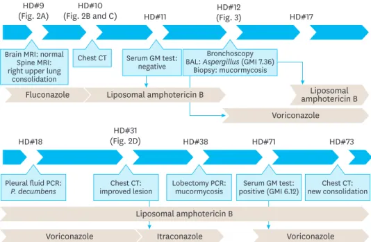
An 18-year-old man with relapsed precursor T-cell lymphoblastic leukemia was admitted with neutropenic fever and bacteremia caused by Klebsiella pneumoniae. Catheter-related blood stream infection was suspected and cefepime was administered. His fever subsided within 24 hours, and a follow-up blood culture was negative. The initial antimicrobial agent was continued along with the preexisting fluconazole prophylaxis. On the 5th hospital day, he developed right shoulder pain and paresthesia and numbness in the right arm. In addition, he showed right arm motor weakness and developed a fever of 38.3°C. Spine and brain magnetic resonance imaging (MRI) performed on the 9th hospital day revealed no significant abnormality in the bony structure of his spine and brain but showed edematous change and increased signal intensity at the right brachial plexus, suggesting brachial plexus impingement (Figs. 1 and 2). Furthermore, pulmonary consolidation in the right upper lobe with interstitial thickening was observed on MRI (Fig. 2A). On the 10th hospital day, chest computed tomography (CT) (Fig. 2B and C) revealed right upper lobe pneumonia. The patient was treated with liposomal amphotericin B (Fig. 1). On the same day, we performed a serum galactomannan (GM) assay using an ELISA kit (IBL International, Hamburg, Germany) targeting Aspergillus fumigatus IgG, which yielded negative results. However, his
HD#9
(Fig. 2A) (Fig. 2B and C)HD#10 HD#11 (Fig. 3)HD#12 HD#17
Brain MRI: normal Spine MRI: right upper lung
consolidation
Fluconazole amphotericin BLiposomal
Voriconazole Chest CT Serum GM test:
negative
Bronchoscopy BAL: Aspergillus (GMI 7.36)
Biopsy: mucormycosis
HD#18 (Fig. 2D)HD#31 HD#38 HD#71 HD#73
Pleural fluid PCR:
P. decumbens improved lesionChest CT: Lobectomy PCR:mucormycosis positive (GMI 6.12)Serum GM test: new consolidationChest CT:
Voriconazole Itraconazole Voriconazole
Liposomal amphotericin B Liposomal amphotericin B
Fig. 1. Flow chart of the diagnostic methods and antibiotic use during the clinical course.
Abbreviations: HD, hospital day; MRI, magnetic resonance imaging; CT, computed tomography; GM, galactomannan; BAL, bronchoalveolar lavage; GMI, Galactomannan index, PCR, polymerase chain reaction.
bronchoalveolar lavage (BAL) sample collected on the 12th hospital day later yielded a positive galactomannan index (GMI) of 7.36. Therefore, liposomal amphotericin B was switched to voriconazole for the treatment of the suspected pulmonary aspergillosis (Fig. 1). On the 17th hospital day, the histopathology of bronchial tissue collected at the time of bronchoscopy identified mucormycosis (Fig. 3). Based on this finding, liposomal amphotericin B was
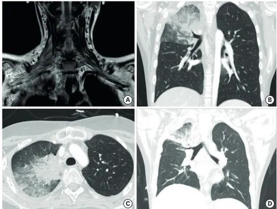
B A
D C
Fig. 2. (A) Spine magnetic resonance imaging showing pulmonary consolidation in the right upper lobe, with edematous change and increased signal intensity at the right brachial plexus indicating brachial plexus impingement. (B, C) Chest CT images showing airspace consolidation, ground glass opacity, intralobular septal thickening in the right upper lobe of the lung and pleural effusion. (D) Chest CT image showing reduced consolidation and ground glass opacity in the right upper lobe after antifungal treatment.
Abbreviation: CT, computed tomography.
Fig. 3. Histopathologic examination of a bronchial tissue section in the right upper lobe and mediastinal lymph node showing non-septate broad filamentous fungi branching nearly 90° (arrow), suggesting mucormycosis.
resumed to cover both aspergillosis and mucormycosis. On the 18th hospital day, a large volume of pleural effusion was noted and was drained. Fungal Sanger sequencing using the BigDye Terminator Cycle Sequencing Kit 3.1 on an ABI prism 3730 Genetic Analyzer (Thermo Fisher Scientific, Waltham, MA, USA) targeting fungus-specific internal transcribed spacer (ITS) regions covering ITS1, 5.8S, and ITS2 from the pleural fluid detected Penicillium decumbens. Multiple follow-up serum GM assay results were all negative for almost 3 months. Therefore, we considered the possibility of false-positive Aspergillus antigen assay result caused by P. decumbens infection. However, we could not completely rule out simultaneous multiple fungal infections and continued both liposomal amphotericin B and voriconazole (Fig. 1).
The patient's condition stabilized and the fever subsided. Lung lesions were resolved on chest CT on the 31st hospital day (Fig. 2D). On the 34th hospital day, voriconazole was switched to itraconazole (Fig. 1) because prolonged liposomal amphotericin B and voriconazole combination treatment is not reimbursed by the Korean national health insurance system. On the 38th hospital day, we performed a lobectomy of the right upper lobe to effectively decrease his fungal burden, with a plan to proceed to chemotherapy for his relapsed leukemia as soon as the fungal infection resolved. On the 48th hospital day, histologic examination of the lung tissue revealed fungal presence and DNA Sanger sequencing identified Rhizomucor species (R. pusillus and R. tauricus), and Aspergillus was not detected. Therefore, itraconazole was discontinued (Fig. 1). His shoulder and arm pain aggravated. The patient's bone marrow biopsy revealed lymphoblasts on the 51st hospital day, and chemotherapy was restarted to treat leukemia. Liposomal amphotericin B was continued to treat mucormycosis.
However, on the 73rd hospital day, follow-up chest radiography and CT showed a newly developed consolidation in the right lower and left upper lobes. Simultaneously, his serum GMI was positive at 6.12 for the first time during his fungal infection course. Voriconazole was reintroduced on the 71st hospital day because aspergillosis could not be ruled out and his GM assay results were positive ever since. He developed neutropenic fever and bacteremia caused by methicillin-resistant Staphylococcus epidermidis on the 87th hospital day. Thereafter, his condition rapidly deteriorated and he died of septic shock and multiorgan failure without responding to inotropes.
This study was conducted with approval of Samsung Medical Center Institutional Review Board (No. 2020-06-145).
DISCUSSION
We present the case of an 18-year-old man with T-cell lymphoblastic leukemia who experienced IFI presumably caused by multiple fungal species. Although we treated the patient with combinational antifungal therapy and surgical resection to control the persistent multiple fungal infections, the patient did not survive.
Early diagnosis of IFI remains a challenge. Currently, the gold standard for diagnosis of IFI is direct observation or culture tests from tissue samples for pathogens.4) In patients with IFI, radiologic studies, including CT imaging, are useful diagnostic tools.5)
Our patient developed Pancoast syndrome that led to the further radiologic imaging work-up, which in turn led to the diagnosis of pulmonary IFI. Pancoast syndrome is characterized by
shoulder and arm pain that radiates to the ulnar aspect of the arm and forearm. It is caused by the involvement of the spinal nerve roots from the 8th cervical spinal nerve to the 2nd thoracic spinal nerve of the brachial plexus. Apical lung cancer and other malignancies are predominant causes of Pancoast syndrome. Furthermore, infective conditions such as fungal infection (as in this case) caused by Aspergillus, Cryptococcus, and Mucor are reported to cause Pancoast syndrome.6) Shoulder and arm pain in immunocompromised patients could be the first symptom of pulmonary infections such as IFIs. Pulmonary infections should additionally be considered when a patient presents with the symptoms of Pancoast syndrome.
Biomarkers are additional tools that can help diagnose IFI. At our center, sensitivity and specificity to serum GM assay among pediatric patients with cancer were 91.3% and 81.7%, respectively.7) Several studies have assessed the utility of GM assays as a diagnostic tool in children with symptoms potentially indicative of IFI. A systematic review reported the pooled sensitivity and specificity at 89% and 85%, respectively.8) Sensitivity and specificity to GM in BAL fluid seems higher than that in the serum among immunocompromised adult patients; however, such studies in pediatric patients are lacking.4)
P. decumbens is found abundantly in natural sources and commonly in soil worldwide. P. decumbens was first identified in 1992 on an indirect fluorescent antibody test for a patient with pneumonia.9) Recently, Penicillium species have emerged as opportunistic pathogens in immunocompromised hosts and have been reported to cause coinfections with the Mucor species.10,11)P. decumbens alone can cause pulmonary IFI, similarly to other fungi. Studies have reported cross-reactions between Aspergillus and Penicillium or other fungal species on GM assays.12) In our patient, the GM assay was positive on 2 different periods. We speculate that the first GM-positive identification with the BAL sample on the 12th hospital day was likely a false positive caused by P. decumbens. However, at that time, we could not ascertain whether it was an Aspergillus infection that was present from the beginning of the course or a Penicillium infection presenting as GM cross-reactivity. Therefore, we continued voriconazole although the pleural fluid returned positive for P. decumbens. The second positive result from the blood sample on the 72nd hospital day was considered to be a true infection by Aspergillus despite the patient being on liposomal amphotericin B.13) We excluded the conditions that would cause false-positives for the following condition. In this patient, a newly identified lesion was observed on CT and consecutive GM values were significantly high. Antibiotics that can potentially cause false-positive results were not used, and we confirmed no issues with the sample collection process.
Mucormycosis can occur in various presentations such as rhinocerebral, pulmonary, cutaneous, and gastrointestinal diseases, and tends to disseminate. Surgery is the treatment of choice for mucormycosis, when complete removal of the fungus is feasible.14) We
considered surgical resection from the beginning of the patient's course. However, the patient's condition was not stable for surgery. When blast cells appeared again in peripheral blood, surgery was performed to achieve a more aggressive reduction of the fungal burden so chemotherapy could be initiated.
If more than one pathogen is found in the same anatomic region, mixed fungal infections should be considered. Such infections have high rates of mortality and morbidity.2,3) In addition, comparatively rare and opportunistic species are found in higher proportions in mixed infections.15) Therefore, early detection and appropriate antifungal therapy are crucial. Although uncommon, Mucor and Aspergillus coinfections are well-reported in the literature.
Table 1 lists several reported cases of Aspergillus and Mucor coinfections. These patients had fungal infections in the orofacial region, lung, maxillary sinus, paranasal sinus, brain, spleen, and kidney. They all had an underlying disease and their clinical courses differed.16-20) We report a case of IFI caused by multiple fungi species including Rhizomucor (proven infection), P. decumbens (polymerase chain reaction [PCR] positive only), and Aspergillus (probable infection) as a symptom of Pancoast syndrome.
REFERENCES
1. Kontoyiannis DP, Lewis RE. How I treat mucormycosis. Blood 2011;118:1216-24.
PUBMED | CROSSREF
2. Jaya S, Vipparti H. Mixed fungal lung infection with Aspergillus fumigatus and Candida albicans in a
immunocomprimised patient. J Clin Diagn Res 2014;8:DD08-10.
PUBMED
3. Xiao H, Tang Y, Cheng Q, Liu J, Li X. Risk prediction and prognosis of invasive fungal disease in hematological malignancies patients complicated with bloodstream infections. Cancer Manag Res 2020;12:2167-75.
PUBMED | CROSSREF
4. Mohammadi S, Khalilzadeh S, Goudarzipour K, Hassanzad M, Mahdaviani A, Aarabi N, et al. Bronchoalveolar galactomannan in invasive pulmonary aspergillosis: a prospective study in pediatric patients. Med Mycol 2015;53:709-16.
PUBMED | CROSSREF
5. Lehrnbecher T, Robinson P, Fisher B, Alexander S, Ammann RA, Beauchemin M, et al. Guideline for the management of fever and neutropenia in children with cancer and hematopoietic stem-cell transplantation recipients: 2017 update. J Clin Oncol 2017;35:2082-94.
PUBMED | CROSSREF
6. Das A, Choudhury S, Basuthakur S, Das SK, Mukhopadhyay A. Pancoast's syndrome due to fungal abscess in the apex of lung in an immunocompetent individual: a case report and review of the literature. Case Rep Pulmonol 2014;2014:581876.
PUBMED | CROSSREF
7. Choi SH, Kang ES, Eo H, Yoo SY, Kim JH, Yoo KH, et al. Aspergillus galactomannan antigen assay and
invasive aspergillosis in pediatric cancer patients and hematopoietic stem cell transplant recipients. Pediatr Blood Cancer 2013;60:316-22.
PUBMED | CROSSREF
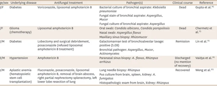
Table 1. Case reports of invasive fungal infections caused by multiple fungal species
Age/sex Underlying disease Antifungal treatment Pathogen(s) Clinical course Reference 53/F Diabetes Voriconazole, liposomal amphotericin B Bacterial culture of bronchial aspirate: Klebsiella
pneumoniae Dead Gupta et al.
16)
Fungal stain of bronchial aspirate: Aspergillus,
Mucor
Fungal culture of bronchial aspirate: Aspergillus 17/F Glioma
(chemotherapy) Liposomal amphotericin B Oral swab: Candida albicans, Candida parapsilosisNasal swab: Aspergillus flavus Dead Chermetz et al.17) Maxillary sinus biopsy: Rhizomucor
52/M Diabetes Lobectomy and surgical debridement, posaconazole (refused liposomal amphotericin B treatment)
Galactomannan test of bronchoalveolar lavage:
positive (5.09) Remission Lin et al. 18)
Bronchial pathogen: Aspergillus, Mucor,
Actinomycetes
68/M Hypertension Amphotericin B Paranasal sinus biopsy: A. flavus, Rhizopus
arrhizus (no mention Discharged
of recovery) Vaidya et al.19) 10/M Aplastic anemia (hematopoietic stem cell transplantation)
Fluconazole, posaconazole, liposomal amphotericin B, removal of brain abscess, right partial nephrectomy splenectomy, left lower lobe resection of lung
Lung needle biopsy: Rhizopus Recovered Weng et al.20) Pus culture from brain, spleen, kidney: A.
fumigatus
8. Huppler AR, Fisher BT, Lehrnbecher T, Walsh TJ, Steinbach WJ. Role of molecular biomarkers in the diagnosis of invasive fungal diseases in children. J Pediatric Infect Dis Soc 2017;6:S32-44.
PUBMED | CROSSREF
9. Yoshida K, Hiraoka T, Ando M, Uchida K, Mohsenin V. Penicillium decumbens. A new cause of fungus ball.
Chest 1992;101:1152-3.
PUBMED | CROSSREF
10. Ramírez I, Hidrón A, Cardona R. Successful treatment of pulmonary invasive fungal infection by
Penicillium non-marneffei in lymphoblastic lymphoma: case report and literature review. Clin Case Rep
2018;6:1153-7.
PUBMED | CROSSREF
11. Choi S, Son HJ, Jung J, Kim MJ, Chong YP, Lee SO, et al. 1703. Bacterial or fungal co-infection in patients with mucormycosis. Open Forum Infect Dis 2019;6:S623-4.
CROSSREF
12. Hesse SE, Luethy PM, Beigel JH, Zelazny AM. Penicillium citrinum: Opportunistic pathogen or idle
bystander? A case analysis with demonstration of galactomannan cross-reactivity. Med Mycol Case Rep 2017;17:8-10.
PUBMED | CROSSREF
13. Van Der Linden JW, Warris A, Verweij PE. Aspergillus species intrinsically resistant to antifungal agents.
Med Mycol 2011;49 Suppl 1:S82-9.
PUBMED | CROSSREF
14. Cornely OA, Alastruey-Izquierdo A, Arenz D, Chen SC, Dannaoui E, Hochhegger B, et al. Global guideline for the diagnosis and management of mucormycosis: an initiative of the European confederation of medical mycology in cooperation with the mycoses study group education and research consortium. Lancet Infect Dis 2019;19:e405-21.
PUBMED | CROSSREF
15. Gawaz A, Weisel G. Mixed infections are a critical factor in the treatment of superficial mycoses. Mycoses 2018;61:731-5.
PUBMED | CROSSREF
16. Gupta V, Rajagopalan N, Patil M, Shivaprasad C. Aspergillus and mucormycosis presenting with normal
chest X-ray in an immunocompromised host. BMJ Case Rep 2014;2014:bcr2014204022.
PUBMED | CROSSREF
17. Chermetz M, Gobbo M, Rupel K, Ottaviani G, Tirelli G, Bussani R, et al. Combined orofacial aspergillosis and mucormycosis: fatal complication of a recurrent paediatric glioma-case report and review of literature. Mycopathologia 2016;181:723-33.
PUBMED | CROSSREF
18. Lin L, Xue D, Lin TY, Wu YX, Jiang YT, Chen LM. Pulmonary aspergillosis, mucormycosis, and actinomycosis co-infection presenting as a cavitary lesion in a patient with diabetes. Chin Med J (Engl) 2019;132:2512-3.
PUBMED | CROSSREF
19. Vaidya D, Shah P. Coinfection by Aspergillus and zygomycetes species in a case of acute rhinosinusitis. Case
Rep Otolaryngol 2011;2011:382473.
PUBMED | CROSSREF
20. Weng TF, Ho MW, Lin HC, Lu MY, Peng CT, Wu KH. Successful treatment of disseminated mixed invasive fungal infection after hematopoietic stem cell transplantation for severe aplastic anemia. Pediatr Transplant 2012;16:E35-8.
PUBMED | CROSSREF
요약
소아암 환자에서 발생하는 침습성 진균 감염은 사망과 후유증에 이르는 중대한 감염이다. 18세 남자 환자가 호중구감소 기 간 동안 입원하여 치료받던 중 우측 견관절과 우측 팔에 감각이상과 신경쇠약을 호소하였고, 우측폐상엽의 폐렴이 진단되 었다. 기관지의 조직학적 소견과 폐 수술검체에서 시행한 polymerase chain reaction (PCR)로 털곰팡이증을 확진하였으며, 흉수액의 PCR로 페니실리움 디쿰벤스 감염, 갈락토마난 항원법으로 아스페르길루스증을 추정하였다. 환자는 백혈병이 치 료되지 못하고 Staphylococcus epidermidis 패혈증이 합병되어 사망하였다. 본 증례에서는 판코스트 증후군의 증상을 보인 환자 에서 진단된 다발성 폐진균증을 보고 하는 바이다.