INTRODUCTION
Packaging materials has been considered as one of the biggest single markets responsible for the polymer con-sumption (Siracusa et al. 2008). The various conventional polymers such as polyethylene terephthalate (PET), polyvi-nylchloride (PVC), polyethylene (PE), polypropylene (PP), and polystryrene (PS) have been widely used as the packag-ing materials due to their good mechanical properties and their wide availability at low cost. However, their use has been environmentally restricted because their disposal pro-blems for post-consumption of wastes (Siracusa et al. 2008). With growing environmental awareness, the biodegrad-ability of polymers has been not only a functional require-ment but also an important environrequire-mental attribute. Thus, a variety of biodegradable polymers such as
poly(3-hydroxy-butyrate), poly(ε-caprolactone) (PCL), poly(glycolic acid), poly(butylene adipate-co-terephthalate) (PBAT), and poly (lactic acid) (PLA), have been widely investigated over the last few decades as alternatives to non-degradable polymers currently employed in film production for biomedical appli-cations and packaging materials (Gross and Kalra 2002; Mohanty and Nayak 2010).
To maintain the safety and quality of end-use products, the increasing demands of food packaging and biomedical materials with the capability of preventing infectious patho-genic microorganisms has stimulated the development of antibacterial materials (Yoksan and Chirachanchai 2010; Fortunati et al. 2012). Thus, the incorporation of antibacte-rial agents into polymers such as antifouling molecules and metal nanoparticles has been explored via various techni-ques including mixing, grafting, and coating (Vermeiren et al. 1999). Among these, the metal nanoparticles (NPs)-based method has been generally preferred due to its good antibacterial property and easy preparation (Sondi and Sondi 2004; Kim et al. 2007; Kong and Jang 2008; Li et al.
─ ─ 141 ──
Fabrication of Antibacterial Biodegradable Films Using
a Radiation-induced Reduction Method
Chan-Hee Jung*, Yong-Jun Cho, Jin-Mook Jung and In-Tae Hwang Advanced Radiation Technology Institute, Korea Atomic Energy Research Institute
Abstract -- The simple and facile radiation technique of the preparation of antibacterial
bio-degradable polymer films containing silver nanoparticles (Ag NPs) was described. The biodegrad-able poly(butylene adipate-co-terephthalate) (PBAT) films containing silver trifluoroacetate (Ag TFA) were prepared by a solvent casting method, and then the films were irradiated by electron beams at the various doses ranging from 20 to 200 kGy to form Ag NPs in the biodegradable polymers. The results of UV-vis and FE-SEM/EDX analyses revealed that the Ag NPs were successfully formed in the PBAT matrix during the electron beam irradiation, and their amounts were dependant on the absorbed dose and Ag TFA concentrations. Furthermore, on the basis of the results of the antibacterial test through disk diffusion and colony counting test, the irradiated PBAT/Ag TFA films exhibited the antibacterial property due to the formation of Ag NPs. Key words : Electron beam, Silver nanoparticle, Poly(butylene adipate-co-terephthalate),
Anti-bacterial properties
* Corresponding author: Chan-Hee Jung, Tel. +82-63-570-3064, Fax. +82-63-570-3090, E-mail. [email protected]
2010). In particular, the silver (Ag) NPs and their derivatives have been extensively studied owing to their strong anti-bacterial property and low toxicity.
The antibacterial biodegradable polymers containing Ag NPs have been commonly synthesized via the chemical, thermal, or photochemical synthesis of Ag NPs and post solution blending with the polymers (Yoksan and Chira-chanchai 2009; Bozanic et al. 2011). However, up to our best knowledge, the fabrication of the antibacterial polymer films has been firstly reported via the direct radiation-induc-ed rradiation-induc-eduction of the Ag NPs precursors in the solid-state PBAT films.
In this study, the simple and green methodology for the fabrication of antibacterial biodegradable polymer films was described via in-situ electron beam-induced reduction of silver trifluoroacetate (Ag TFA) precursors incorporated in the solid-state PBAT (PBAT/Ag TFA) films. The forma-tion of Ag NPs in the PBAT/Ag TFA films via electron beam irradiation at various absorbed doses was investigated using UV-Vis and SEM equipped with EDX. Moreover, the antibacterial activities of the prepared PABT films contain-ing Ag NPs against various bacteria were studied.
MATERIALS AND METHODS
1. Materials
Poly(butylene adipate-co-terephthalate) (PBAT, trade name: Enpol, Grade: G8060, Ire Chemical (Korea)) was used as a matrix. Silver trifluoroacetate (Ag TFA) purchas-ed from Sigma Aldrich (USA) was utilizpurchas-ed as a precursors of silver nanoparticle. Chloroform was purchased from Showa Chemical (Japan). Phosphate buffer saline (PBS) (pH==7.4) was supplied by Gibco, Invitrogen (USA). The bacterial species of Gram-positive bacteria Staphylococcus aureus (12214), Gram-negative bacteria Escherichia coli (11234) were purchased from KCCM, KOREA Republic. All reagents in this research were used without further puri-fication.
2. The preparation of PBAT/Ag TFA films
In order to prepare the PBAT/Ag TFA films at different compositions shown in Table 1, the PBAT with a concen-tration of 10 wt% was homogeneously dissolved in
chloro-form with a continuous stirring for 6 h. Next, the certain amounts of Ag TFA were added into the solution, and then completely dissolved with a stirring for 30 min. The pre-pared PBAT/Ag TFA solutions were cast on well-cleaned glass substrates, and subsequently, dried in air at room temperature for a slow evaporation. The dried films were further dried in a vacuum oven for 24 h to remove the re-maining solvent. All steps were performed in the dark at the room temperature. The thickness of the resulting PBAT films was around 150μm.
3. The formation of Ag NPs in the PBAT/Ag TFA films by electron beam irradiation
The prepared PBAT films were put into aluminum pouches and the pouches thermally sealed after purging with nitro-gen gas. The pouches were irradiated by using an ELV-3 e-beam accelerator installed at EB-Tech (Daejeon, Korea). The total absorbed dose ranged from 20 to 200 kGy. The energy and current density of the electron beams were 2 MeV and 3.7 mA/cm2, respectively. In order to investigate the forma-tion of silver nanoparticles (Ag NPs) in the PBAT matrix by electron beam irradiation, the pure PBAT and the irra-diated PBAT/Ag TFA at various absorbed doses was analyz-ed by ultraviolet and visible spectrometer (UV-vis, sinco S-3100 (Korea)). The elemental composition and morpho-logies of the irradiated PBAT/Ag TFA at various absorbed doses were investigated using a field emission scanning elec-tron microscope (FE-SEM, S-4800, Hitach, JAPAN) with an energy dispersive X-ray spectrometer (EDX).
4. Study on antibacterial activity of the irradiated PBAT/Ag TFA films
The bacteria used in this study were Staphylococcus aur-eus (12214) as Gram-positive bacteria, and Escherichia coli (11234) as Gram-negative bacteria. Antibacterial activity of the pure PBAT and irradiated PBAT/Ag TFA flims was in-vestigated by colony forming technique. The pure PBAT
Table 1. Formulation for pure PBAT and PBAT/Ag TFA films
Sample index Concentration of Concentration of
PBAT (wt%) Ag TFA (wt%)
PBAT 100 0
1 wt% Ag TFA 99 1
film and the irradiated PBAT/Ag TFA films (3 cm×3 cm, � 0.25 g) were placed in flask containing 99 ml of buffer solution and 1 ml of bacterial inoculums, respectively. After 24 h incubation at 37�C, the 100μl of resulting bacte-rial solutions were collected and sebacte-rially diluted with by placing in tubes containing 900μl of phosphate buffer solution 3 times. Afterward, 100μl of diluted bacterial solu-tion was transferred to agar plate media and incubated dur-ing incubation period from 1 h to 24 h at 37�C. The colony forming units (CFU) was determined by counting the colo-nies on plate. The percentage of microbial cells reduction (R, %) was calculated using the following equation:
R, %==(CFUcontrol-CFUsample) / CFUcontrol×100,
where CFUcontrol and CFUsample are the numbers of colony forming units for bacterial samples incubated with pure PBAT and PBAT/Ag TFA films prepared at different absorbed doses and concentration of Ag precursor, respectively.
Antibacterial susceptibility studies were also performed by the disk diffusion method. This method was performed on solid agar petri dish. The pure PBAT and PBAT/Ag TFA films were cut into a round disc shape with 7 mm dia-meter and were placed on agar plates at which Staphylo-coccus aureus, and Escherichia coli were cultured at 37�C. After 24 h, the diameters of inhibition zones surrounding the film discs were measured.
RESULTS AND DISCUSSION
To confirm the formation of Ag NPs in the PBAT films,
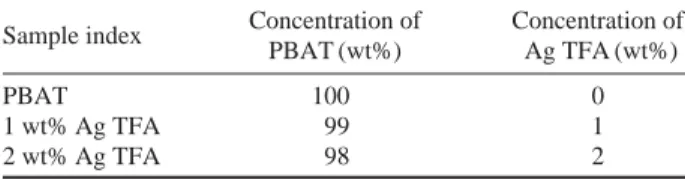
the pure PBAT and the PBAT containing different amounts of Ag TFA irradiated at the absorbed dose of 100 kGy were investigated by the UV-vis analysis and the results are shown in Fig. 1(a). In case of the pure PBAT and the PBAT con-taining different amounts of Ag TFA before the irradiation at the absorbed dose of 100 kGy, there was no characteristic absorption band for the formation of Ag NPs in the PBAT films. On the other hand, for all the irradiated PBAT films containing Ag TFA, the characteristic bands associated to the typical surface plasmon resonance of Ag NPs were observed obviously. The wavelengths and intensities of the characteristic bands were red-shifted from 463 nm to 480 nm with increasing amounts of Ag TFA due to the increase in the size and numbers of Ag NPs (Kobayashi et al. 2001; Callegari et al. 2003). This result verifies that Ag NPs were
Fig. 1. UV-visible absorption spectra of pure PBAT and PBAT/Ag TFA film containing 1 and 2 wt% Ag TFA (a) before and (b) after
electron beam irradiation at the absorbed dose of 100 kGy.
Absorbance Absorbance 1.5 1.0 0.5 0.0 1.5 1.0 0.5 0.0 300 400 500 600 700 Wavelength (nm) 300 400 500 600 700 Wavelength (nm) 2 wt% Ag TFA 2 wt% Ag TFA 1 wt% Ag TFA 1 wt% Ag TFA
pure PBAT pure PBAT
(a) (b)
Fig. 2. UV-visible absorption spectra of PBAT/Ag TFA films
con-taining 1 wt% Ag TFA irradiated at the absorbed dose of 0, 100, and 200 kGy. Absorbance 1.5 1.0 0.5 0.0 300 400 500 600 700 Wavelength (nm) 200 kGy 100 kGy 0 kGy
formed in the PBAT films by electron beam irradiation-induced reduction.
In order to investigate the effect of the absorbed does on the reduction of Ag TFA in the PBAT films, the irradiated 1 wt% Ag TFA films at the absorbed doses of 100 and 200 kGy were analyzed using UV-vis and the results were pre-sented in Fig. 2. As shown in Fig. 2, for all the irradiated 1 wt% Ag TFA films, the characteristic absorption band
indicating the formation of Ag NPs was identified at around 480 nm and its intensity was increased with an increasing absorbed dose. This tendency can be attributed to the higher formation of Ag NPs into the PBAT matrix with an increas-ing absorbed dose (Kassaee et al. 2008). Furthermore, after the irradiation, the color of the PBAT/Ag TFA films was changed from colorless to dark brown with an increasing fluence, indicating the formation of highly concentrated Ag Fig. 3. FE-SEM images and their corresponding EDX spectra of PBAT/Ag TFA films containing different content of Ag TFA irradiated at
the different absorbed dose: 1 wt% Ag TFA/100 kGy (a), 1wt% Ag TFA/200 kGy (b), 2 wt% Ag TFA/100 kGy (c), and 2 wt% Ag TFA/200 kGy (d). Atomic % C: 74.05 O: 20.96 Ag: 4.99 Atomic % C: 73.22 O: 20.69 Ag: 6.09 Ag C (a) (b) (c) (d) O O O O C C C Ag Ag Ag Atomic % C: 73.44 O: 21.39 Ag: 5.17 Atomic % C: 72.00 O: 21.69 Ag: 6.31 2 Energy(keV) 2 Energy(keV) 2 Energy(keV) 2 Energy(keV)
NPs as not shown in this paper.
The morphology and elemental analysis of the irradiated PBAT/Ag TFA films at the absorbed doses of 100 kGy and 200 kGy was investigated by FE-SEM and EDX analysis. As shown in the FE-SEM micrographs and their EDX spectra Fig. 3, white Ag NPs were generated on all the surface of the irradiated PBAT/Ag TFAs films and their numbers was relatively increased with the increase in the absorbed dose and the amounts of Ag TFA. Moreover, as presented in the EDX Ag mapping micrographs Fig. 4, the formed Ag NPs were well dispersed in the PBAT matrix for both of 1 wt% and 2 wt% Ag TFA. Thus, these results revealed that the Ag NPs were successfully generated on the PBAT matrix with-out aggregation by electron irradiation under all the given
conditions (Shameli et al. 2010).
The antibacterial activity of the PBAT and irradiated PBAT/Ag TFA films at the various absorbed doses against S. aureus and E. coli was estimated by a colony counting test and the results are presented in Fig. 5. The pure PABT showed no antibacterial activity against both bacteria. The non-irradiated PBAT/Ag TFA films seemed to slightly have the antibacterial activity, in that the silver ion-containing species is broadly regarded as an antibacterial agent (Manee-rung et al. 2008). On the other hand, in comparison to those of the non-irradiated films, the irradiated PBAT/Ag TFA films exhibited more effective antibacterial activity against both bacteria and their effectiveness was significantly en-hanced with increasing absorbed dose. Especially, For the Fig. 4. FE-SEM/EDX Ag-mapping images of the surface of of PBAT/Ag films containing 1 (a) and 2 wt% Ag TFA irradiated at the absorbed
dose of 200 kGy (b).
Fig. 5. Antibacterial activity of pure PBAT and PBAT/Ag TFA containing different content of Ag TFA irradiated at the different absorbed
dose against Escherichia coli (a) and Staphylococcus aureus (b): (◆) PBAT film, and PBAT/Ag TFA films e-beam irradiation with different doses; (□) 1 wt% Ag TFA/ 0 kGy, (■) 2 wt% Ag TFA/ 0 kGy, (○) 1 wt% Ag TFA/ 100 kGy, (●) 2 wt% Ag TFA/ 100 kGy,
(△) 1 wt% Ag TFA/ 200 kGy, and (▲) 2 wt% Ag TFA/ 200 kGy.
Cell death (%) Cell death (%) 100 90 80 70 60 50 40 30 20 10 0 100 90 80 70 60 50 40 30 20 10 0 0 5 10 15 20 25 Reaction time (h) 0 5 10 15 20 25 Reaction time (h) 1 wt% Ag TFA/0 kGy 1 wt% Ag TFA/100 kGy 1 wt% Ag TFA/200 kGy 2 wt% Ag TFA/0 kGy 2 wt% Ag TFA/100 kGy 2 wt% Ag TFA/200 kGy PBAT 1 wt% Ag TFA/0 kGy 1 wt% Ag TFA/100 kGy 1 wt% Ag TFA/200 kGy 2 wt% Ag TFA/0 kGy 2 wt% Ag TFA/100 kGy 2 wt% Ag TFA/200 kGy PBAT (a) (b) (a) (b)
initial duration of 8 h after the their expose, the death rate of both bacteria was remarkably enhanced with increasing absorbed dose and concentration of Ag TFA, but, after 8 h, the bacterial death reached up to 99% for all the irradiated films. The strong antibacterial activity of all the irradiated PBAT/Ag TFA films prepared at the given absorbed doses might be attributed to a large amount of Ag NPs in the PBAT/Ag TFA films formed by electron beam irradiation.
The contact antibacterial property of PBAT/Ag TFA films was also investigated using disk diffusion test and the result are shown in Table 2. The results of the diffusion test also exhibited the almost similar tendency to the above-mention-ed results of the colony counting test. All the irradiatabove-mention-ed PBAT/Ag TFA films exhibited the more effective inhibi-tion activity against S. aureus and E. coli in comparison to those of the non-irradiated films, indicating that they pos-sessed more effective antibacterial activity. Moreover, the inhibition zone surrounding the irradiated PBAT/Ag TFA films became wider gradually with the increase in the absorb-ed doses and Ag TFA concentrations. These results confirm-ed that the irradiatconfirm-ed PBAT/Ag TFA films had the outstand-ing antibacterial property against both of S. aureus and E. coli on account of the formation of Ag NPs in the films during the electron beam irradiation (Feng et al. 2000; Shameli et al. 2010).
CONCLUSION
In this study, the antibacterial biodegradable PBAT films containing Ag NPs was successfully prepared through
in-situ electron beam-induced reduction of silver Ag TFA pcursors incorporated in the solid-state PBAT films. The re-sults of UV-vis and FE-SEM with EDX analyses revealed that the Ag NPs were successfully formed and homogene-ously distributed in the PBAT matrix during electron beam irradiation. On the basis of the results of the antibacterial test through disk diffusion and colony counting test, the irradiated PBAT/Ag TFA films exhibited the antibacterial property against various bacteria because of the generation of the higher amount of antibacterial Ag NPs in the PBAT matrix. Thus, this simple radiation-based technique can be used in the fabrication of antibacterial materials for the applications of food packaging and biomedical devices.
ACKNOWLEDGEMENTS
This work was supported by Radiation Technology R&D program through the National Research Foundation of Korea funded by the Ministry of Science, ICT, & Future Planning.
REFERENCES
Bozanic DK, Brankovic SD, Bibic N, Luyt AS and Djokovic V. 2011. Silver nanoparticles encapsulated in glycogenbio-polymer: morphology, optical and antimicrobial properties. Carbohydr. Polym. 83:883-890.
Callegari A, Tonti D and Chergui M. 2003. Photochemically grown silver nanoparticles with wavelength-controlled size and shape. Nano. Letter. 3:1565-1568.
Feng QL, Wu J, Chen GQ, Cui FZ, Kim TN and Kim JO. 2000. A mechanistic study of the antibacterial effect of silver ions on Escherichia coli and Staphylococcus aureus. J. Biomed. Mater. Res. 52:662-668.
Fortunati E, Armentano I, Zhou Q, Iannoni A, Saino E, Visai L, Berglund LA and Kenny JM. 2012. Multifunctional bio-nanocomposite films of poly (lactic acid), cellulose nano-crystals and silver nanoparticles. Carbohydr. Polym. 87: 1596-1605.
Gross RA and Kalra B. 2002. Biodegradable polymers for the environment. Science. 297:803-807.
Hwang IT, Jung CH, Kuk IS, Choi JH and Nho YC. 2010. Elec-tron beam-induced crosslinking of poly (butylene adipate-co-terephthalate). Nucl. Instrum. Meth. B 268:3386-3389. Kassaee MZ, Akhavan A, Sheikh N and Beteshobabrud R. Table 2. Average inhibition zones of pure PBAT and PBAT/Ag
TFA films containing 1 and 2 wt% Ag TFA irradiated at the absorbed dose of at the absorbed dose of 0, 100, and 200 kGy.
Inhibition zone (mm)
Gram-positive Gram-negative
Samples
bacteria bacteria
Staphylococcus aureus Escherichia coli
PBAT 0.00±0.00 0.00±0.00 1 wt% Ag TFA/0 kGy 0.20±0.52 0.22±0.41 1 wt% Ag TFA/100 kGy 0.47±0.18 0.59±0.16 1 wt% Ag TFA/200 kGy 0.68±0.11 0.94±0.15 2 wt% Ag TFA/0 kGy 0.24±0.71 0.23±0.37 2 wt% Ag TFA/100 kGy 1.12±0.31 1.53±0.07 2 wt% Ag TFA/200 kGy 1.53±0.21 2.25±0.01
2008. γ-Ray synthesis of starch-stabilized silver nanoparti-cles with antibacterial activities. Radiat. Physics. Chem. 77:1074-1078.
Kim JS, Kuk E, Yu KN, Kim JH, Park SJ, Lee HJ, Kim SH, Park YK, Park YH, Hwang CY, Kim YK, Lee YS, Jeong DH and Cho MH. 2007. Antimicrobial effects of silver nano-particles. Nanomed-Nanotechnol. 3:95-101.
Kobayashi Y, Maceira VS and Liz-Marzan LM. 2001. Deposi-tion of sliver nanoparticles on silica spheres by pretreat-ment steps in electroless plating. Chem. Mater. 13:1630-1633.
Kong H and Jang J. 2008. Antibacterial properties of novel poly(methyl methacrylate) nanofiber containing silver nano-particles. Langmuir 24:2051-2056.
Li W, Xie X, Shi Q, Zeng H, OU-Yang Y and Chen Y. 2010. Antibacterial activity and mechanism of silver nanoparti-cles on Escherichia coli. Appl. Microbiol. Biotechnol. 85: 1115-1122.
Liao Y, Wang Y, Feng X, Wang W, Xu F and Zhang L. 2010. Antibacterial surfaces through dopamine functionalization and silver nanoparticle immobilization. Mater. Chem. Phys. 121:534-540.
Maneerung T, Tokura S and Rujiravanit R. 2008. Impregnation of silver nanoparticles into bacterial cellulose for antimicro-bial wound dressing. Carbohydr. Polym. 72:43-51. Mohanty S and Nayak SK. 2010. Aromatic-aliphatic
poly(buty-lene adipate-co-terephthalate) bionanocomposite: Influ-ence of organic modification on structure and properties. Polym. Compos. 31:1194-1204.
Sedlarik V, Galya T, Sedlarikova J, Valasek P and Saha P. 2010. The effect of preparation temperature on the mechani-cal and antibacterial properties of poly(vinyl alcohol)/sil-ver nitrate film. Polym. Degrad. Stab. 95:399-404. Shameli K, Ahmad MB, Yunus WM, Ibrahim NA, Rahman
RA and Jokar M. 2010. Silver/poly (lactic acid) nanocompo-sites: preparation, characterization, and antibacterial activity. Int. J. Nanomed. 5:573-579.
Siracusa V, Roccule P, Romani S and Rosa MD. 2008. Biode-gradable polymers for food packaging: a review. Trends. Food. Sci. Tech. 19:634-643.
Sondi I, Sondi BS and Colloid J. 2004. Silver nanoparticles as antimicrobial agent: a case study on E. coli as a model for Gram-negative bacteria. Interface. Sci. 275:177-182. Vermeiren L, Devlieghere F, van Beest M, de Kruijf N and
De-bevere J. 1999. Developments in the active packaging of foods. Trends. Food. Sci. Tech. 10:77-86.
Yoksan R and Chirachanchai S. 2009. Silver nanoparticles dispersing in chitosan solution: preparation by γ-ray irradia-tion and their antimicrobial activities. Mater. Sci. Phy. 115: 296-302.
Yoksan R and Chirachanchai S. 2010. Silver nanoparticle-loaded chitosan-starch based films: fabrication and evalua-tion of tensile, barrier and antimicrobial properties. Mater. Sci. Eng. C 30:891-897.
Manuscript Received: October 24, 2013 Revised: October 29, 2013 Revision Accepted: November 20, 2013