Approximately 90% of ovarian cancers are derived from the coelomic epithelium or mesothelium1). Nonepithelial malignancies of the ovary account for about 10% of all ovarian cancers. Nonepithelial ovarian cancers include malignancies of germ cell origin, sex cord‐stromal cell origin, metastatic carcinomas to the ovary, and a variety of extremely rare ovarian cancers (e.g., sarcomas, lipoid cell tumors)1). Ovarian granulosa cell tumors(GCTs) arise from the sex‐cord stromal cells of the ovary and represent 2% to 5% of all ovarian cancers. According to their clinical presentation and histologic characteristics, GCTs are divided into two subtypes: juvenile(5%) and adult(95%)2).
The most common clinical symptoms of GCTs are abnormal uterine bleeding and pain due to their large size mass2). Between 80% and 90% of GCTs are confined to the ovary. Advanced metastatic disease with ascites is present in about 10% of cases2). GCTs are characterized by long natural history and their tendency to recur years after the initial diagnosis2).
There have been few reports describing granulosa cell tumor with massive ascites as a initial symptom. This paper reports a case of a juvenile granulosa cell tumor(JGCT) with
massive ascites as main clinical symptom. Other specific clinical features indicating granulosa cell tumor on cytology and image studies were not seen. Although JGCT in early FIGO stage rarely recur, its early recurrence was detected nineteen months after the first surgery.
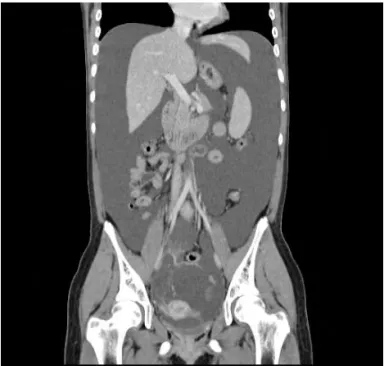
A 13‐year‐old Asian female, gravida 0, para 0, presented in September 2011 with clinical finding of ovarian mass, abdominal distension and dysmenorrhea. She had menarche at age 13 and had last menstrual period in June. Ultrasound, CT revealed massive ascites and large cystic masses (Fig. 1). In the CT imaging, cystic masses were surrounded with ascites. Therefore, it was difficult to distinguish between cyst and ascites, which made hard to make decision about malignant potential. Ascites, smear cytology showed reactive mesothelial cells and result was negative for malignancy. CA‐125 was 37 U/ml.
Pelviscopic right salpingo‐oophorectomy was performed during the first operation. Nine liters of ascites was drained. The salpinx (3.5x1cm in size) is unremarkable grossly. The frozen section diagnosis of the mass was benign. But, in final pathologic report, microfollicles of varying sizes and shapes represented granulosa cell tumor were seen. In addition, abundant cytoplasm, dark and pleomorphic nuclei, but rare nuclear grooves showed that the tumor was Juvenile granulosa cell tumor (Fig. 2a, b). Other pelvic and abdominal peritoneum was normal. Postoperative serum
Granulosa Cell Tumor Presenting with Massive Ascites
: a Case Report and Reviews of the Literature
Jisuk Han
1, Misun Kim
2, Sung‐yob Kim
31,2,3Department of Obstetrics & Gynecology, Jeju National University Hospital (Received April 8, 2014; Revised April 15, 2014; Accepted April 22, 2014)
This case of ovarian granulosa cell tumor is characterized by massive ascites and its early recurrence. It aimed for reviewing the diagnosis and management of granulosa cell tumor from the reported literature. A 13‐year‐old Asian female was presented with abdominal distension and massive ascites. Pathologic report was a juvenile granulosa cell tumor of the ovary, FIGO stage 1A. The patient treated with pelviscopic right salpingo‐oophorectomy. After eighteen months follow up, recurrence was detected. Granulosa cell tumors have known diagnostic features on cytology and image studies. However those features were not helpful in this case. (J Med Life Sci 2014;11(1):8-12)
Key Words
: Ovarian Tumor, Juvenile Granulosa Cell Tumor, AsciteIntroduction
Correspondence to : Sung-yob Kim
Department of Obstetrics & Gynecology, Jeju National University Hospital, Aran 13‐gil(Ara‐1 Dong), Jeju‐si, Jeju 690‐767, South Korea
Abstract
estrogen level was normal. The level of CA‐125 had been normalized since 3 month later after surgery.
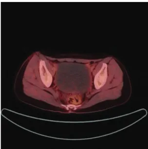
Checking CA‐125 level was performed every 3 months and the results were consistently normal. Whole Body FED Fusion PET follow‐up was done every 9 months. First PET imaging had no evidence of recurrence or metastasis. Eighteen months after surgery Ultrasound revealed a large cystic mass. Whole body FDG fusion PET revealed large cystic lesion without FDG uptake in pelvic cavity suggesting benign (Fig. 3) Enhanced pelvis MR revealed a 9.8cm sized cystic mass with septation and 1.2cm, 0.9cm solid portion in right upper part of septum in pelvic cavity suggesting ovarian epithelial tumor with malignant potential(Fig. 4). CA‐125 was 22.20 U/ml.
Nineteen months after the first operation, she underwent the laparoscopic surgery for the ovarian cyst. A large left ovarian cyst was seen. Also, 1cm sized and 2cm sized pelvic
wall masses with pelvic adhesion suspicious for malignancy were noted. During laparoscopic exploration, 1.1×1×0.8cm sized omental mass in right paracolic area near appendix and 0.6×0.5×0.6cm sized omental nodule in subhepatic area were found. Pelviscopic left ovarian cystectomy and peritonectomy was performed for septated left ovarian cyst and pelvic side wall masses. Also, omentectomy with omental masses was done. Frozen biopsy result was serous cyst with tumor involvement. Pelvic side wall masses and omental masses also reported tumor involvement in frozen biopsy. In the final pathologic report, the ovarian cystic mass, 2 pelvic wall masses and 2 omental masses presented granulosa cell tumor. For ovarian cyst fluid, smear cytology showed negative for malignancy. She is scheduled to be treated with six cycles of bleomycin 15 Unit/m2 on Day 1 and etoposide 100mg/m2 and cisplatin 20mg/m2 on Day 1 to 5 every 3 weeks.
Figure 1. Preoperative Computed Tomography(CT) images
demonstrating the massive ascites. CT scan shows ovarian cysts with massive ascites.
Figure 2. Pathologic report for ovarian mass after first
surgery (a) (Hemotoxylin & Eosin
stain, 100) Microfollicles of varing sizes and shapes are separated by cellular areas diffusely. (b) (Hemotoxylin & Eosin stain, 400) Abundant cytoplasm, dark & pleomorphic nuclei, and numerous mitoses are shown. Grooved nuclei usually shown in adult granulose cell tumor are not seen.
Ovarian neoplasms consist of several histopathological entities; treatment depends on the specific tumor type. Epithelial ovarian cancer comprised the majority of malignant ovarian neoplasms(about 90%)3-6). Nonepithelial malignancies of the ovary, including sex cord‐stromal tumors, account for about 10% of all ovarian cancers1).
Compared to epithelial ovarian cancers, GCTs have different biology and clinical presentation due to their retained ability to produce estrogen7-8). However, it is difficult to distinguish GCTs from epithelial ovarian tumors preoperatively. The accuracy of intraoperative pathologic diagnosis is also not good. The diagnosis of granulosa cell tumor is often not made until after surgery6). Therefore the management of epithelial ovarian cancer are recommended as the initial therapy of GCTs. Also, staging system used for GCTs is that applied for epithelial ovarian cancer (FIGO staging system)2,9).
The most common clinical symptom of GCTs is abnormal uterine bleeding and pain due to their large size2). GCT is usually associated with a mass on pelvic examination which is subsequently confirmed with imagine techniques2). At CT and ultrasound, the tumor appears as a multilocular mass with thick, irregular septations and solid component10). The more distinctive apprearance of GCTs is often seen on MR imaging. GCTs appear solid masses with a various amount of cystic components and hemorrhage on MRI11). The most
common forms of primary GCTs are large multi‐septated cystic mass and medium solid mass with internal cysts on CT and MRI11-12).
Hormonally active GCTs which secrete estrogen can cause menstruation problems in the reproductive age group2,13-14). Because of the endogenous estrogen effect, approximately 25 –50% of GCTs are associated with endometrial hyperplasia, whereas 5–13% are associated with an endometrial carcinoma2). The other symptoms and signs of GCTs are nonspecific and the same as most ovarian malignancies. Ascites is present in about 10% of cases15). Recently, Ashnagar A. et al reported a young girl who presented massive ascites as the only clinical manifestation in the JGCT16). In this case, the patient also presented mainly massive ascites and abdominal distension as main clinical symptom.
Ascites is a term that describes a pathologic accumulation of fluid within the peritoneal cavity. It can develop as a result of liver disease, congestive heart failure or nephrotic syndrome, and is not an uncommon complication accompanying some gynecologic diseases17). Epithelial ovarian cancer is possibly the most common type of cancer leading to malignant ascites17-18). GCTs rarely can cause the ascites and advanced metastatic disease with ascites is present in about 10% of cases2). In retrospective review by Parsons et al., ovarian cancer was the commonest cause of ascites with far better prognosis than patients with GI cancers18-19).
Discussion
Figure 3. Whole body Fludeoxyglucose uptake Positron
Emission Tomography image eighteen months after first surgery. Image scan shows ovarian cyst without FDG uptake.
Figure 4. Enhanced pelvis Magnetic Resonance scan
eighteen months after first surgery. This image shows a 9.8 cm cystic mass with septation (arrow).
presentation. As a result, 80‐90% of GCTs are classified into stage I at the time of detection13-14). If the patient with stage I is reproductive age woman, a more conservative unilateral salpingo‐oophorectomy is indicated3-6). GCTs tend to run an indolently malignant course and most recurrences occur within 5–10 years following initial therapy2). The juvenile subtype behaves less aggressively than the adult type15). If it diagnosed at an early stage, juvenile granulosa cell tumor (JGCT) has a favorable prognosis and its recurrences are uncommon20). Once JGCTs recur it is typically within the first year21-24). These recurrences are usually rapid, leading to death within 13 to 16 months24).
There is controversial about the detection of prognostic factors except stage. In most studies, stage is accepted as the most important prognostic factor. Published ten‐year survival rates are 84‐95% in stage I, 50‐65% in stage II, and 17‐33% in stages III‐IV in studies with long follow‐up periods22-23). In retrospective review of 113 patients, Lee and colleagues evaluate the clinicopathologic characteristics and prognostic factors of GCTs. Positive cytology (P=0.099) was marginally significant, but bilateral disease, ascites, tumor rupture, and capsule invasion were not associated with differences in recurrence rates7).
In conclusion, this report of JGCT demonstrates that known diagnostic features of GCTs were not enough to distinguish GCTs from epithelial ovarian tumors preoperatively. Although laboratory and image studies were performed, the diagnosis of granulosa cell tumor was not made preoperatively. Because of its rarity, limited data are available in the literature to guide diagnosis and clinical management of GCTs.
This review also indicates the need for clinicians to examine possible GCTs in female patients presenting with massive ascites and pelvic mass. Clinicians should attempt to synthesize the whole situation, including detailed history, physical examination and even uncommon clinical symptom of GCTs.
Therefore, further studies are required in order to identify the patients of JGCTs who was presented massive ascites as main clinical symptom. It also needed to confirm massive ascites as the prognostic factor affecting early recurrence. In the future, a prospective multicenter study with uniform protocols may be necessary to confirm the known diagnostic features of GCTs and prognostic factors.
1) Scully RE, Young RH, Clement PB. : Tumors of the ovary, maldeveloped gonads, fallopian tube, and broad ligament. In: Atlas of tumor pathology. Washington, DC: Armed Forces Institute of Pathology, 1998; Fascicle 23, 3rd series.
2) D. Pectasides *, E. Pectasides, A. Psyrri : Granulosa cell tumor of the ovary : Cancer Treatment Reviews 2008;34:1–12.
3) Chan JK, Cheung MK, Husain A, Teng NN, West D, Whittemore AS, Berek JS, Osann K: Patterns and progress in ovarian cancer over 14 years: Obstet Gynecol 2006;108(3 Pt 1):521‐8.
4) Prat J.: New insights into ovarian cancer pathology : Ann Oncol 2012;23 Suppl 10:x111‐117.
5) Jelovac D, Armstrong DK. : Recent progress in the diagnosis and treatment of ovarian cancer. : CA Cancer J Clin 2011;61:183‐203.
6) Ovarian Cancer Guideline (Version 2. 2013). NCCN Clinical Practice Guidelines in Oncology. http://www.nccn.org/ professionals/physician_gls/pdf/ovarian.pdf.
7) Lee IH, Choi CH, Hong DG, Song JY, Kim YJ, Kim KT, Lee KW, Park IS, Bae DS, Kim TJ : Clinicopathologic characteristics of granulosa cell tumors of the ovary: a multicenter retrospective study : J Gynecol Oncol 2011;22(3):188‐95.
8) McNatty KP, Makris A, DeGrazia C, Osathanondh R, Ryan KJ. : The production of progesterone, androgens, and estrogens by granulosa cells, thecal tissue, and stromal tissue from human ovaries in vitro. : J Clin Endocrinol Metab 1979;49:687‐99.
9) Segal R, DePetrillo AD, Thomas G.: Clinical review of adult granulosa cell tumors of the ovary: Gynecol Oncol 1995;56(3):338–44.
10) E K Outwater, B J Wagner, C Mannion, J K McLarney and B Kim : Sex cord‐stromal and steroid cell tumors of the ovary : November 1998 RadioGraphics, 18, 1523‐1546.
11) Kim SH, Kim SH Granulosa cell tumor of the ovary : common findings and unusual appearances on CT and MR. J:Comput Assist Tomogr 2002;26:756–761.
12) Sung Eun Rha, Soon Nam Oh, Seung Eun Jung, et al : Recurrent ovarian granulosa cell tumors : clinical and imaging features : Abdom Imaging 2008;33:119–125. 13) Schweppe KW, Beller FK. Clinical data of granulosa cell
tumors. J Cancer Res Clin Oncol 1982;104:161‐9. 14) Fox H, Agrawal K, Langley FA. A clinicopathologic study
Conclusion
of 92 cases of granulosa cell tumor of the ovary with special reference to the factors influencing prognosis. Cancer 1975;35:231‐41.
15) Berek & Novak’s Gynecology, 14th edition. Edited by Berek, Jonathan S. Lippincott Williams & Wilkins; 2007;1520‐1523.
16) Ashnagar A, Alavi S, Nilipour Y, Azma R, Falahati F. : Massive ascites as the only sign of ovarian juvenile granulosa cell tumor in an adolescent : a case report and a review of the literature. : Case Rep Oncol Med. 2013;2013:386725.
17) Cheng MH, Yen MS, Chao KC, Sheu BC, Wang PH. : Differential diagnosis of gynecologic organ‐related diseases in women presenting with ascites : Taiwan J Obstet Gynecol. 2008;47(4):384‐90.
18) Ayantunde AA, Parsons SL. : Pattern and prognostic factors in patients with malignant ascites: a retrospective study : Ann Oncol. 2007;18(5):945‐9. 19) Parsons SL, Lang MW, Steele RJC. : Malignant ascites:
a 2 year review from a teaching hospital : Eur J Surg Oncol 1996;22:237–239.
20) R. Young, G. Dickerson, R. Scully : Juvenile granulosa cell tumor of the ovary : a clinicopathologic analysis of 125 cases Am J Surg Pathol, 8 (1984), pp. 575–596 21) G. Calaminus, R. Wessalowski, D. Harms, U. Gobel :
Juvenile granulosa cell tumors of the ovary in children and adolescents : results from 33 patients registered in a prospective cooperative study:Gynecol Oncol, 65 (1997), pp. 447–452
22) Ringenberg QS, Doll DC, Loy TS, Yarbro JW. : Malignant ascites of unknown origin.: Cancer 1989;64:753–755.
23) Greenway B, Johnson PJ, Williams R. : Control of malignant ascites with spironolactone. : Br J Surg 1982;69:441–442.
24) Stephen D. Frausto MS, John P. Geisler MD, Mavis S. Fletcher MD, Anil K. Sood MD* : Late recurrence of juvenile granulosa cell tumor of the ovary : American Journal of Obstetrics and Gynecology (2004) 191, 366e7