Abstract
BACKGROUND: Soil salinity causes reduction of crop productivity. Rhizosphere microbes have metabolic capa-bilities and ability to adaptation of plants to biotic and abiotic stresses. Plant growth-promoting bacteria (PGPB) could play a role as elicitors for inducing tolerance to stresses in plants by affecting resident microorganism in soil. This study was conducted to demonstrate the effect of selected strains on rhizosphere microbial community under salinity stress. METHODS AND RESULTS: The experiments were con-ducted in tomato plants in pots containing field soil. Bacterial suspension was inoculated into three-week-old tomato
plants, one week after inoculation, and -1,000 kPa-balanced salinity stress was imposed. The physiological and bio-chemical attributes of plant under salt stress were monitored by evaluating pigment, malondialdehyde (MDA), proline, soil pH, electrical conductivity (EC) and ion concentrations. To demonstrate the effect of selected Bacillus strains on rhi-zosphere microbial community, soil microbial diversity and abundance were evaluated with Illumina MiSeq sequencing, and primer sets of 341F/805R and ITS3/ITS4 were used for bacterial and fungal communities, respectively. As a result, when the bacterial strains were inoculated and then salinity stress was imposed, the inoculation decreases the stress sus-ceptibility including reduction in lipid peroxidation, en-hanced pigmentation and proline accumulation which sub-sequently resulted in better plant growth. However, bacterial inoculations did not affect diversity (observed OTUs, ACE, *Corresponding author: Mee Kyung Sang
Phone: +82‐63-238-3055; Fax: +82‐63-238-3834; E-mail: [email protected]
Korean J Environ Agric. 2021;40(1):49‐59. Korean Online ISSN: 2233-4173
Published online 2021 March 29. https://doi.org/10.5338/KJEA.2021.40.1.6 Print ISSN: 1225-3537
토마토에 염류 내성을 유도하는 바실러스 균주 처리 후
근권 미생물 군집 구조 연구
유성제
1,2, 이신애
1, 원항연
1, 송재경
1, 상미경
1*1농촌진흥청 국립농업과학원 농업생물부 농업미생물과,
2상주시농업기술센터 미래농업과
Assessment of Rhizosphere Microbial Community Structure in Tomato Plants after
Inoculation of Bacillus Species for Inducing Tolerance to Salinity
Sung-Je Yoo1,2, Shin Ae Lee1, Hang-Yeon Weon1, Jaekyeong Song1 and Mee Kyung Sang1* (1Division of Agricultural Microbiology, Department of Agricultural Biology, National Institute of Agricultural Sciences, Rural Development Administration, Wanju 55365, Korea, 2Current Address: Future Agriculture Division, Sangju Agricultural Technology Center, Sangju 37154, Korea
Received: 4 January 2021/ Revised: 26 February 2021/ Accepted: 22 March 2021 Copyright ⓒ 2021 The Korean Society of Environmental Agriculture
This is an Open-Access article distributed under the terms of the Creative Commons Attribution Non-Commercial License (http://creativecommons.org/licenses/by-nc/3.0) which permits unrestricted non-commercial use, distribution, and reproduction in any medium, provided the original work is properly cited.
ORCID Sung-Je Yoo https://orcid.org/0000-0001-7031-4214 Shin Ae Lee https://orcid.org/0000-0002-2107-8750 Hang-Yeon Weon https://orcid.org/0000-0002-9084-4316 Jaekyeong Song https://orcid.org/0000-0002-3553-6660 Mee Kyung Sang
https://orcid.org/0000-0001-9032-7013
49
Chao1 and Shannon) and structure (principle coordinate analysis) of microbial communities under salinity stress. Furthermore, relative abundance in microbial communities had no significant difference between bacterial treated- and untreated-soils under salinity stress.
CONCLUSION: Inoculation of Bacillus strains could affect plant responses and soil pH of tomato plants under salinity stress, whereas microbial diversity and abundance had no significant difference by the bacterial treatments. These findings demonstrated that Bacillus strains could alleviate plant’s salinity damages by regulating pigments, proline, and MDA contents without significant changes of microbial community in tomato plants, and can be used as effective biostimulators against salinity stress for sustainable agriculture. Key words: Bacillus, Microbial diversity, Salinity, Tomato
서 론
세계 주요 재배 채소 중 하나인 토마토는 연간 1억 6천만 톤이 생산되는 고부가가치 작물로 영양분과 비타민, 미네랄이 풍부하다[1,2]. 토마토는 18.3-32.2℃의 온도 범위와 토양 pH 가 6.0-6.5, 전기전도도가 0.25-0.50 mS/cm 일 때 가장 최적 으로 생육되지만, 이상기후에 의한 극심한 온도 변화, 오랜 기 간 과도한 비료 사용에 의한 염류집적 등에 의해 생육에 영향 을 받아 생산량이 감소할 수 있다[3-5]. 특히, 염류집적에 의한 스트레스는 작물의 수확량을 감소시키는 대표적인 비생물적 스트레스 중 하나로써 과도한 화학비료의 사용과 부적절한 재 배 관행이나 관개지 관리로 인하여 발생하며, 농경지의 20%와 관개지의 33%가 이미 높은 염류로 피해를 받았으며, 2050년 에는 피해 그 면적이 50%까지 증가할 것으로 보인다[6,7]. 이와 같은 비생물적 스트레스로부터 작물을 보호하기 위하 여 육종을 통해 새로운 내성 품종을 개발하고자 하는 연구가 수행되어 왔다. Moreno-Limon 등은 높은 염류 조건에서 뿌 리 생육과 양분 흡수 능력이 높은 콩(Phaseolus vulgaris)을 개발하였고, Zongsuo 등은 수분 흡수율이 높은 유채(Brassica napus)와 갓(B. juncea) 등 건조 스트레스 내성 작물을 개발하 였다[8,9]. 또한 근래에는 유전자 도입 기법을 이용한 노력도 이루어지고 있는데, 신호전달 체계와 호르몬 생합성 기작을 조 절하여 염류 및 건조 스트레스에 강한 벼와 토마토가 연구되기 도 하였다[10,11]. 하지만 이러한 전략은 시간이 오래 걸릴 뿐 만 아니라 아직까지 전 세계 많은 국가에서 유전자 변형 작물 에 대한 우려로 인하여 다른 다양한 방안이 모색되고 있다. 그 중에서 작물의 병 억제와 작물 생육증진에 효과가 있는 토양의 유용 미생물을 비생물적 스트레스에 적용하려는 연구가 이루 어지고 있다[12].식물생육촉진세균(Plant growth-promoting bacteria, PGPB) 은 식물의 생육을 증진시킬 뿐만 아니라 직⋅간접적으로 염류 스트레스에 의한 식물의 피해를 감소시킬 수 있다[13]. 식물생육 촉진세균은 근권에 정착하여 식물과 상호작용하면서, 스트레 스 조건에서 작물의 엽록소와 카로티노이드(Carotenoid) 같 은 색소의 함량을 유지시키거나, 프롤린(Proline), 유리당, 단 백질, 아미노산과 같은 삼투물질의 함량을 조절하여 세포막의 안정성을 높여 식물이 스트레스 환경에 잘 적응을 할 수 있도 록 돕는다[14,15]. 또한 식물의 항산화 효소 활성을 높여 활성 산소종(Reactive oxygen species, ROS)의 수치를 낮추고, 세포막의 지질과산화(Lipid peroxidation)를 막아 염류 스트 레스에 대한 피해를 막아주는 것으로 알려져 있다[16]. 식물생육촉진세균의 주요 서식처인 근권은 수많은 미생물 의 활력 지대로 지구의 복잡한 생태계 중 하나에 속하며[17], 주위 미생물에 영향을 주면서 고염류, 건조, 양분 부족 등의 스 트레스 환경에서도 식물의 적응에 도움을 주는 것으로 알려져 있다[7,18]. 특히 미생물의 다양성이나 상대 빈도수는 지상과 지하에 서식하는 생명체의 다양성 및 기능에 영향을 주기 때문 에 생태계를 이해하기 위한 대표적인 군집구조의 지표로 사용 되고 있다[19]. 또한 미생물 군집에 대한 연구를 통해 식물과 미생물, 미생물과 미생물의 상호작용 및 식물에 유용한 미생물 이 생태학적으로 주는 영향에 대한 이해를 돕기 때문에, 전 세 계적으로 다양한 환경 변화에 따른 토양 미생물 군집의 변화에 대한 관심이 증가하고 있다[7,20]. 이전의 연구에서 토마토의 염류 스트레스 피해를 감소시키는 작물 유용 세균 Bacillus aryabhattai H19-1과 B. mesonae H20-5를 선발하였고, 선발한 세균을 처리한 토마토 식물이 염 류 스트레스를 받았을 때 생리학적, 생화학적, 분자생물학 변 화를 검정하였다[21]. 본 연구에서는 이전의 연구에서 선발한 바실러스 H19-1과 H20-5 균주를 염류 스트레스 조건의 밭 토 양에 처리하였을 때 토마토 작물의 생리적 변화, 토양화학성과 근 권 미생물의 군집에 어떠한 영향을 주는지 규명하고자 하였다.
재료 및 방법
재료 본 연구는 전라북도 완주군 이서면 국립농업과학원 내 포장 (35°49'33''N 127°02'38''E) 내 임의로 지정한 세 지점에서 깊 이 10-30 cm의 토양을 확보하여 자연건조 시킨 뒤[22], 5 mm 체를 통해 이물질을 제거한 후 사용하였다[23,24]. 식물은 ‘슈 퍼도태랑’품종(Koregon, Korea)의 토마토를 사용하였고, 종 자는 2% Sodium hypochlorite로 3분간 표면 소독을 실시한 뒤, 멸균수로 5번 세척한 후 건조시켰다. 포장 토양과 상토 (Bunong, Korea)를 90℃에서 1시간씩 2회 멸균시킨 뒤, 1:1 (v/v)로 섞어 128공 트레이(2 × 2 × 2 cm/hole)에 담은 후 종자를 파종하였고 상온의 유리온실에서 2주간 재배하였다. 토 마토 유묘는 자연건조 시킨 비멸균 포장 토양(pH: 7.3; EC: 0.8 mS/cm; organic C: 6.2 g/kg; total N: 0.7 g/kg)이 담긴 플라스틱 포트(지름 14 cm, 깊이 8.5 cm)에 정식하여 재 배하였다.유용 세균 및 염류 스트레스 처리
이전의 연구에서 Yoo 등이 선발한 B. aryabhattai H19-1과 B. mesonae H20-5를 tryptic soybean agar (TSA, Difco) 배지에서 28℃, 24시간 동안 배양하여 생성된 단일 콜로니 (colony)를 TSB에 접종하여 48시간 동안 28℃, 150 rpm으로 진탕 배양하였다[25]. 세균 배양액은 6,000 rpm으로 10분간 원심분리하여 상청액(Supernatant)을 제거한 후, 세균의 pellet을 10 mM MgSO4로 OD600 = 0.25가 되도록 현탁하였다. 파종 3주 후, 준비한 세균 현탁액을 토마토 식물에 토양 1 g 당 0.1 mL로 관주 처리하였고, 대조군은 10 mM MgSO4 용 액을 관주하였다. 세균 현탁액 처리 일주일 후, 토마토 식물의 생육과 색소, 유 리당, 아미노산 함량을 감소시키는 것으로 알려진 Polonenko 등에 따른 –1,000 kPa 복합 염용액(125 mM potassium nitrate, 82 mM calcium nitrate, 41 mM magnesium sulfate, 22 mM monopotassium phosphate)을 토양 1 g 당 0.1 mL로 2일 간격으로 3회 관주 처리하였다[21,26]. 대조군은 같은 양 의 멸균수를 식물에 관주하였다. 식물 생리적 변화 검정 모든 식물 검정은 염류 스트레스 처리 후 0, 7, 14, 21일 후 샘플링하여 수행하였고, 식물의 엽록소와 카로티노이드 함량 은 Fernandez 등과 Lichtenthaler의 방법을 이용해 측정하 였다[27,28]. 토마토 잎 500 mg을 샘플링하여 0.5% MgCO3 가 함유된 25 mL의 80% acetone에서 균질화한 후 4℃에서 24시간 동안 진탕하였다. 색소가 추출된 샘플은 4,000 rpm으 로 10분간 원심분리한 뒤 UV/VIS spectrophotometer (Infinite M200 PRO, TECAN, Austria)를 이용하여 상등액 을 흡광도 470, 646, 663 nm에서 측정한 뒤 엽록소 a, b와 카로티노이드 함량을 아래 공식을 이용해 계산하였다.
Chlorophyll a = (12.25 × A663.2) – (2.79 × A646.8) Chlorophyll b = (21.50 × A646.8) – (5.10 ×A663.2) Total chlorophyll contents = Chlorophyll a + Chlorophyll b Carotenoid concentration = [(1,000 × A470) – (1.8 ×
Chlorophyll a) – (85.02 × Chlorophyll b)] / 198 프롤린 함량 측정은 Bates 등의 Ninhydrin 방법을 이용하 였다[29]. 식물 잎 100 mg을 1.2 mL의 3% Aqueous sulfo-salicylic acid에서 균질화한 후, 13,000 × g로 10분간 원심분 리하였고, 상등액 500 µL를 Glacial acetic acid 500 µL, Acidic ninhydrin 500 µL와 혼합하였다. 혼합물은 90℃로 1시간 동안 열처리해준 뒤 Ice bath에서 식혀주었고, 1 mL의 Toluene을 넣어 분리된 위층의 용액을 Spectrophotometer 를 이용하여 520 nm에서 비색정량하였다.
Malondialdehyde (MDA) 측정은 식물 잎 100 mg을 500 µL의 0.1% Trichloroacetic acid (TCA)에 넣어 균질화 한 뒤 13,000 × g로 4℃에서 10분간 원심분리하여 얻은 상등 액을 이용하였다. 상등액 500 µL를 20% TCA가 함유된 0.5% 2-thiobarbituric acid 1.5 mL과 혼합하여 90℃에서 30분간 열처리하였고, Ice bath에서 5분간 식힌 뒤 다시 6,000 rpm으 로 3분간 원심분리하였다. Spectrophotometer를 통해 450, 532, 600 nm에서 상등액의 흡광도를 측정한 뒤 아래 식을 이 용해 MDA의 함량을 계산하였다[30].
MDA content (nmol/g) = C (µmol/L) × V (L) / fresh weight (g) × 1,000 C (µmol/L) = 6.45 × (A532 - A600) - 0.56 × A450 생체중은 식물을 포트에서 꺼내 흙을 조심스럽게 털어낸 후, 흐르는 물에 빠르게 씻어내고 습기를 제거한 뒤 무게를 측정하 였고, 뿌리 활력은 Liu 등의 방법을 이용하여 측정하였다[31]. 토양화학성 분석 토양화학성 분석을 위해 pH, 전기전도도, 유기탄소, 총 질 소, 이온 함량을 측정하였다. 포트에서 토양 샘플을 채취하여 균일하게 혼합 후 2 mm 체를 이용해 이물질을 제거하여 자연 건조 하였고, 유기탄소와 총 질소 함량 분석을 Cho 등의 방법 을 이용하였다[32]. pH와 전기전도도, 이온 함량은 토양과 물 을 1:5 비율로 혼합한 뒤 28℃에서 30분간 진탕한 후 6,000 rpm에서 5분간 원심분리한 후 상등액을 분리하여 pH, EC, 이온 측정기(LAQUAtwin, Japan)를 이용해 측정하였다. 토양 미생물 군집 분석 근권토양 시료는 염류 스트레스 처리 0, 21일 뒤 각각 채취 하여 분석하였다. 포트를 뒤집은 후 조심히 흔들어 뿌리 표면 에 붙은 부분을 제외한 모든 토양을 제거한 뒤, 근권토양(뿌리 표면에서 1 mm 이하)을 포함한 식물 뿌리를 50 mL의 멸균 Phosphate buffer saline (PBS)에 넣고 150 rpm으로 30분 간 진탕한 후, 뿌리를 제거한 현탁액을 10,000 rpm으로 15분 간 원심분리하여 상등액을 제거하였다. 수거한 근권토양은 액 체질소에 얼린 후 -80℃에 보관하였다[22,33].
토양 시료의 Total genomic DNA는 FastDNA® spin kit (MP biomedicals, USA)를 사용하여 추출하였고, NanoDrop 2000 Spectrophotometer (Thermo, USA)로 DNA 농도를 측정하여 Illumina MiSeq Sequencing system (Illumina Inc., San Diego, CA, USA)을 통해 ㈜마크로젠에 의뢰해 분 석하였다. 세균은 16S rRNA의 V3-V4 구역을 Primer 341F (5’-CCTACGGGNGGCWGCAG-3’)와 805R (5’-GACTA CHVGGGTATCTAATCC-3’)을 사용해 증폭하였고, 곰팡이 는 primer ITS3 (5’-GCATCGATGAAGAACGCAGC-3’) 와 ITS4 (5’-TCCTCCGCTTATTGATATGC-3’)를 이용해 ITS 구간의 PCR을 수행하였다. PCR 조건은 Initial denatu-ration: 95℃, 3분; Denatudenatu-ration: 95℃, 30초 (25 cycles); annealing: 55℃, 30초; Extension: 72℃, 30초; Final ex-tension: 72℃, 5분 조건으로 수행하였다. Raw sequence 분 석은 Mothur (ver. 1.40) 프로그램을 이용하여[34] 유사수준 97%의 Operational taxonomic units (OTUs)로 군집화하
여 다양성, 상대 빈도수(Relative abundance), 주좌표 분석 (Principle coordinate analysis, PCoA)을 하였다. 통계분석
통계분석은 SAS (ver. 9.1.3, SAS Institute Inc., Cary, NC, USA)와 R-forge community ecology package를 이 용하였고, 모든 데이터는 평균±표준오차로 나타내었다. 식물 생체중, 색소, 프롤린, MDA 함량과 토양화학성 및 미생물 군 집 cluster 간의 종 풍부도(OTU 수, ACE, Chao1)와 다양성 (Shannon), 상대 빈도수는 Tukey 검정을, 주좌표 분석은 ANOSIM (analysis of similarities) 검정을 통해 P<0.05 수 준에서 분석하였다. 미생물 군집 분석은 3개의 sub-repli-cation을 하나로 통합하여 총 3반복으로 수행하였다[35].
결과 및 고찰
염류 스트레스 조건에서 Bacillus H19-1과 H20-5 균주 처리에 의한 식물의 생리적 변화 H19-1과 H20-5 균주를 처리한 토마토에서 잎의 색소 함량 중 엽록소의 경우 스트레스 처리 0, 21일 후, 카로티노이드는 21일 후에 그 함량이 더 높았다(Fig. 1A와 1B, 엽록소: 0 daysafter salinity stress, DAS[36]: P = 0.0126; 21 DAS: P = 0.0277, 카로티노이드: 21 DAS: P = 0.0167). 식물의 엽록소 는 광합성과, 카로티노이드는 빛 흡수, 광 보호 및 식물 호르몬 앱시스산(Abscisic acid, ABA)의 생성과 연관된 색소로 알려 져 있는데[37,38], 식물이 염류 스트레스를 받게 되면 잎의 색 소 함량이 감소하여 식물의 생육이 줄어들며, 이러한 현상은 염류 농도가 높아질수록 더 증가한다고 보고되었다[21,39]. 따 라서 두 균주 H19-1과 H20-5을 처리한 토마토 식물은 염류 스 트레스로부터 엽록소와 카로티노이드의 손실을 줄여 염류 스 트레스에 의한 식물 생육 감소를 완화시키는 것으로 생각된다. 식물은 스트레스 환경에 노출되면 아미노산을 축적하여 외 부 환경 변화에 대응하며, 아미노산 중 하나인 프롤린은 삼투 균형, 세포막 안정, ROS 수치 안정화에 기여하여 식물의 내성 을 유도하고, DNA와 단백질의 안정화, 탄소 및 질소 양분 제 공을 통해 환경 스트레스로부터 피해를 막아준다[40,41]. 본 실험에서는 스트레스 처리 7일 후부터 모든 식물에서 급격하 게 프롤린 함량이 증가하기 시작하였고, H19-1을 처리한 식물 은 스트레스 처리 14일 후, H20-5 처리 식물은 14, 21일 후 무처리 식물에 비해 더 높은 함량으로 프롤린이 축적되었다 (Fig. 1C). 이는 두 균주가 식물의 프롤린 함량을 조절하여 스 트레스 내성을 유도하였으며, 특히 H20-5 균주는 H19-1보다
Days after stress treatment
0 7 14 21 C h lo rophy ll con te nt ( m g /g FW ) 0 100 200 300 400 500 600 Control H19-1 H20-5
Days after stress treatment
0 7 14 21 C a ro tenoi d cont e n t (m g/ g FW ) 0 20 40 60 80 100 120 140 Control H19-1 H20-5
(A)
(B)
**
**
**
(C)
(D)
Days after stress treatment
0 7 14 21 P rol in e c o nt ent (m g/ g F W ) 0.0 0.5 1.0 1.5 2.0 Control H19-1 H20-5
Days after stress treatment
0 7 14 21 M D A co nt ent ( n m o l/g) 0 2 4 6 8 10 12 Control H19-1 H20-5
**
*
*
*
**
Fig. 1. (A) Chlorophyll, (B) carotenoid, (C) proline and (D) MDA contents in leaves of tomato plants treated with bacterial suspensions at 0, 7, 14 and 21 days after salinity stress. Asterisks on the bar mean statistical difference by Tukey’s test (P < 0.05) and error bars indicate standard error of 20 replications from two experiments.
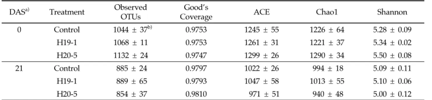
더 오랜 시간 동안 지속적으로 프롤린의 축적을 증가시켜 식물 을 보호하는 것으로 예상된다. 식물은 염류 스트레스를 받게 되면, 활성산소종(ROS)이 증 가하는데 이는 세포막의 지질과산화와 유동성 및 선택성 감소 를 유도하여 식물의 피해를 일으킨다[42]. 이러한 지질과산화 반응에서 MDA와 같은 짧은 지질 파편이 생성되는데 이는 식 물이 스트레스로부터 받은 피해 정도를 검정하기 위한 bio-marker로 사용될 수 있다[43]. 염류 스트레스 처리 14일 후 모 든 식물에서 MDA 함량이 증가하였지만, 세균을 처리한 식물은 대조군에 비해 통계적으로 수치가 더 낮았다(Fig. 1D). 특히 H20-5 균주가 처리된 식물은 스트레스 처리 0, 7일 후에도 MDA 함량이 낮은 것으로 나타났다(0 DAS: P = 0.0854; 7 DAS: P = 0.0255; 14 DAS: P = 0.0158). 이는 두 균주를 처리한 식물에서 염류 스트레스로 인한 식물 세포막의 지질과산화가 감 소되었으며, 특히 H20-5 균주를 처리한 경우 H19-1에 비해 더 빠르고 안정적으로 세포막 손상이 감소하는 것으로 생각된다. 생체중의 경우 염류 스트레스 처리 직후에는 통계적 유의성 이 없었지만 7, 14, 21일 후에는 작물에 유용한 바실러스 (H19-1, H20-5)를 처리한 토마토의 생체중이 대조구 식물에 비해 더 높았으며(Fig. 2, 7 DAS: P = 0.0009; 14 DAS: P = 0.0190; 21 DAS: P = 0032), 스트레스 21일 후의 뿌리 활력 에 있어서도 H19-1 처리구가 229 µg TTF/g/h, H20-5 처리 구가 252 µg TTF/g/h로 대조구의 125 µg TTF/g/h보다 높았다. 이는 균주에 의한 염류 스트레스 피해 완화 효과로 인 해 생체중과 뿌리 활력이 증가한 것으로 생각된다. 염류 스트레스 조건에서 Bacillus H19-1과 H20-5 균주 처리에 의한 토양화학성 영향 염류 스트레스 조건에서 두 선발 균주의 처리에 의한 토양 화학성 안정화 효과를 검정하기 위해, 스트레스 처리 0, 21일 후 토마토 식물 포트의 토양 시료를 채취한 후 pH, EC, 유기 탄소, 총 질소 함량과 이온 농도를 측정하여 Table 1에 나타내 었다. 토양 pH는 식물이 필요한 영양분의 가용성과 식물 뿌리 의 원형질, 토양미생물 활성에 직간접적으로 영향을 줄 수 있 는데[44-46], 염류 스트레스 처리 21일 후 EC, total N 및 K+, Ca2+, Mg2+ 이온 함량이 처리 직후(0 DAS)에 비해 감소하였고, H19-1과 H20-5를 처리한 토양의 pH는 무처리 식물의 토양보 다 감소하였으나(P = 0.0221) 나트륨흡착비(Sodium adsorption ratio, SAR), 교환성 나트륨퍼센트(Exchangeable sodium percentage, ESP)에서 통계적 차이를 확인할 수 없었다. 이를 통해 복합염용액 처리 직후 식물이 높은 염류로 스트레스를 받 았으며, H19-1과 H20-5균주가 토양의 이화학성 중 pH를 감 소시킴으로써 식물의 피해를 완화시킬 것으로 생각된다. 염류 스트레스 조건에서 Bacillus H19-1과 H20-5 균주 처리에 의한 미생물 군집 영향 세균과 곰팡이의 다양성(diversity)은 풍부도(richness)를 의미하는 OTU 수, ACE 및 Chao1 지수와 균등성(eveness)을 나타내는 Shannon 지수로 예측하였고, 그 결과를 Table 2와 Table 3에 각각 나타내었다. 시료 대표 지수인 Good’s Coverage 는 세균에서 0.9747-0.9810, 곰팡이에서 0.9993-0.997로 시료를 아주 잘 대표하는 것을 확인하였다. 분석 결과, 염류 스트레스 처리 0일 후와 21일 후의 미생물 풍부도와 균등성은 세균 (observed OTUs: p < 0.0001; ACE: p < 0.0001; Chao1: Days after stress treatment
0 7 14 21 Fres h w eigh t (g ) 0 5 10 15 20 Control H19-1 H20-5
**
**
**
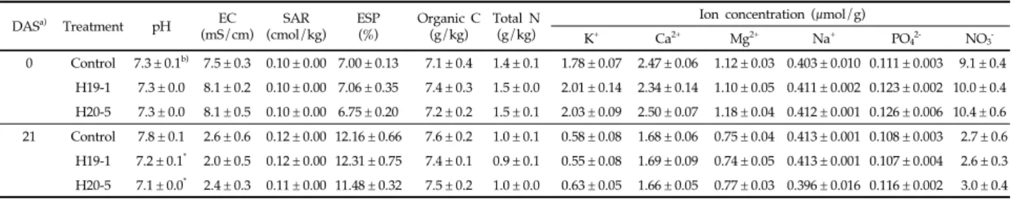
Fig. 2. Total fresh weights of plants treated with bacterial suspensions or 10 mM MgSO4 (control) at 0, 7, 14 and 21 days after salinity stress. Asterisks on the bar mean statistical difference by Tukey’s test (P < 0.05) and error bars indicate standard error of 20 replications from two experiments. DASa) Treatment pH EC (mS/cm) SAR (cmol/kg) ESP (%) Organic C (g/kg) Total N (g/kg)
Ion concentration (μmol/g)
K+ Ca2+ Mg2+ Na+ PO42- NO3 -0 Control 7.3 ± 0.1b) 7.5 ± 0.3 0.10 ± 0.00 7.00 ± 0.13 7.1 ± 0.4 1.4 ± 0.1 1.78 ± 0.07 2.47 ± 0.06 1.12 ± 0.03 0.403 ± 0.010 0.111 ± 0.003 9.1 ± 0.4 H19-1 7.3 ± 0.0 8.1 ± 0.2 0.10 ± 0.00 7.06 ± 0.35 7.4 ± 0.3 1.5 ± 0.0 2.01 ± 0.14 2.34 ± 0.14 1.10 ± 0.05 0.411 ± 0.002 0.123 ± 0.002 10.0 ± 0.4 H20-5 7.3 ± 0.0 8.1 ± 0.5 0.10 ± 0.00 6.75 ± 0.20 7.2 ± 0.2 1.5 ± 0.1 2.03 ± 0.09 2.50 ± 0.07 1.18 ± 0.04 0.412 ± 0.001 0.126 ± 0.006 10.4 ± 0.6 21 Control 7.8 ± 0.1 2.6 ± 0.6 0.12 ± 0.00 12.16 ± 0.66 7.6 ± 0.2 1.0 ± 0.1 0.58 ± 0.08 1.68 ± 0.06 0.75 ± 0.04 0.413 ± 0.001 0.108 ± 0.003 2.7 ± 0.6 H19-1 7.2 ± 0.1* 2.0 ± 0.5 0.12 ± 0.00 12.31 ± 0.75 7.4 ± 0.1 0.9 ± 0.1 0.55 ± 0.08 1.69 ± 0.09 0.74 ± 0.05 0.413 ± 0.001 0.107 ± 0.004 2.6 ± 0.3 H20-5 7.1 ± 0.0* 2.4 ± 0.3 0.11 ± 0.00 11.48 ± 0.32 7.5 ± 0.2 1.0 ± 0.0 0.63 ± 0.05 1.66 ± 0.05 0.77 ± 0.03 0.396 ± 0.016 0.116 ± 0.002 3.0 ± 0.4 a) Soil physicochemical properties were determined at 0 and 21 DAS (days after salinity stress).
b) The values are mean ± standard errors of three soil replications by pooling three sub replicates into one. An asterisk indicates a statistical difference (P < 0.05) in a column by the Tukey’s test.
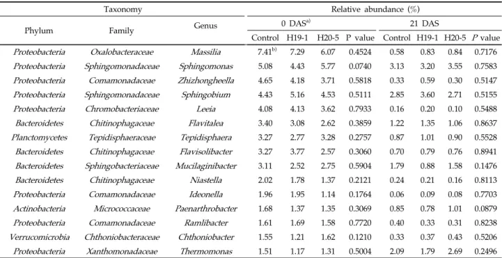
p < 0.0001; Shannon: p = 0.0005)과 곰팡이(observed OTUs: p = 0.0442; ACE: p = 0.0168; Chao1: p = 0.0161; Shannon: p = 0.0211)에서 각각 유의한 차이를 보였지만 H19-1과 H20-5 균주 처리에 의한 효과는 없었다. 스트레스 처리 후 주 좌표 분석(PCoA)을 통해 선발 균주의 처리가 근권의 세균 및 곰팡이 군집구조에 영향을 주는지 분석하였다. Bray-curtis similarity를 이용해 미생물 군집 염기서열 간의 유사성을 분 류하였으며, 분석 결과 염류 스트레스 처리 직후와 21일 후에 서도 H19-1과 H20-5의 처리가 세균(Fig. 3A와 3B) 및 곰팡이 (Fig. 3C와 3D)의 군집구조에 유의한 영향을 미치지 않았고, Rifampicin-resistant mutant를 통해 H19-1은 3.27 log CFU/g, H20-5는 3.50 log CFU/g로 처리 21일까지 토양에 분포함을 확인하였다. 위 결과를 통해 염류 스트레스 조건에서 바실러스 H19-1과 H20-5 균주에 의한 토마토 근권의 전체적인 미생물 군집 변화는 미미함을 알 수 있다. 이와 유사하게, Nautiyal 등은 Bacillus amyloliquefaciens가 벼에서 스트레스와 관련 된 유전자 발현을 조절함으로써 염류 스트레스 내성을 유도하 지만, 근권 미생물 군집의 다양성과 풍부도에는 큰 영향이 없 었으며, 주성분 분석(Principal component analysis, PCA) 에서도 PC1과 PC2의 뚜렷한 상관관계를 확인할 수 없었고, 이를 통해 처리한 균주가 Stimulator로써 작용하여 식물의 염 류 스트레스 내성을 유도할 것이라 보고하였다[47]. 미생물의 상대 빈도수는 토양 생태계 기능에 영향을 주며, 낮 은 상대 빈도수를 가지는 미생물이더라도 병원균이나 환경 변화 로부터 식물을 보호할 수 있는 환경을 조성하는데 중요한 역할 을 수행하기도 한다[48,49]. 염류 스트레스 조건에서, 처리한 세 균에 따른 근권 미생물의 상대 빈도수 검정 결과, 먼저 문 (Phylum) 수준에서는 세균의 경우 우점종으로 Proteobacteria (0 DAS: 51.41-52.16%; 21 DAS: 44.32-5.94%)가 나타났고 그 외 Bacteroidetes, Acidobacteria, Actinobacteria, Verrucomicrobia, Planctomycetes, Cyanobacteria, Gemmatimonadetes, Firmicutes, Armatimonadetes, Chloroflexi 순으로 나타났다(Fig. 4A). 곰팡이 군집에서는 Ascomycota (0 DAS: 23.33-27.92%; 21 DAS: 19.65-23.38%)가 우점종으로 나타났으며, Mortierellomycota, Basidiomycota, Chytridiomycota 순으로 높게 나타났다(Fig. 4B). 하지만 균주 처리에 의한 세균과 곰팡이 군집의 상대 빈도수에 서 유의한 차이는 없었고, 이를 통해 스트레스 처리 후 21일간 문 수준에서 균주에 의한 미생물 군집의 상대 빈도수 변화는 미미하다는 것을 알 수 있었다. 염류 스트레스 처리 직후 토양을 기준으로, 가장 OTU 수가 높은 15개 속(genus)의 상대 빈도수를 비교한 결과, 세균 군집 에서는 H19-1과 H20-5 균주 처리에 의한 차이를 보이지 않았
DASa) Treatment Observed
OTUs
Good’s
Coverage ACE Chao1 Shannon
0 Control 1044 ± 37b) 0.9753 1245 ± 55 1226 ± 64 5.28 ± 0.09 H19-1 1068 ± 11 0.9753 1261 ± 31 1221 ± 37 5.34 ± 0.02 H20-5 1132 ± 24 0.9747 1299 ± 26 1290 ± 34 5.50 ± 0.08 21 Control 885 ± 24 0.9797 1022 ± 26 994 ± 18 5.09 ± 0.11 H19-1 889 ± 65 0.9793 1047 ± 58 1013 ± 55 5.10 ± 0.06 H20-5 854 ± 37 0.9810 971 ± 51 940 ± 48 5.00 ± 0.12
a) Alpha-diversity indices were determined at 0 and 21 DAS (days after salinity stress).
b) The values are mean ± standard errors of three soil replications by pooling three sub replicates into one, and an asterisk indicates a statistical difference (P < 0.05) in a column by the Tukey’s test.
Table 2. Bacterial diversity estimates of the rhizosphere soil
DASa) Treatment Observed
OTUs
Good’s
Coverage ACE Chao1 Shannon
0 Control 274 ± 14b) 0.9997 279 ± 17 278 ± 17 2.77 ± 0.22 H19-1 272 ± 7 0.9996 278 ± 7 279 ± 7 2.64 ± 0.26 H20-5 276 ± 7 0.9997 280 ± 9 283 ± 10 3.02 ± 0.27 21 Control 294 ± 11 0.9993 306 ± 12 310 ± 13 2.46 ± 0.28 H19-1 302 ± 19 0.9993 315 ± 17 317 ± 15 2.49 ± 0.24 H20-5 278 ± 7 0.9993 291 ± 8 290 ± 7 2.29 ± 0.21
a) Alpha-diversity indices were determined at 0 and 21 DAS (days after salinity stress).
b) The values are mean ± standard errors of three soil replications by pooling three sub replicates into one, and an asterisk indicates a statistical difference (P < 0.05) in a column by the Tukey’s test.
지만(Table 4), 곰팡이 군집에서는 스트레스 처리 0일 후 Papiliotrema의 상대 빈도수가 H19-1과 H20-5 처리 토양에 서 각각 0.02%, 0.03%로 대조군(0.12%)에 비해 통계적으로 감 소하였다(Table 5). Papiliotrema 속 중 하나인, Papiliotrema
terrestris는 토양과 일부 식물에서 분리되어 보고되었지만, 이들 이 식물과 어떤 상호작용을 하는지는 아직 알려지지 않았다[50]. 본 연구에서는 토마토의 염류 스트레스를 완화하는 바실러 스 H19-1과 H20-5를 처리하였을 때 토양화학성 및 근권 미생
(A)
(B)
(C)
(D)
Fig. 3. Principal coordinate analysis (PCoA) of bacterial community at (A) 0 and (B) 21 DAS and fungal community at (C) 0 and (D) 21 DAS in the rhizosphere soils of bacteria-treated or untreated tomato plants.
(A) (B)
Fig. 4. Relative abundances of major bacterial (A) and fungal (B) phyla in the rhizosphere soils of bacteria-treated and untreated tomato plants at 0 and 21 days after salinity stress. Each bar indicates the relative mean abundance of three replications by pooling three sub replicates into one; asterisk means statistical difference at P < 0.05 by Tukey’s test.
물 군집의 변화를 검정하였다. 두 균주는 염류 스트레스에 의 한 식물의 생체중과 색소(엽록소 및 카로티노이드) 함량의 감 소를 완화시켰고 프롤린 축적과 지질과산화 반응 완화를 통해 토마토의 염류 스트레스 피해를 감소시켰다. 또한 염류에 의한 토양 알칼리화를 다소 감소시켜 토양 화학성에 일부 영향을 주 었으나, H19-1과 H20-5의 처리에 의한 근권 미생물 군집의 다
Taxonomy Relative abundance (%)
Phylum Family Genus
0 DASa) 21 DAS
Control H19-1 H20-5 P value Control H19-1 H20-5 P value Proteobacteria Oxalobacteraceae Massilia 7.41b) 7.29 6.07 0.4524 0.58 0.83 0.84 0.7176 Proteobacteria Sphingomonadaceae Sphingomonas 5.08 4.43 5.77 0.0740 3.13 3.20 3.55 0.7583 Proteobacteria Comamonadaceae Zhizhongheella 4.65 4.18 3.71 0.5818 0.33 0.59 0.30 0.5147 Proteobacteria Sphingomonadaceae Sphingobium 4.43 5.16 4.53 0.5111 2.85 3.60 2.71 0.5155 Proteobacteria Chromobacteriaceae Leeia 4.08 4.13 3.62 0.7933 0.16 0.20 0.10 0.5488 Bacteroidetes Chitinophagaceae Flavitalea 3.40 3.08 2.62 0.3859 1.22 1.35 1.06 0.8637 Planctomycetes Tepidisphaeraceae Tepidisphaera 3.27 2.77 3.28 0.2757 0.87 1.01 0.90 0.5528 Bacteroidetes Chitinophagaceae Flavisolibacter 3.27 3.77 2.57 0.3060 0.70 0.79 0.76 0.8941 Bacteroidetes Sphingobacteriaceae Mucilaginibacter 3.11 2.52 2.75 0.5904 1.79 0.88 1.58 0.1476 Bacteroidetes Chitinophagaceae Niastella 2.02 1.78 1.37 0.2121 0.24 0.21 0.16 0.8113 Proteobacteria Comamonadaceae Ideonella 1.96 1.95 1.14 0.1764 0.06 0.09 0.08 0.7703 Actinobacteria Micrococcaceae Paenarthrobacter 1.68 1.37 1.35 0.3069 0.85 0.78 1.01 0.0879 Proteobacteria Comamonadaceae Ramlibacter 1.61 1.69 1.58 0.7720 0.40 0.33 0.31 0.8238 Verrucomicrobia Chthoniobacteraceae Chthoniobacter 1.55 1.21 1.62 0.1210 0.33 0.37 0.43 0.5206 Proteobacteria Xanthomonadaceae Thermomonas 1.51 1.17 1.31 0.5004 2.09 1.79 2.69 0.2496 a) Relative abundances were determined in rhizosphere soil at 0 and 21 DAS (days after salinity stress).
b) The values are the mean of three replications by pooling three sub replicates into one; asterisks indicate statistical difference (P < 0.05) compared to control by the Tukey’s test.
Table 4. Relative abundances of 15 most abundant bacterial OTUs in the rhizosphere soil
Taxonomy Relative abundance (%)
Phylum Family Genus 0 DAS
a) 21 DAS
Control H19-1 H20-5 P value Control H19-1 H20-5 P value Ascomycota Ascobolaceae Ascobolus 3.51b) 2.08 3.15 0.6497 1.53 1.74 1.46 0.8655 Mortierellomycota Mortierellaceae Mortierella 2.80 2.70 3.58 0.5828 1.62 2.02 1.69 0.7128 Basidiomycota Ceratobasidiaceae Ceratobasidium 2.66 2.20 1.73 0.8857 1.56 1.35 1.85 0.9594 Ascomycota Aspergillaceae Aspergillus 1.18 1.03 0.71 0.9068 0.18 0.15 0.14 0.6343
Ascomycota Nectriaceae Fusarium 1.06 1.15 1.32 0.8492 2.05 2.26 2.35 0.8118
Ascomycota Pleosporaceae Curvularia 0.67 0.49 0.37 0.4461 1.01 0.74 0.86 0.7887 Chytridiomycota Spizellomycetaceae Spizellomyces 0.24 0.16 0.21 0.8178 0.15 0.13 0.09 0.9024 Ascomycota Stachybotryaceae Myrothecium 0.23 0.20 0.26 0.8123 0.54 0.62 0.54 0.9318 Basidiomycota Serendipitaceae Serendipita 0.18 0.19 0.31 0.4751 0.12 0.14 0.13 0.5785 Ascomycota Didymosphaeriaceae Pseudopithomyces 0.14 0.10 0.11 0.9026 0.12 0.11 0.09 0.4060 Basidiomycota Rhynchogastremataceae Papiliotrema 0.12 0.02* 0.03* 0.0003 0.06 0.06 0.05 0.9700 Ascomycota Didymellaceae Epicoccum 0.12 0.08 0.10 0.7073 0.09 0.10 0.08 0.8505 Ascomycota Trichocomaceae Talaromyces 0.10 0.13 0.14 0.8236 0.12 0.10 0.10 0.8793 Ascomycota Lasiosphaeriaceae Cladorrhinum 0.06 0.03 0.07 0.3288 0.02 0.03 0.03 0.7479 Ascomycota Hypocreaceae Trichoderma 0.01 0.04 0.07 0.0786 0.03 0.02 0.02 0.7562 a) Relative abundances were determined in rhizosphere soil at 0 and 21 DAS (days after salinity stress).
b) The values are the mean of three replications by pooling three sub replicates into one; asterisks indicate statistical difference (P < 0.05) compared to control by the Tukey’s test.
양성과 상대 빈도수 영향은 미미했다. 이는 H19-1과 H20-5 균 주가 염류 스트레스 조건의 토양에서 근권 미생물 군집의 뚜렷 한 영향보다는 식물 자체에 직접적으로 영향을 주어 식물의 피 해를 완화시키는 Biostimulator 역할을 할 것으로 생각된다.
Note
The authors declare no conflict of interest.
Acknowledgment
This work was supported by the National Institute of Agricultural Sciences (Project No. PJ014758002) of the Rural Development Administration, Republic of Korea.
References
1. Willcox JK, Catignani GL, Lazarus S (2003) Tomatoes and cardiovascular health. Critical Reviews in Food Science and Nutrition, 43, 1-18.
https://doi.org/10.1080/10408690390826437.
2. Gerszberg A, Hnatuszko-Konka K, Kowalczyk T, Kononowicz AK (2015) Tomato (Solanum lycopersicum L.) in the service of biotechnology. Plant Cell, Tissue and Organ Culture (PCTOC), 120, 881-902.
https://doi.org/10.1007/s11240-014-0664-4.
3. Ayyogari K, Sidhya P, Pandit MK (2014) Impact of climate change on vegetable cultivation - a review. International Journal of Agriculture, Environment and Biotechnology, 7(1), 145-155.
https://doi.org/10.5958/j.2230-732X.7.1.020.
4. Shamshiri RR, Jones JW, Thorp KR, Ahmad D, Man HC, Taheri S (2018) Review of optimum temperature, humidity, and vapour pressure deficit for microclimate evaluation and control in greenhouse cultivation of tomato: A review. International Agrophysics, 32, 287-302. https://doi.org/10.1515/intag-2017-0005.
5. Putranta H, Permatasari AK, Sukma TA, Suparno, Dwandaru WSB (2019) The effect of pH, electrical conductivity, and nitrogen (N) in the soil at Yogyakarta special region on tomato plant growth. TEM Journal, 8(3), 860-865.
6. Shrivastava P, Kumar R (2015) Soil salinity: A serious environmental issue and plant growth promoting bacteria as one of the tools for its alleviation. Saudi Journal of Biological Sciences, 22(2), 123-131. https://doi.org/10.1016/j.sjbs.2014.12.001.
7. Bharti N, Barnawal D, Maji D, Kalra, A (2015)
Halotolerant PGPRs prevent major shifts in indigenous microbial community structure under salinity stress. Microbial Ecology, 70(1), 196-208.
https://doi.org/10.1007/s00248-014-0557-4.
8. Moreno-Limon S, Maiti RK, Nunez-Gonzalez A, Star JV, Foroughbakhch R, Gamez-Gonzalez H (2000) Genotypic variability in bean cultivars (Phaseolus vulgaris L.) for resistance to salinity at the seedling stage. Indian Agriculturist, 44, 1-12.
9. Zongsuo L, Zhongrong D, Shaotang W (1992) Study on types of water stress adaptation in both Brassica napus L. and Brassica juncea L. Acta Botanica Boreali- occidentalia Sinica, 01.
10. Li HW, Zang BS, Deng XW, Wang XP (2011) Overexpression of the trehalose-6-phosphate synthase gene OsTPS1 enhances abiotic stress tolerance in rice. Planta, 234(5), 1007-1018.
https://doi.org/10.1007/s00425-011-1458-0.
11. Muñoz-Mayor A, Pineda B, Garcia-Abellán JO, Antón T, GarciaSogo B, Sanchez-Bel P, Flores FB, Atarés A, Angosto T, PintorToro JA, Moreno V, Bolarin MC (2012) Overexpression of dehydrin tas14 gene improves the osmotic stress imposed by drought and salinity in tomato. Journal of Plant Physiology, 169, 459-468. https://doi.org/10.1016/j.jplph.2011.11.018.
12. Chen L, Liu Y, Wu G, Veronican Njeri K, Shen Q, Zhang N, Zhang R (2016) Induced maize salt tolerance by rhizosphere inoculation of Bacillus amyloliquefaciens SQR9. Physiologia Plantarum, 158(1), 34-44.
https://doi.org/10.1111/ppl.12441.
13. Bhattacharyya PN, Jha DK (2012) Plant growth-promoting rhizobacteria (PGPR): Emergence in agriculture. World Journal of Microbiology and Biotechnology, 28(4), 1327-1350.
https://doi.org/10.1007/s11274-011-0979-9.
14. El-Esawi MA, Alaraidh IA, Alsahli AA, Alzahrani SM, Ali HM, Alayafi A, Ahmad M (2018) Serratia liquefaciens KM4 improves salt stress tolerance in maize by regulating redox potential, ion homeostasis, leaf gas exchange and stress-related gene expression. International Journal of Molecular Science, 19, 3310. https://doi.org/10.3390/ijms19113310.
15. Silini A, Cherif-Silini H, Yahiaoui B (2016) Growing varieties durum wheat (Triticum durum) in response to the effect of osmolytes and inoculation by Azotobacter chroococcum under salt stress. African Journal of Microbiology Research, 10(12), 387-399.
https://doi.org/10.5897/AJMR2015.7723.
Saleem S, Riaz S (2016) Deciphering Staphylococcus sciuri SAT-17 mediated anti-oxidative defense mechanisms and growth modulations in salt stressed maize (Zea mays L.). Frontiers in Microbiology, 7, 867.
https://doi.org/10.3389/fmicb.2016.00867.
17. Mendes R, Garbeva P, Raaijmakers JM (2013) The rhizosphere microbiome: significance of plant beneficial, plant pathogenic, and human pathogenic microorganisms. FEMS Microbiology Reviews, 37(5), 634-663.
https://doi.org/10.1111/1574-6976.12028.
18. Fu L, Penton CR, Ruan Y, Shen Z, Xue C, Li R, Shen Q (2017) Inducing the rhizosphere microbiome by biofertilizer application to suppress banana fusarium wilt disease. Soil Biology and Biochemistry, 104, 39-48. https://doi.org/10.1016/j.soilbio.2016.10.008.
19. Xue L, Ren H, Li S, Leng X, Yao X (2017) Soil bacterial community structure and co-occurrence pattern during vegetation restoration in karst rocky desertification area. Frontiers in Microbiology, 8, 2377.
https://doi.org/10.3389/fmicb.2017.02377.
20. Bharti N, Barnawal D, Wasnik K, Tewari SK, Kalra A (2016) Co-inoculation of Dietzia natronolimnaea and Glomus intraradices with vermicompost positively influences Ocimum basilicum growth and resident microbial community structure in salt affected low fertility soils. Applied Soil Ecology, 100, 211-225. https://doi.org/10.1016/j.apsoil.2016.01.003.
21. Yoo SJ, Weon HY, Song J, Sang MK (2019) Induced tolerance to salinity stress by halotolerant bacteria Bacillus aryabhattai H19-1 and B. Mesonae H20-5 in tomato plants. Journal of Microbiology and Biotechnology, 29(7), 1124-1136.
https://doi.org/10.4014/jmb.1904.04026.
22. Vacheron J, Moenne-Loccoz Y, Dubost A, Goncalves- Martins M, Muller D, Prigent-Combaret C (2016) Fluorescent Pseudomonas strains with only few plant-beneficial properties are favored in the maize rhizosphere. Frontiers in Plant Science, 7, 1212. https://doi.org/10.3389/fpls.2016.01212.
23. Piromyou P, Buranabanyat B, Tantasawat P, Tittabutr P, Boonkerd N, Teaumroong N (2011) Effect of plant growth promoting rhizobacteria (PGPR) inoculation on microbial community structure in rhizosphere of forage corn cultivated in Thailand. European Journal of Soil Biology, 47(1), 44-54.
https://doi.org/10.1016/j.ejsobi.2010.11.004.
24. Chihaoui SA, Trabelsi D, Jdey A, Mhadhbi H, Mhamdi R. (2015) Inoculation of Phaseolus vulgaris with the nodule-endophyte Agrobacterium sp. 10C2
affects richness and structure of rhizosphere bacterial communities and enhances nodulation and growth. Archives of Microbiology, 197, 805-813.
https://doi.org/10.1007/s00203-015-1118-z.
25. Yoo SJ, Shin DJ, Weon HY, Song J, Sang MK (2018) Selection of bacteria for enhancement of tolerance to salinity and temperature stresses in tomato plants. Korean Journal of Organic Agriculture, 26, 463-475. 26. Polonenko DR, Mayfield CI, Dumbroff EB (1981)
Microbial responses to salt-induced osmotic stress. Plant and Soil, 59, 269-285.
27. Fernandez O, Theocharis A, Bordiec S, Feil R, Jacquens L, Clément C, Fontaine F, Ait Barka E (2012) Burkholderia phytofirmans PsJN acclimates grapevine to cold by modulating carbohydrate metabolism. Molecular Plant-Microbe Interactions, 25, 496-504.
https://doi.org/10.1094/MPMI-09-11-0245.
28. Lichtenthaler HK (1987) Chlorophyll fluorescence signatures of leaves during the autumnal chlorophyll breakdown. Journal of Plant Physiology, 131, 101-110. https://doi.org/10.1016/S0176-1617(87)80271-7. 29. Bates LS, Waldren RP, Teare I (1973) Rapid determination
of free proline for water-stress studies. Plant and Soil, 39, 205-207.
https://doi.org/10.1007/BF00018060.
30. Tiwari S, Prasad V, Chauhan PS, Lata C (2017) Bacillus amyloliquefaciens confers tolerance to various abiotic stresses and modulates plant response to phytohormones through osmoprotection and gene expression regulation in rice. Frontiers in Plant Science, 8, 1510.
https://doi.org/10.3389/fpls.2017.01510.
31. Liu RX, Zhou ZG, Guo WQ, Chen BL, Oosterhuis DM (2008) Effects of N fertilization on root development and activity of water-stressed cotton (Gossypium hirsutum L.) plants. Agricultural Water Management, 95, 1261-1270.
32. Cho HS, Seo MC, Kim JH, Sang WG, Shin P, Baek JK (2018) The changes of soil carbon as affected by several kinds of organic material in upland soil. Korean Journal of Soil Science and Fertilizer, 51, 586-595.
33. Qiao Q, Wang F, Zhang J, Chen Y, Zhang C, Liu G, Zhang J (2017) The variation in the rhizosphere microbiome of cotton with soil type, genotype and developmental stage. Scientific Reports, 7, 3940. https://doi.org/10.1038/s41598-017-04213-7.
34. Schloss PD, Westcott SL, Ryabin T, Hall JR, Hartmann M, Hollister EB, Lesniewski RA, Oakley BB, Parks DH, Robinson CJ, Sahl JW, Stres B, Thallinger GG, Van
Horn DJ, Weber CF (2009) Introducing mothur: open-source, platform-independent, community supported software for describing and comparing microbial communities. Applied and Environmental Microbiology, 75, 7537-7541.
35. Badri DV, Chaparro JM, Zhang RF, Shen QR, Vivanco JM (2013) Application of natural blends of phytochemicals derived from the root exudates of Arabidopsis to the soil reveal that phenolic-related compounds predominantly modulate the soil microbiome. Journal of Biological Chemistry, 288, 4502-4512.
https://doi.org/10.1074/jbc.M112.433300.
36. Kong XQ, Wang T, Li WJ, Tang W, Zhang DM, Dong, HZ (2016) Exogenous nitric oxide delays salt-induced leaf senescence in cotton (Gossypium hirsutum L.). Acta Physiologiae Plantarum, 38, 61. 37. Turan S, Tripathy BC (2015) Salt-stress induced
mo-dulation of chlorophyll biosynthesis during de-etiolation of rice seedlings. Physiologia Plantarum, 153, 477-491. https://doi.org/10.1111/ppl.12250.
38. Ruiz-Sola MÁ, Arbona V, Gómez-Cadenas A, Rodríguez- Concepción M, Rodríguez-Villalón A (2014) A root specific induction of carotenoid biosynthesis contributes to ABA production upon salt stress in Arabidopsis. PLoS ONE, 9, e90765.
https://doi.org/10.1371/journal. pone.0090765.
39. Jamil M, Lee KJ, Kim JM, Kim HS, Rha, ES (2007) Salinity reduced growth PS2 photochemistry and chlorophyll content in radish. Scientia Agricola, 64, 111-118.
https://doi.org/10.1590/S0103-90162007000200002.
40. Hayat S, Hayat Q, Alyemeni MN, Wani AS, Pichtel J, Ahmad A (2012) Role of proline under changing environments: A review. Plant Signaling and Behavior, 7, 1456-1466.
https://doi.org/10.4161/psb.21949.
41. Ben Rejeb K, Abdelly C, Savoure A (2014) How reactive oxygen species and proline face stress together. Plant Physiology and Biochemistry, 80, 278-284. https:// doi.org/10.1016/j.plaphy.2014.04.007.
42. Sevengor S, Yasar F, Kusvuran S, Ellialtioglu S (2011) The effect of salt stress on growth, chlorophyll content, lipid peroxidation and antioxidative enzymes of pumpkin seedling. African Journal of Agricultural Research, 6, 4920-4924.
43. Tsikas D (2017) Assessment of lipid peroxidation by measuring malondialdehyde (MDA) and relatives in biological samples: Analytical and biological challenges. Analytical Biochemistry, 524, 13-30.
https://doi.org/10.1016/j.ab.2016.10.021.
44. Soti PG, Jayachandran K, Koptur S, Volin JC (2015) Effect of soil pH on growth, nutrient uptake, and mycorrhizal colonization in exotic invasive Lygodium microphyllum. Plant Ecology, 216, 989-998.
https://doi.org/10.1007/s11258-015-0484-6.
45. De Boer W, Kowalchuk G (2001) Nitrification in acid soils: Micro-organisms and mechanisms. Soil Biology and Biochemistry, 33, 853-866.
https://doi.org/10.1016/S0038-0717(00)00247-9.
46. Nicol GW, Leininger S, Schleper C, Prosser JI (2008) The influence of soil pH on the diversity, abundance and transcriptional activity of ammonia oxidizing archaea and bacteria. Environmental Microbiology, 10, 2966-2978.
https://doi.org/10.1111/j.1462-2920.2008.01701.x.
47. Nautiyal CS, Srivastava S, Chauhan PS, Seem K, Mishra A, Sopory SK (2013) Plant growth-promoting bacteria Bacillus amyloliquefaciens NBRISN13 modulates gene expression profile of leaf and rhizosphere community in rice during salt stress. Plant Physiology and Biochemistry, 66, 1-9.
https://doi.org/10.1016/j.plaphy.2013.01.020.
48. Dawson W, Hör J, Egert M, Kleunen VM, Pester M (2017) A small number of low-abundance bacteria dominate plant species-specific responses during rhizosphere colonization. Frontiers in Microbiology, 8, 975.
https://doi.org/10.3389/fmicb.2017.00975.
49. Jousset A, Bienhold C, Chatzinotas A, Gallien L, Gobet A, Kurm V, Küsel K, Rillig MC, Rivett DW (2017) Where less may be more: how the rare biosphere pulls ecosystems strings. The ISME Journal, 11, 853-862.
https://doi.org/10.1038/ismej.2016.174.
50. Yurkov A, Guerreiro MA, Sharma L, Carvalho C, Fonseca A (2015) Multigene assessment of the species boundaries and sexual status of the basidiomycetous yeasts Cryptococcus flavescens and C. Terrestris (tremellales). PLoS One, 10, e0120400.