ardiomyopathy (CMP) is a heterogeneous group of myocardial diseases with multifactorial etiologies. With the development of molecular biologic tech-niques, there has been remarkable improvement in the under-standing of the pathophysiology of CMP, and several types of CMPs are now regarded as diseases with other causes. CMP is still one of the most common diseases requiring cardiac transplantation. With improvement of the results of organ transplantation and the advances in other therapeutic strategies epidemiologic data on CMP are very important.
Several studies have reported on CMP in pediatric age groups with regard to incidence and prevalence across ethnic groups. We suspect that the results can vary because of the heterogeneity of the subgroups of CMP influenced by genetic factors. Accordingly, the clinical characteristics and results
on CMPs reported in Asia are somewhat different from those of other ethnic groups. Furthermore, many patients with idio-pathic CMPs are initially diagnosed in pediatric age groups, so studies of pediatric primary CMPs are considered very important.
To date, a few studies have reported on idiopathic CMPs in several ethnic groups, with even fewer in Asian pediatric age groups. Therefore, the aim of the present study was to assess the clinical features and epidemiologic data of Korean children with primary CMPs.
Methods
Subjects
As a retrospective epidemiologic study, medical records of
Received January 13, 2011; revised manuscript received April 3, 2011; accepted May 2, 2011; released online July 12, 2011 Time for primary review: 17 days
Department of Pediatrics, The Catholic University of Korea, Seoul (J.H.O.); Department of Pediatrics, School of Medicine, Ewha Womans University, Seoul (Y.M.H., S.S.); Division of Pediatric Cardiology, Severance Cardiovascular Hospital, Yonsei University, Seoul (J.Y.C.); Department of Pediatrics, Sejong General Hospital, Bucheon (S.J.K.); Department of Pediatrics, Ajou University, Suwon (J.W.J.); Department of Pediatrics, Kyungpook National University, Daegu (M.C.H.); Department of Pediatrics, Seoul National Univer-sity, Seoul (C.I.N.); Department of Pediatrics, Korea UniverUniver-sity, Seoul (J.W.L.); and Department of Pediatrics, Asan Medical Center, University of Ulsan, Seoul (I.S.P.), Republic of Korea
Mailing address: Young Mi Hong, MD, PhD, Department of Pediatrics, School of Medicine, Ewha Womans University, 911-1, Mokdong, YangCheon-Ku, Seoul, Korea. E-mail: [email protected]
ISSN-1346-9843 doi: 10.1253/circj.CJ-11-0051
All rights are reserved to the Japanese Circulation Society. For permissions, please e-mail: [email protected]
Idiopathic Cardiomyopathies in Korean Children
– 9-Year Korean Multicenter Study –
Jin Hee Oh, MD, PhD; Young Mi Hong, MD, PhD; Jae Young Choi, MD, PhD;
Soo Jin Kim, MD; Jo Won Jung, MD, PhD; Sejung Sohn, MD, PhD;
Myung Chul Hyun, MD, PhD; Chung Il Noh, MD, PhD;
Joo Won Lee, MD, PhD; In Sook Park, MD, PhD
Background: Idiopathic cardiomyopathies (CMPs) are an important heterogeneous group of diseases. With the advance of therapeutic strategies, epidemiologic data on CMP have become very important, but only a few have been reported in Asian children. We conducted a retrospective epidemiologic study of primary CMP in Korean children.
Methods and Results: Using a multicenter survey, we studied primary CMP among Korean children from January 1998 to December 2006 based on classification (2006) of CMP by the American Heart Association. A total of 277 primary CMP patients were reported from 17 cardiovascular centers. The average annual occurrence of new cases of primary CMP was 0.28 per 100,000 Korean children younger than 15 years of age (95% confi-dence interval (CI) 0.24–0.31). Dilated CMP (DCMP) was 66.43%, hypertrophic CMP (HCMP) 23.47%, restrictive CMP (RCMP) 6.50% and others 3.61%. The point prevalence of primary CMP at the end of the study was esti-mated as 2.11/100,000 (95%CI 1.83–2.43), DCMP 1.39/100,000, HCMP 0.51/100,000, RCMP 0.16/100,000 and others 0.04/100,000. Survival rates over 9 years were 69.8% in DCMP, 90.3% in HCMP, and 47.2% in RCMP. Conclusions: Recent point prevalence of childhood primary CMP in Korea was estimated as 2.11/100,000. Fur-ther epidemiologic study with a nationwide survey is necessary. (Circ J 2011; 75: 2228 – 2234)
Key Words: Cardiomyopathy; Epidemiology; Pediatrics; Primary disease
C
patients ranging in age from the first day of life to 15 years presented to pediatric cardiologists with CMPs between January 1, 1998 and December 31, 2006 were reviewed in 17 centers in Korea. Of the potential 289 cases entered into the data base collected from replies to the survey, a total of 277 patients were definitely identified as having primary CMPs (143 males, 134 females) during the period between 1998 and 2006, excluding duplicated data or patients with secondary CMPs, inappropriate diagnosis, or endomyocar-dial fibroelastosis. In this study, patients without autopsy and whose initial presentation was sudden cardiac death were not included. In addition to 242 newly diagnosed cases, 35 pa-tients who had been diagnosed before 1998 and who were younger than 15 years were also included in calculation of the prevalence rate (Table 1).
Eligibility Criteria
For this study, among patients with CMPs, we confined eligi-ble subjects to those having primary CMPs of which the pathologic finding is found mainly in heart muscles. Second-ary (myocardial involvement as part of a systemic disorder) or acquired CMPs, for example, congenital heart disease, ischemic CMP, cardiotoxic agent induced CMP, and other systemic diseases, including endocrine or metabolic disorders affecting myocardial involvement, arrhythmia-induced CMPs, and endocardial fibroelastosis, were all excluded in this study. Several years ago in Korea, a large number of CMP patients in their late teens were initially presented to adult cardiologists. As this study covered approximately 10 years, only patients younger than 15 years of age at diagnosis were included in order to minimize the number of missing potential adolescent cases who were taken care of by other physicians or chest surgeons, not by pediatric cardiologists.
Included patients should have one of the following: echo-cardiographic evidence of CMP, including left ventricular measurements exceeding 2 SD for age and body surface area, an echocardiographic pattern of CMP with localized ventricu-lar hypertrophy or restrictive CMP, a pathological diagnosis of CMP at autopsy or endomyocardial biopsy, or other clini-cal evidence of CMP provided by pediatric cardiologists. Patients in this study were categorized according to con-temporary definitions and classification of CMP from an American Heart Association Scientific Statement from the Council on Clinical Cardiology, Heart Failure and Transplan-tation Committee (2006).1
Statistical Analysis
Average annual occurrences were calculated with the arrhyth-mic mean of the following: observed new cases with CMP of each year divided by the mid-year population in groups of the same age and sex among the general Korean population for each year between 1998 and 2006. Point prevalence rates
for each subgroup of CMP were calculated by dividing the total number of cases by the number of individuals in the age-specific population on December 31, 2006. For non-paramet-ric samples, we used a Wilcoxon rank-sum test for compari-son of 2 samples and a Kruskal-Wallis test for more than 3 samples. Adjustment was made in multiple comparisons with Bonferroni correction and corrected P values were obtained. We obtained information regarding general population from data published by the Korean National Statistical Office. The 95% confidence intervals (CI) were estimated from the Poisson distribution. P<0.05 was considered statistically significant.
Results
Incidence and Prevalence of CMP
Among the 277 patients with primary CMPs, dilated CMP (DCMP) was the most common and diagnosed in 184 cases (66.4%) (94 males, 90 females), hypertrophic CMP (HCMP) in 65 (23.5%) (34 males, 31 females), restrictive CMP (RCMP) in 18 (6.5%) (10 males, 8 females), and other types of CMP in 10 (3.6%) (5 ion channelopathies and 5 mitochondrial CMP) (Table 1).
The average annual occurrence of new cases of primary CMP between 1998 and 2006 was 0.28 per 100,000 popu-lation (95%CI 0.24–0.31) (Table 2). For each subgroup, the average annual occurrence of DCMP was 0.18/100,000 (95%CI 0.15–0.21), HCMP was 0.07/100,000 (95%CI 0.05– 0.086), RCMP was 0.02/100,000 (95%CI 0.01–0.03) and other CMP was 0.01/100,000 (95%CI 0.00–0.02). The annual occurrence of new cases of DCMP did not increase annually during the study period; however, when the study period was divided into 3 parts, the annual occurrence of new cases of RCMP showed an increasing trend toward the last 3 years (data not shown). The average numbers of patients newly diagnosed each year during the study were 81 male and 78 female patients with DCMP, 30 males and 28 females with HCMP, 8 males and 8 females with RCMP, and 4 males and 5 females with other CMP (Table 2).
On December 31, 2006, the number of patients with pri-mary CMPs was estimated as 193 of 9,128,397 age-specific (<15 years old in 2006) members of the Korean population, resulting in a point prevalence of 2.11/100,000 (95CI 1.83– 2.43) (Table 3). For each subgroup, the point prevalence of DCMP was 1.39/100,000 (95%CI 1.16–1.66), HCMP was 0.51/100,000 (95%CI 0.38–0.68), RCMP was 0.16/100,000 (95%CI 0.09–0.27), and other CMP was 0.04/100,000 (95%CI 0.01–0.11) (Table 3).
Clinical Characteristics
Among patients with primary CMPs, family history was doc-umented in 5 cases (2.7%) among 184 patients with DCMP, 14 cases (21.5%) among 65 patients with HCMP, 1 patient Table 1. Clinical Characteristics of Pediatric Patients With CMP
DCMP HCMP RCMP Other CMPs Total patients
No. of patients (%) 184 (66.4%) 65 (23.5%) 18 (6.5%) 10 (3.6%) 277 (100%) Sex (M/F) 90/94 31/34 8/10 5/5 134/143 Median age at diagnosis (months) 12.0 (0.0–190.0) 9.0 (0.0–168.0) 49.5 (0.0–180.0) 135.0 (6.0–172.0) Family history/total patients (% among each subgroup) 5/184 (2.7) 14/65 (21.5) 1/18 (5.6) 3/10 (30.0) 23/277 (8.3) Fatal cases at the end of study 48 (26.1%) 6 (9.2%) 2 (11.1%) 4 (40.0%) 60 (26.7%) CMP, cardiomyopathy; DCMP, dilated cardiomyopathy; HCMP, hypertrophic cardiomyopathy; RCMP, restrictive cardiomyopathy.
(5.6%) among 18 patients with RCMP, and 3 patients (30%) among 10 other CMP patients with ion channelopathy (40%) and mitochondrial myopathy (20%) (Table 1).
Some primary CMP patients had a history of previous in-fection within 3 months of diagnosis with CMP and that was also found in 32 patients (17.4%) among the patients with DCMP, 5 patients (7.7%) with HCMP, and 3 patients (16.7%) with RCMP.
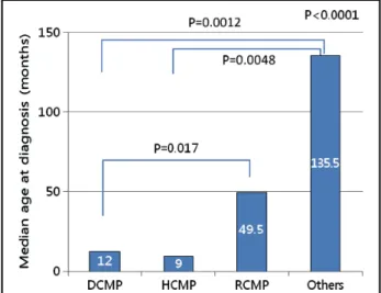
Age at diagnosis with CMP differed significantly among the 4 types of CMPs (P<0.001) (Figure 1). For each subgroup, the median age at diagnosis of DCMP was 12 months (range, 1 day after birth to 190 months), HCMP 9 months (range, 1 day after birth to 168 months), RCMP 49.5 months (range,
Table 2. Average Annual Occurrence of New Cases of Primary CMP in Korean Children 1998–2006 Age Male No./100,000 Female No./100,000 Total No./100,000Total CI
Lower Upper DCMP 0–4 61 0.44 55 0.43 116 0.43 0.36 0.52 5–9 10 0.06 14 0.10 24 0.08 0.05 0.12 10–14 10 0.06 9 0.06 19 0.06 0.04 0.10 Total 81 0.18 78 0.19 159 0.18 0.15 0.21 HCMP 0–4 22 0.16 21 0.17 43 0.16 0.12 0.22 5–9 1 0.01 1 0.01 2 0.01 0.00 0.02 10–14 7 0.04 6 0.04 13 0.04 0.02 0.07 total 30 0.07 28 0.07 58 0.07 0.050 0.086 RCMP 0–4 5 0.04 4 0.03 9 0.03 0.015 0.064 5–9 1 0.01 4 0.03 5 0.02 0.01 0.04 10–14 2 0.01 0 0.00 2 0.01 0.00 0.02 Total 8 0.02 8 0.02 16 0.02 0.01 0.03 Other CMP* 0–4 0 0.00 1 0.01 1 0.00 0.00 0.02 5–9 0 0.00 3 0.02 3 0.01 0.00 0.03 10–14 4 0.03 1 0.01 5 0.02 0.01 0.04 Total 4 0.01 5 0.01 9 0.01 0.00 0.02 All CMP 0–4 88 0.63 81 0.64 169 0.63 0.54 0.73 5–9 12 0.07 22 0.15 34 0.11 0.08 0.15 10–14 23 0.15 16 0.11 39 0.13 0.09 0.18 Total 123 0.27 119 0.29 242 0.28 0.24 0.31 *Includes 5 patients with ion channelopathies (2 with Brugada syndrome, 3 with long QT syndrome (1 KCNQ1; Jervell-Lange-Nielson syndrome) and 5 patients with mitochondrial myopathies. CI, confidence interval. Other abbreviations see in Table 1.
Table 3. Point Prevalence of Primary CMP in Korean Children on December 31, 2006
Subgroup No. of cases prevalence/ Point 100,000 CI Lower Upper DCMP 127 1.39 1.16 1.66 HCMP 47 0.51 0.38 0.68 RCMP 15 0.16 0.09 0.27 Others 4 0.04 0.01 0.11 Total 193 2.11 1.83 2.43 Age-specific Korean population aged 0 to <15 years at the end of 2006 was 9,128,397. Abbreviations see in Tables 1,2. Figure 1. Median ages at diagnosis with primary CMPs were significantly different with each other in all 4 subgroups of CMPs (P<0.001). DCMP group was diagnosed significantly younger than those of RCMP (Pcor =0.017) and other groups
(Pcor =0.0012). The HCMP group was also diagnosed younger
than the other groups (Pcor =0.0048). CMP, cardiomyopathy;
DCMP, dilated CMP; HCMP, hypertrophic CMP; RCMP, re-strictive CMP; Others, other types of CMP.
1 day after birth to 180 months), and other CMP 135.5 months (range, 6–172 months). In the Wilcoxon rank sum 2-sample test for each pair of subgroups of primary CMPs, age at diag-nosis with DCMP was significantly younger than the age at diagnosis with RCMP (corrected P (Pcor) =0.0174) and not with HCMP (Pcor >0.05). Age at diagnosis of HCMP did not differ significantly from that of DCMP or RCMP (Pcor >0.05). Age of RCMP was significantly older than the age of diagno-sis with DCMP (Pcor =0.017) and age of other CMP was sig-nificantly older than those of patients diagnosed with DCMP and HCMP (Pcor =0.0012, Pcor =0.0048 each), but not sig-nificantly different from age of RCMP. A case of RCMP was diagnosed with echocardiography during the fetal period.
Initial symptoms of patients with DCMP at the time of diagnosis included dyspnea (45.1% of total DCMP cases), coughing (42.9%), feeding difficulty (28.8%), dyspnea on ex-ertion (DOE) (17.4%), poor weight gain (14%), and syncope (1%); those of HCMP included dyspnea (32.3%), coughing (20.0%), feeding difficulty (18.4%), DOE (7.7%), poor weight gain (12.3%), and syncope (4.6%); and those of RCMP in-cluded dyspnea (55.6%), coughing (33.3%), feeding difficulty (16.7%), DOE (33.3%), and poor weight gain (11.1%); none of the patients presented with syncope, and other CMP in-cluded syncope (50.0%), DOE (33.3%), and dyspnea (40.0%). Asymptomatic patients with DCMP showed initial signs of cardiomegaly (64.1% of total DCMP patients), tachy-pnea (23.4%), murmur (22.8%), arrhythmia (10.9%) and cyanosis (7.6%); those with HCMP showed cardiomegaly (23.1%), tachypnea (18.4%), murmur (36.9%), arrhythmia (3.1%) and cyanosis (4.6%); and those with RCMP showed cardiomegaly (61.1% of RCMP patients), tachypnea (22.2%), murmur (27.8%), arrhythmia (16.7%) and cyanosis (11.1%) (Table 4).
Diagnosis
Among the total number of patients with a primary CMP, diagnostic catheterization was performed in 51 cases (18.4%). For each subgroup, 27 cases (14.7%) among 184 patients with DCMP, 15 cases (23.1%) among 65 patients with HCMP, and 9 cases (50.0%) among 18 patients with RCMP underwent catheterization.
Endomyocardial biopsy was performed in 33 cases (11.9%) of primary CMP patients, and in each subgroup, 23 cases (12.5%) (range 2 months to 175 months) of DCMP, 6 cases (9.2%) of HCMP, and 5 cases (27.8%) of RCMP patients. Among patients with other CMP, 1 patient was diagnosed with Jervell-Lange-Nielson syndrome (KCNQ1/11p) on elec-trophysiologic study. Two patients with mitochondrial myop-athies each showed MERRF and MELAS.
Treatment
In addition to medical therapy, 3 patients were treated with a ventricular assist device (VAD), 2 patients with subaortic septal myomectomy, 2 with alcohol ablation for HCMP, and 1 with an intraaortic balloon pump (IABP); finally, 8 patients underwent cardiac transplantation.
Figure 2. Kaplan-Meier survival curves for the subgroups of primary CMPs and log-rank test for survival rates after 1, 2, 5 and 9 years after being diagnosed within each subgroup of CMPs (P=0.017). CMP, cardiomyopathy; DCMP, dilated CMP; HCMP, hypertrophic CMP; RCMP, restrictive CMP; Others, other types of CMP.
Table 4. Symptoms and Signs of Korean CMP Patients at Initial Presentation
Symptoms* and signs at presentation No. of patients (%) in subgroup DCMP (n=184) HCMP (n=65) RCMP (n=18) Symptomatic patients
(% among total patients of each subgroup)
Dyspnea 83 (45.1%) 21 (32.3%) 10 (55.6%) Coughing 79 (42.9%) 13 (20.0%) 6 (33.3%) Feeding difficulty 53 (28.8%) 12 (18.4%) 3 (16.7%) DOE 32 (17.4%) 5 (7.7%) 6 (33.3%) Poor weight gain 26 (14.1%) 8 (12.3%) 2 (11.1%) Syncope 2 (1.1%) 3 (4.6%) 0 (0.00%) Asymptomatic patients
(% among total patients of each subgroup)
Cardiomegaly 118 (64.1%) 15 (23.1%) 11 (61.1%) Tachypnea 43 (23.4%) 12 (18.4%) 4 (22.2%) Murmur 41 (22.8%) 24 (36.9%) 5 (27.8%) Arrhythmia 29 (10.9%) 2 (3.1%) 3 (16.7%) Cyanosis 14 (7.6%) 3 (4.6%) 2 (11.1%) *A single patient often had multiple symptoms at presentation. DOE, dyspnea on exertion. Other abbreviations see in Table 1.
Cardiac transplantation was performed in 6 patients with DCMP and 2 patients with HCMP. There was a case of a sib-ling with DCMP, both of whom successfully underwent car-diac transplantation independently within a few years. Prognosis
At the end of the study, a total of 60 patients (21.7%) with primary CMP had died and 217 patients (78.3%) survived. A total of 48 patients (26.1%) of DCMP, 6 patients (9.2%) of HCMP, 2 patients (11.1%) of RCMP and 4 patients (40.0%) of other CMP died. In particular, the mortality of 10 patients of other CMP was as high as 25.0% for ion channelopathy and 60% for mitochondrial CMP.
The 9-year survival rate was estimated as 69.8% in DCMP, 90.3% in HCMP, 47.2% in RCMP and 42.0% in other CMP patients with ion channelopathy and mitochondrial CMP (P=0.017) (Figure 2).
Prognosis with 1-year survival rate of DCMP was 80.2%, the 2-year survival rate was 77.7%, the 5-year survival rate was 72.6%, and the 9-year survival rate was 69.8% using the log-rank test (Figures 2,3). Survival rates at 1, 2, 5 and 9 years after diagnosis did not differ statistically from each other, whether patients were diagnosed with DCMP before or after 1 or 2 years of age (P>0.05). The 9-year survival rate of HCMP was 90.3% (Figures 2,3), and in all cases of fatal HCMP, the period from diagnosis to death was less than 6 months. Survival rates of HCMP patients did not differ sta-tistically from each other, whether patients were diagnosed before or after 1 or 2 years of age (P>0.05). Regarding RCMP, the 9-year survival rate was estimated as 94.4%. The 1-year survival rate of other CMP was 78.8%, and the 9-year sur-vival rate was 42.0%. All of the fatal cases in this group were cardiac death within 24 months of diagnosis (Figure 3).
Comparison of the survival rates of children with a family history and those of patients without found no statistically significant differences between the groups in all CMP sub-groups of subjects enrolled in this study (data not shown).
Discussion
Because of the multifactorial etiologies, the pathophysiology of each subgroup of CMP is not fully understood. Roughly every 10 years, a new classification of the category of CMP is announced.
In this retrospective study, the eligible criteria were based on rigorous definitions of the AHA (2006) on primary CMPs, which resulted in a relatively smaller study population than expected. In 2000, Cheon et al reported national survey data on idiopathic CMP in Korean children during a study period between 1988 and 1997, according to WHO classification criteria (1995).2 At that time, they reported the total number of new patients with CMP for 10 years as 277, rendering the average annual incidence as 0.265/100,000, which was very similar to our result (n=242), with 0.28/100,000 (1998–2006), because of the change in the corresponding age-specific popu-lation in Korea. Numbers and clinical features of patients in each subgroup during both study periods were similar and the incidence and prevalence were considered not significantly changed.
Primary CMP, previously known as idiopathic CMP, is rare and associated with a high prevalence of family history, which implies that genetic factors might affect the incidence and might explain why the incidence of CMPs in every re-port vary widely according to the ethnicity of the patients.2–8 Reports on the incidence of primary CMP in pediatric age groups are rare and reports on the incidence of primary CMP are extremely rare in Asian children. Among Asian countries, Korea is uniquely comprised of a relatively homogeneous ethnic group, compared with other countries, and it would be interesting to study the incidence and prevalence of sub-groups of CMPs in Korean children.
According to an Australian report on patients younger than 10 years of age, the annual incidence was 1.24/100,000 children, with 314 new cases for 10 years. The number of new cases for 10 years in our subjects with primary CMP was 242, rendering the annual incidence as low as 0.28/100,000 because of a much larger corresponding age population
Figure 3. No difference in survival rates according to timing of diagnosis, whether patients had been diagnosed with dilated cardiomyopathy (DCMP) before or after infancy (<1 year of age after birth).
younger than 15 years than in Australia.3
With the exclusion of patients older than 15 years of age, a total of 193 patients (0.69% of Korean primary CMP) out of a total of 277 patients with primary CMP in Korea at the end of survey were included in the calculation of the preva-lence rate, with the same percentage as that of the Finnish report, with 0.69% of total primary CMP patients at the end of 1997. However, the age-specific population in Korea at the end of 2006 was approximately 7-fold larger than that of the Finnish population, which resulted in a much lower preva-lence rate of Korean CMP patients.6 The point prevalence of DCMP on December 31, 2006 was 1.39/100,000 (95%CI 1.16–1.66) (Table 3), which was very similar to the result of Japanese children in 1998, 1.4/100,000 in 10–19-year-old Japanese patients with DCMP.7 These findings might imply that the prevalence of primary CMP is significantly lower in Asian children than in other ethnic groups.
Age at diagnosis with CMPs differed significantly among the 4 types of CMPs (P<0.001). In the Wilcoxon rank sum 2-sample test for each pair of subgroups of primary CMPs, age at diagnosis with DCMP was significantly younger than age at diagnosis with RCMP, but not with HCMP.
In the Australian report on primary CMPs in children under the age of 10 years, lymphocytic myocarditis accounted for 25 patients out of 62 children with DCMP, which seems to be a very high percentage.3 In our study, 17.4% of patients with DCMP had a history of prior infection, although not all of them had been confirmed with endomyocardial biopsy.
Many patients in our study were already diagnosed with primary CMP at an early age and this reflects a change in the practices of family care, with children of affected families being screened much earlier than before. Usually, HCMP does not fully present until adolescence and is rarely seen during infancy. Some reports of HCMP include isolated and secondary forms of HCMP as well, with variable ages of pa-tients included, leading to a relatively high annual incidence.9 In this study, because we confined eligible patients to those younger than 15 years, the prevalence of HCMP differs slight-ly from results in other populations. The annual occurrence of new cases of DCMP did not increase during the study period, whereas the annual occurrence of new cases of RCMP showed an increasing trend toward the last 3 years of the study (data not shown). Understanding of RCMP in pediatric patients could also have shown improvement over that time.
The familial tendency of HCMP (21.5%) and of other types of CMP (30%) with mitochondrial and channelopathy (40%) was very high, as predicted; however, the incidence of a family history of HCMP in Korean children appears to be lower than in Western populations.10,11 According to the report by Wilkinson et al regarding pediatric CMP in the USA, study of familial isolated CMP demonstrated that children with HCMP who underwent metabolic study were 6.4-fold as likely to have a causal diagnosis established than patients without such testing.12 With advances in genetic study, the percentages of CMPs with a familial tendency are expected to change in the future.
The mortality of other types of CMP (mitochondrial and channelopathy) was very high (40.0%), comprising the over-all mortality of primary CMP as 26.7%. In particular, some cases of these types of CMP were extremely fatal, leading to cardiac death within 24 months after diagnosis, as reported in other studies.13–17 Regarding the prognosis of patients with CMP with or without a family history in prior research studies, familial history of sudden death is a well-known risk factor for patients with poor prognosis. In this study,
com-parison of the survival curves of each patient belonging to each subgroup of CMP regarding family history found no statistically significant difference between them. According to a recent report on survival of patients with HCMP with a family history, multiple family history of sudden death was an ominous risk factor with a strong impact.18 However, most of the enrolled patients in our study were younger than 15 years and most of the fatal cases were patients without a family history of either CMP or sudden cardiac death. They could have been relatively young during the study period to draw a conclusion of long-term prognosis or early screening of patients with family members with CMP in each sub-group might have had an impact on the survival of patients in pediatric age groups in Korea.
Regarding the therapeutic trials of patients with CMPs, among the enrolled patients, 8 patients underwent cardiac transplantation, 3 had a VAD and 1 had IABP during the study period. A study of the epidemiologic characteristics of idio-pathic CMP from the pediatric CMP registry in the USA revealed that approximately 5% of patients underwent car-diac transplantation, 2% IABP, 2% ECMO, and 1% VAD; however, the medical outcomes in these children did not show improvement in the previous several decades.19 There have been reports on various therapeutic trials in addition to medical therapy of CMP prior to cardiac transplantation.20–22 Although we could not collect all of the data on CMP patients nationwide, the numbers of patients treated with surgical mechanical support have shown an increase in recent years and these therapeutic trials and experience might have an impact on the prognosis of primary CMP in the future. In this study, because nationwide data collection was not feasible, the prevalence and incidence rate might be underestimated. However, most patients from major tertiary centers were enrolled in this study and the clinical features should not be significantly different from the actual characteristics of pri-mary CMP in Korea.
Conclusions
In conclusion, the point prevalence of CMP was estimated as 2.11/100,000 in this multicenter epidemiologic study of childhood primary CMP in Korea. Most DCMP and HCMP cases were identified at an early age. Clinical characteristics of primary CMP in Korea did not show significant change, compared with the results of previous Korean reports, and some patients were successfully treated with mechanical sup-port and cardiac transplantation. Further epidemiologic study with a nationwide survey is necessary.
Acknowledgments
We are deeply grateful to the following doctors who replied to the ques-tionnaires and made this study possible (major contributors are listed in alphabetical order).
Choi Jae Young (Severance Cardiovascular Hospital, Yonsei Uni-versity), Choi Jong Woon (Daejin Medical Center, Bundang Jesaeng Hospital), Hong Young Mi (Ewha Womans University Hospital), Hyun Myung Chul (Kyungpook National University Hospital), Jung Jo Won (Ajou University Medical Center), Kim Soo Jin (Sejong Heart Insti-tute), Kim Yeon Woo (Cheju National University), Kwon Tae Chan (Keimyung University Dongsan Medical Center), Lee Hae Yong (Yonsei University Wonju Christian Hospital), Lee Hyoung Doo (Pusan National University Hospital), Lee Joo Won (Korea University Medical Center), Lee Soon Ju (Catholic University of Korea St. Mary’s Hospital), Noh Chung Il (Seoul National University Hospital), Oh Jin-Hee (The Catholic University of Korea, St. Vincent’s Hospital), Park In Sook (Asan Medi-cal Center), Park Yong Won (Inje University Seoul Paik Hospital), Sohn Chang Sung (Korea University Anam Hospital), Sohn Sejung (Ewha Womans University Mokdong Hospital), and many other doctors from
each institution.
We also want to extend special thanks to Wan Sook Jang, RN, a Co-ordinator of the pediatric cardiologic department of Asan Medical Center for her dedication to this study and for collecting a large amount of pre-cious data that made this study possible.
References
1. Maron BJ, Towbin JA, Thiene G, Antzelevitch C, Corrado D, Arnett D, et al. Contemporary definitions and classification of the cardiomyopathies: An American Heart Association Scientific State-ment from the Council on Clinical Cardiology, Heart Failure and Transplantation Committee; Quality of Care and Outcomes Research and Functional Genomics and Translational Biology Interdisci-plinary Working Groups; and Council on Epidemiology and Pre-vention. Circulation 2006; 113: 1807 – 1816.
2. Cheon EJ, Kang IS, Bae EJ, Lee JK, Gil HR, Yoon HS, et al. Idio-pathic cardiomyopathies in Korean children: A nationwide study.
Korean Circ J 2000; 30: 635 – 645.
3. Nugent AW, Daubeney PE, Chondros P, Carlin JB, Cheung M, Wilkinson LC, et al. The epidemiology of childhood cardiomy-opathy in Australia. N Engl J Med 2003; 348: 1639 – 1646. 4. Lipshultz SE, Sleeper LA, Towbin JA, Lowe AM, Orav EJ, Cox
GF, et al. The incidence of pediatric cardiomyopathy in two regions of the United States. N Engl J Med 2003; 348: 1647 – 1655. 5. Matsumori A, Furukawa Y, Hasegawa K, Sato Y, Nakagawa H,
Morikawa Y, et al. Epidemiologic and clinical characteristics of car-diomyopathies in Japan: Results from nationwide surveys. Circ J 2002; 66: 323 – 336.
6. Arola A, Jokinen E, Ruuskanen O, Saraste M, Pesonen E, Kuusela AL, et al. Epidemiology of idiopathic cardiomyopathies in children and adolescents: A nationwide study in Finland. Am J Epidemiol 1997; 146: 385 – 393.
7. Miura K, Nakagawa H, Morikawa Y, Sasayama S, Matsumori A, Hasegawa K, et al. Epidemiology of idiopathic cardiomyopathy in Japan: Results from a nationwide survey. Heart 2002; 87: 126 – 130. 8. El-Menyar AA, Bener A, Numan MT, Morcos S, Taha RY,
Al-Suwaidi J. Epidemiology of idiopathic cardiomyopathy in Qatar during 1996–2003. Med Princ Pract 2006; 15: 56 – 61.
9. Colan SD, Lipshultz SE, Lowe AM, Sleeper LA, Messere J, Cox GF, et al. Epidemiology and cause-specific outcome of hyper-trophic cardiomyopathy in children: Findings from the pediatric cardiomyopathy registry. Circulation 2007; 115: 773 – 781. 10. Brito D, Richard P, Komajda M, Madeira H. Familial and sporadic
hypertrophic myopathy: Differences and similarities in a geno-typed population: A long follow-up study. Rev Port Cardiol 2008;
27: 147 – 173.
11. Morales A, Cowan J, Dagua J, Hershberger RE. Family history: An essential tool for cardiovascular genetic medicine. Congest Heart
Fail 2008; 14: 37 – 45.
12. Wilkinson JD, Sleeper LA, Alvarez JA, Bublik N, Lipshultz SE. The Pediatric Cardiomyopathy Registry: 1995–2007. Prog Pediatr
Cardiol 2008; 25: 31 – 36.
13. Jons C, Moss AJ, Goldenberg I, Liu J, McNitt S, Zareba W, et al. Risk of fatal arrhythmic events in long QT syndrome patients after syncope. J Am Coll Cardiol 2010; 55: 783 – 788.
14. Wedekind H, Smits JP, Schulze-Bahr E, Arnold R, Veldkamp MW, Bajanowski T, et al. De novo mutation in the SCN5A gene asso-ciated with early onset of sudden infant death. Circulation 2001;
104: 1158 – 1164.
15. Strauss A, Lock JE. Pediatric cardiomyopathy: A long way to go.
N Engl J Med 2003; 348: 1703 – 1705.
16. Ruiter JS, Berkenbosch-Nieuwhof K, van den Berg MP, van Dijk R, Middel B, van Tintelen P. The importance of the family history in caring for families with long QT syndrome and dilated cardio-myopathy. Am J Med Genet 2010; 152: 607 – 612.
17. Song MK, Baek JS, Kwon BS, Kim GB, Bae EJ, Noh CI, et al. Clinical spectrum and prognostic factors of pediatric ventricular tachycardia. Circ J 2010; 74: 1951 – 1958.
18. Dimitrow P, Chojnowska L, Rudzinski T, Piotrowski W, Ziólkowska L, Wojtarowicz A, et al. Sudden death in hypertrophic cardiomyopa-thy: Old risk factors re-assessed in a new model of maximalized follow-up. Eur Heart J 2010; 31: 3084 – 3093.
19. Harmon WG, Sleeper LA, Cuniberti L, Messere J, Colan SD, Orav EJ, et al. Treating children with idiopathic dilated cardiomyopathy (from the Pediatric Cardiomyopathy Registry). Am J Cardiol 2009;
104: 281 – 286.
20. Sugiyama H, Hoshiai M, Naitoh A, Kadono T, Suzuki S, Sugita K. Outcome of non-transplant surgical strategy for end-stage dilated cardiomyopathy in young children. Circ J 2009; 73: 1045 – 1048. 21. Chang BC, Lim CY, Park PW, Park KY, Lee YT, Kim YJ. Volume
reduction surgery for end-stage heart failure: Experience in Korea.
J Card Surg 2001; 16: 159 – 164.
22. Saito K, Ibuki K, Yoshimura N, Hirono K, Watanabe S, Watanabe K, et al. Successful cardiac resynchronization therapy in a 3-year-old girl with isolated left ventricular non-compaction and narrow QRS complex: A case report. Circ J 2009; 73: 2173 – 2177.