DOI : 10.3341/jkos.2009.50.11.1639 = 증례보고 = 접수번호 : 50-11-02-08
세 종류의 비구면 인공수정체 간의 술 후 구면수차,
대비감도 및 초점심도 정도의 비교
배형원⋅김응권⋅김태임 연세대학교 의과대학 안과학교실, 시기능 개발 연구소 목적: 세 가지 종류의 비구면 인공수정체 삽입 후 구면수차, 대비감도 및 초점심도를 평가하고자 하였다.대상과 방법: Akreos adapt Advanced Optics (AO) (18안), AcrySof IQ SN60WF (20안), Tecnis Acrylic IOL ZA9003 (18안), 총 48명 56안을 대상으로 술 후 3개월째 고위수차 및 구면수차를 포함한 안구 전체 및 안구 내 수차와 대비감도를 측정하고, 원거리시력을 교정 한 상태에서 33 cm 및 1 m 시력을 이용한 초점심도를 평가하였다. 결과: 전체 구면수차 및 안구 내 구면수차에 있어 AO를 삽입 받은 군이 다른 두 군에 비해 다소 높은 구면수차 값을 보였으나, 결과적 으로 대비감도 및 초점심도 정도에 있어서는 세 군간에 있어 유의한 차이는 발견되지 않았다. 결론: 세 군간 대비감도 및 초점심도에 유의한 차이를 보이지 못한 것은 세 군간 구면수차의 차이가 크지 않고 구면수차를 제외한 다른 요인들 간에 차이가 없어 대비감도 및 초점심도에 큰 영향을 주지 못한 것으로 생각된다. <대한안과학회지 2009:50(11):1639-1644> ■ 접 수 일: 2009년 2월 10일 ■ 심사통과일: 2009년 7월 21일 ■ 책 임 저 자: 김 태 임 서울시 서대문구 신촌동 134 연세대학교 의과대학 연세의료원 안과 Tel: 02-2228-3570, Fax: 02-312-0541 E-mail: [email protected] * 본 논문의 요지는 2008년 대한안과학회 제99회 춘계학술대회에서 구연으로 발표되었음.
* This work was supported by the National Research Foundation of Korea (NRF) grant funded by the Korea government (MEST) (No.M1AQ19, 2009-0082186) 현대 사회에서 삶의 질이 점차 중요시됨에 따라 백내장 수술 또한 수술 기법과 인공수정체의 발전을 통해 일차적 인 백내장의 제거 수준에서 시력의 질 향상까지 고려하는 방향으로 발전하고 있다.1 수술 받지 않은 눈에 있어서 양의 구면수차 값을 가진 각 막에 대해 젊었을 때는 수정체가 음의 값을 가짐으로써 구 면수차가 서로 상쇄되어 비교적 좋은 시력의 질을 유지할 수 있으나, 나이가 듦에 따라 수정체가 양의 구면수차 값으 로 증가하게 됨으로써 시력의 질이 떨어지게 된다.2-6따라 서 양의 구면수차 값을 갖는 기존의 구면 인공수정체는 백 내장 수술 후에도 구면수차의 증가가 그대로 유지되어 눈 부심, 빛번짐 등의 증상을 야기하는 반면에,7-9최근의 비구 면 인공수정체는 백내장 수술 후 구면수차 값을 상쇄시켜 시기능을 향상시키는 효과를 나타내고 있다.10 반면 구면수차의 감소는 망막에 명확한 상을 맺게 함으 로써 원거리시력의 질적 향상을 가져올 수는 있으나, 초점 심도(depth of focus)는 오히려 낮아져 근거리시력은 더욱 나빠진다는 여러 보고들도 제기되고 있다.11,12,22,30,31백내장 수술을 받은 환자라 할지라도 동공 및 각막, 또는 삽입된 인공 수정체의 구면수차 등에 의해 어느 정도의 근거리시력을 유지 할 수 있는데, 구면수차가 낮아지면 초점심도(depth of focus) 가 얕아지기 때문에 근거리시력은 더욱 저하된다는 것이다. 따라서 구면수차의 감소가 원거리시력의 질을 향상시키긴 하지만 근거리시력의 저하를 함께 유발할 수도 있기 때문에 비구면 인공수정체로 인한 보다 전반적인 시기능 평가를 위해 서는 수차 및 대비감도와 함께 초점심도를 아울러 비교하는 것이 필요할 것으로 여겨진다.
Akreos adapt Advanced Optics (AO) (Bausch & Lomb, Inc., Rochester, NY) 인공수정체는 일체형 형태의 친수성 아크릴 재질의 인공수정체로서 인공수정체의 전면과 후면 모두를 비구면화 함으로써 0 μm의 구면수차 값을 갖도록 고안된 인공수정체이고, AcrySof IQ SN60WF (Alcon, Inc., Forth Worth, TX) 인공수정체는 일체형 형태의 소수성 아 크릴 재질로서 후면 비구면 표면을 가짐으로써 -0.2 μm의 음의 구면수차 값을 가진 인공수정체이다. 3-piece 형태의 Tecnis Acrylic IOL ZA9003 (AMO, Inc., Santa Ana, CA) 인공수정체는 소수성 아크릴 재질로서 전면부 표면을 변형 하여 -0.27 μm의 음의 구면수차 값을 가진다(Table 1). 본 연구는 세 종류의 비구면 인공수정체 간의 술 후 고위 수차와 구면수차를 비롯한 수차 및 대비감도를 비교하고, 구면수차 차이로 인한 초점심도의 차이를 비교하고자 하였다.
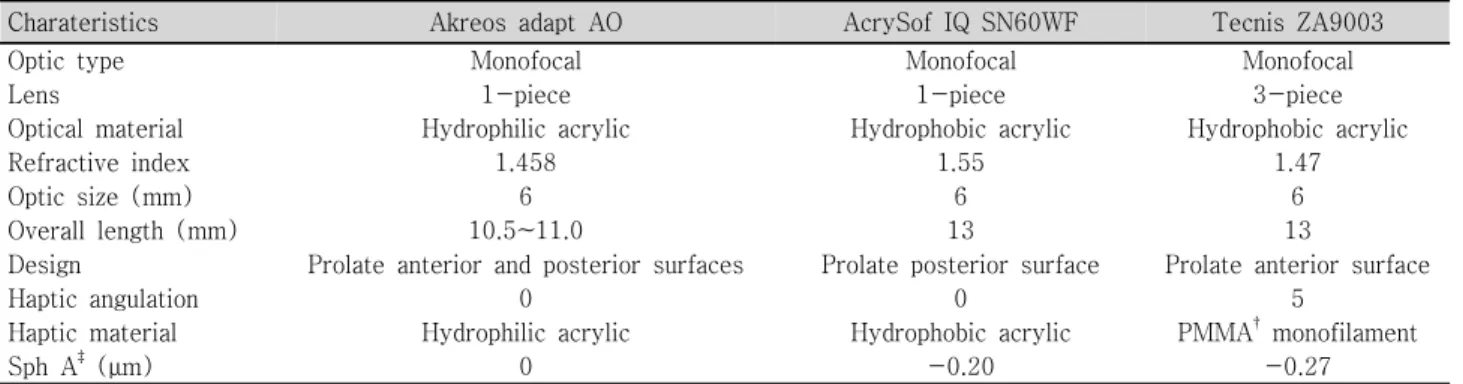
Table 1. Optical characteristics of the aspherical IOLs* used in the study
Charateristics Akreos adapt AO AcrySof IQ SN60WF Tecnis ZA9003
Optic type Monofocal Monofocal Monofocal
Lens 1-piece 1-piece 3-piece
Optical material Hydrophilic acrylic Hydrophobic acrylic Hydrophobic acrylic
Refractive index 1.458 1.55 1.47
Optic size (mm) 6 6 6
Overall length (mm) 10.5~11.0 13 13
Design Prolate anterior and posterior surfaces Prolate posterior surface Prolate anterior surface
Haptic angulation 0 0 5
Haptic material Hydrophilic acrylic Hydrophobic acrylic PMMA†monofilament
Sph A‡(μm) 0 -0.20 -0.27
*
IOLs=intraocular lens; †PMMA=polymethylmethacrylate; ‡Sph A=spherical aberration.
대상과 방법
2007년 8월부터 2007년 12월까지 본원에서 백내장 초음 파유화술 및 인공수정체 안내삽입술을 시행 받은 환자 48명 56안을 대상으로 무작위 할당을 통하여 Akreos adapt Ad-vanced Optics (AO)를 삽입 받은 군(I 군), AcrySof IQ SN60WF 를 삽입 받은 군(II 군), Tecnis Acrylic IOL ZA9003를 삽입 받은 군(III 군)의 세 군으로 구분하고 전향적인 연구를 진행 하였다. 각막 난시가 2D가 넘지 않는 노인성 백내장 환자를 대상으로, 각막혼탁, 약시, 녹내장, 망막질환 등 눈에 다른 질환 을 가진 사람과 이전에 굴절교정수술 받은 사람 및 수술 과정 에서 후낭 파열이 발생한 경우, 그리고 수술 후 경과 관찰시 인공수정체 이탈 소견이 보이는 경우는 연구 대상에서 제외 하였다. 수술은 단일 술자에 의해 시행되었고, 점안 마취 후 이측 또는 비측 투명각막절개를 통해 전방 내 점탄물질을 주입한 후 26 gauge의 주사침을 이용하여 약 5.5 mm 크기의 수정 체낭 원형절개를 시행하였다. 평형염액을 사용하여 수력분리 술 및 수력분층술을 시행한 후 초음파를 이용한 수정체유화 술로 수정체 핵을 제거하였으며 관류흡입기로 남아있는 수정 체 피질을 제거하였다. 그 후 카트리지를 이용하여 인공수정 체를 수정체 낭 내에 삽입하였으며 남아있던 점탄물질을 관류 흡입기로 제거하고 평형염액을 사용하여 안구의 긴장도를 유지하였다. 모든 환자는 술 후 3개월째 수차 및 대비감도를 측정하였 으며, 초점심도를 비교하기 위하여 원거리시력 및 원거리교 정된 상태에서의 중간거리, 근거리시력이 측정되었다. 수차검사는 산동된 상태에서 동공 중심부 6 mm 영역에 대 하여 시행되었다. iTrace®(Tracey Tech., Houston, TX)를 사용하여 RMS (Root mean square) 총합, 고위수차, 구면 수차, 코마수차, 트레포일수차를 각각 측정하였고, 안구 전체 의 수차(total ocular aberration)와 각막 수차를 제외한 안
구 내 수차(internal ocular aberration)를 나누어 분석하였다. 대비감도는 FACT 차트 방식의 Optec 6500®(Stereo Optical Co, Inc., Chicago, IL)을 이용하여 굴절이상을 교정한 상태 로 시행하였으며, 명소시(photopic condition, 85 cd/m2)와 박명시(mesopic condition, 3 cd/m2) 조건하에서 각각 1.5, 3, 6, 12,18 cpd (cycle perdegree)의 주파수에 대해서 측정 하였다. 초점심도는 원거리시력을 교정한 상태(distance-corrected) 에서의 중간거리시력과 근거리시력의 평균을 비교함으로써 판단하였다.11 초점심도에 영향을 줄 수 있는 인자인 동공 크기 및 각막난시도 함께 고려하였으며,11-13동공크기는 명 소시(85 cd/m2)와 박명시(3 cd/m2) 상태에서 각각 측정하였 고, 각막난시는 Auto Ref-keratometer RK-3 (Canon,Inc., Tokyo, Japan)를 사용하여 측정하였다. 원거리시력은 교정된 상태에서 4 m에서 ETDRS 시력표를 사용하여 측정하였고, 중간거리시력은 원거리시력이 교정된 상태에서 ETDRS 1 m 시력측정법으로 측정하였다. 이 경우 중간거리시력이 ETDRS 1 m 시력측정법으로 측정가능 한 최대값인 0.5 이상으로 측정되면 1 m 거리에서 로젠바움 (Rosenbaum, Cleveland, OH) 근거리 시력표를 이용하여 측정 한 값의 3배 값을 중간거리시력으로 하였다. 근거리시력 역시 원거리시력이 교정된 상태로 33 cm 거리에서 로젠바움 근 거리 시력표를 사용하여 측정하였다.
통계학적 분석은 통계프로그램 SPSS 12.0 for Windows (SPSS Inc., Chicago, IL)를 사용하였으며, 통계학적 유의 성의 기준은 p<0.05로 하였다.
결
과
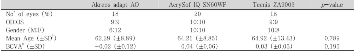
환자 군은 총 48명 56안이었고, I 군은 18안, II 군은 20 안, III 군은 18안이었다. 연령, 성별 및 술 후 3개월째 최대 교정시력에 있어서 세 군 간의 유의한 차이는 보이지 않았
Table 2. Demographics of study groups
Akreos adapt AO AcrySof IQ SN60WF Tecnis ZA9003 p-value
No* of eyes (%) OD:OS Gender (M:F) Mean Age (±SD†) BCVA‡(±SD) 18 9:9 6:12 62.29 (±8.89) -0.02 (±0.12) 20 10:10 10:10 64.21 (±8.85) 0.04 (±0.06) 18 9:9 10:8 64.92 (±13.43) 0.03 (±0.05) 0.789 0.195 All values are displayed as logMAR visual acuity.
*No=number; †SD=standard deviation; ‡BCVA=best corrected visual acuity.
Table 3. Total ocular aberrations (μm) of three groups measured by iTrace® (Mean±Standard deviation)
IOL groups RMS* total HO A† Sph A‡ Coma 7A Coma 8A Trefoil 6A Trefoil 9A
AO 1.46±0.38 0.80±0.33 0.26±0.20 -0.09±0.24 0.12±0.31 0.12±0.29 -0.04±0.28
IQ 1.46±0.64 0.63±0.26 0.00±0.14 0.07±0.23 0.06±0.26 0.12±0.40 -0.07±0.25
Tecnis 1.09±0.39 0.56±0.21 -0.01±0.13 -0.01±0.23 -0.02±0.23 -0.09±0.29 0.07±0.21
p-value 0.107 0.071 0.000§ 0.143 0.385 0.198 0.351
*RMS=root mean square; †HO A=higher-order aberration; ‡Sph A=spherical aberration; §Statistically significant (p<0.05).
Table 4. Internal aberrations (μm) of three groups measured by iTrace® (Mean±Standard deviation)
IOL groups RMS* total HO A† Sph A‡ Coma 7A Coma 8A Trefoil 6A Trefoil 9A
AO 1.54±0.69 0.90±0.44 -0.02±0.17 -0.14±0.34 0.13±0.27 0.20±0.36 -0.28±0.31
IQ 1.69±0.70 0.80±0.40 -0.22±0.16 0.06±0.28 0.05±0.22 0.16±0.44 -0.08±0.27
Tecnis 1.44±0.70 0.60±0.24 -0.28±0.10 -0.09±0.19 0.01±0.27 0.10±0.33 -0.02±0.25
p-value 0.605 0.134 0.000§ 0.116 0.455 0.810 0.072
*
RMS=root mean square; †HO A=higher-order aberration; ‡Sph A=spherical aberration; §Statistically significant (p<0.05).
Table 5. Visual acuities and depth of focus (Mean±Standard deviation)
AO IQ Tecnis p-value Far* Intermediate† Near† Depth of focus‡ -0.02±0.12 0.26±0.17 0.55±0.29 0.40±0.21 0.04±0.06 0.26±0.15 0.53±0.13 0.39±0.09 0.03±0.05 0.31±0.12 0.56±0.22 0.43±0.15 0.195 0.681 0.936 0.812 All values are displayed as logMAR visual acuity.
*
Best corrected visual acuity; †Distance-corrected visual acuity; ‡Mean of intermediate visual acuity and near visual acuity.
다(Table 2).
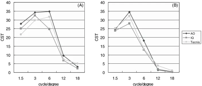
술 후 3개월째 6 mm 동공 크기로 산동된 상태에서 iTrace® 로 측정한 수차를 살펴볼 때 RMS 총합, 고위수차, 코마수차, 트레포일 수차의 경우에 있어서는 안구 전체 수차(total ocular aberration) 및 안구 내 수차(internal ocular aberration) 모 두에서 세 군 간에 유의한 차이가 발견되지 않았지만, 구면 수차에 있어서는 I 군이 다른 두 군에 비해 통계적으로 유의 하게 다소 높은 값을 나타냈다(p<0.000)(Table 3, 4). Optec 6500®으로 측정된 대비감도에서는 명소시와 박명 시 모두 모든 주파수 영역에서 세 군 간에 유의한 차이가 관찰되지 않았다(Fig. 1). 또한 세 군 간에 동공 크기 및 각막 난시는 유의한 차이 를 보이지 않은 상태에서 세 군 간에 원거리 교정된 상태의 중간거리 및 근거리시력의 평균은 통계적으로 유의한 차이 를 보이지 않아, 세 군간 초점심도에서도 유의한 차이를 관 찰할 수 없었다(Table 5).
고
찰
최근 백내장 수술의 목표는 백내장 치료의 목적 외에도 수술을 통해 굴절 이상의 교정 및 시력의 질을 향상시키는 개념으로 점차 발전하고 있다.1,14,15즉, 시력의 회복뿐만 아 니라 시기능의 향상이 백내장 수술에 있어 중요한 요소가 되고 있으며, 대비감도검사와 수차 분석은 이러한 시기능을 수치화하여 분석하는데 수단으로 사용되고 있다.7,9,16,17 비구면 인공수정체는 백내장 수술에 있어 구면 인공수정 체 삽입 시 발생할 수 있는 구면수차의 증가를 감소시킴으 로써 구면 인공수정체에 비해 보다 나은 대비감도를 얻을 수Figure 1. Contrast sensitivity test of three groups at photopic condition (A) and mesopic condition (B). There was no statistically significant difference among three groups.
있다는 것이 이미 기존의 국내외 많은 연구에서 입증된 바 있으며,16,18-19각 회사들은 이러한 연구들을 기반으로 더 좋은 시력의 질을 얻기 위해 다양한 비구면 인공수정체를 양산해 내고 있다. 본 연구는 이렇게 서로 다른 비구면 인공수정체 간에 술 후 결과의 차이가 존재하는지에 대해 알아보고자 하였다. 본 연구에서는 세 종류의 비구면 인공수정체를 대상으로 하였다. Akreos adapt Advanced Optics (AO) 인공수정체는 전면과 후면 모두에 비구면 처리를 함으로써 인공수정체 자 체의 구면수차를 없앴으며, 이는 기존의 구면 인공수정체로 인해 발생할 수 있는 구면수차의 증가를 막을 수 있는 동시 에, 인공수정체 중심부 이탈 등의 상황에서 인공수정체 자체 로 인해 발생할 수 있는 수차의 유발 가능성을 줄이는 장점 을 갖는다.23,24실제로 Altmann et al23의 연구에서 인공수정 체 중심부 이탈 시 구면수차 값이 없는 비구면 렌즈는 음의 구면수차 값을 갖는 비구면 인공수정체에 비해 더 나은 시기 능을 보이는 것이 보고되었으나, 본 연구에서는 인공수정체 가 중심부 이탈된 경우 연구에서 제외하였기 때문에 이에 대한 분석은 시행하지 않았다. AcrySof IQ SN60WF 인공수정체는 후면 비구면 표면을 가짐으로써 -0.2 μm의 음의 구면수차 값을 가진 비구면 인공수정체로써 평균 각막의 구면수차 값을 +0.27~0.28 μm 정도라고 할 때24-26시력이 좋은 젊은 사람 눈의 평균 구면수차를 +0.1 μm 정도로 보고 술 후 안구 전체 의 구면수차를 이와 유사하게 유지하는 것을 목표로 한다.27 반면 전면부 비구면 표면을 갖는 Tecnis Acrylic IOL ZA9003 인공수정체는 -0.27 μm의 인공수정체 구면수차를 갖고 백 내장 수술 후 눈 전체 구면수차를 0 μm으로 만들고자 계획 되었다. 본 연구에서, 안구 내 구면수차, 즉 인공수정체의 구면수 차는 각 회사가 계획했던 것과 유사한 값(I 군 -0.02±0.17 μm, II 군 -0.22±0.16 μm, III 군 -0.28±0.10 μm)을 나 타내었다(Table 4). 그러나 안구 전체의 구면수차에 있어 서는 I 군 및 III 군에는 의도된 값과 유사한 반면, II 군은 기대했던 +0.1 μm 보다 약간 낮은 0.00±0.14 μm의 구면 수차 값을 보였다. AcrySof IQ SN60WF 인공수정체 삽입 후 구면수차를 측정한 기존의 여러 다른 연구들을 살펴볼 때, +0.01 μm 부터 +0.09 μm 까지 다양한 구면수차 값을 나타냈지만 전반적으로 예상 값보다 낮은 구면수차 값을 보였는데,18,28,29AcrySof IQ SN60WF 인공수정체 삽입 시 눈 전체 구면수차가 기대 했던 값보다 조금 낮게 측정되는 이유에 대해서는 추가적인 연구가 필요할 것으로 생각된다. 구면수차의 감소는 시력의 질의 향상을 가져오게 되고, 이는 대비감도의 증가로 나타나게 된다.16,18,19,28즉, 구면수 차가 낮을수록 대비감도가 증가해야하나 본 연구에서는 구 면수차가 높은 I 군의 대비감도는 다른 두 군에 비해서 통 계적 차이가 없었다. 이는 Johansson et al30이 연구한 AO 인공수정체와 Tecnis Z9000 비구면 인공수정체 간의 비교 연구에서도 유사한 결과를 보였는데, 비록 기존의 여러 다른 연구에서 구면수차가 낮은 비구면 수정체는 구면 수정체에 비하여 우수한 시기능을 보였지만, AO 인공수정체 또한 다른 구면 수정체에 비해 더 좋은 시기능을 보이는 비구면 인공 수정체 이기 때문에 비구면 인공수정체 군 간의 크지 않은 구면수차의 차이는 전체적인 대비감도의 결과에 유의한 영 향을 끼치지 못하는 것으로 생각된다. 게다가 Dietze and
Cox31의 연구에 의하면 구면수차 자체가 전체 수차에 미치는 영향 또한 크지 않을 수 있기 때문에 이러한 비구면 인공수 정체 간의 작은 구면수차의 차이는 결과적으로 시력의 질의 차이에 큰 영향을 미치지 못할 가능성이 있다고 하겠다. 한편 많은 연구에서 비구면 인공수정체는 구면수차가 낮 기 때문에 원거리시력의 질은 향상되지만, 초점심도(depth of focus)가 낮아져 근거리시력은 더욱 나빠진다는 단점이 제기되고 있다.11,12,22,30,31 즉, 초점심도는 구면수차가 클수 록 깊어지는데, 초점심도가 깊을수록 상대적으로 우수한 근 거리시력을 가질 수 있기 때문이다. 초점 심도에 영향을 줄 수 있는 요인으로는 구면수차 이외에도 동공 크기, 각막 난 시, 인공수정체 이탈 등이 있으나,11-13본 연구에서는 인공 수정체가 이탈된 경우에는 연구 대상에서 제외하였으며, 동 공 크기 및 각막 난시에 있어서도 세 군 간에 유의한 차이는 보이지 않았다. 따라서 다른 요인들이 배제된 상태에서 원 거리시력이 교정된 상태(distance-corrected)의 중간거리 (1 m) 및 근거리(33 cm)시력의 평균을 측정함으로써 세 비 구면 인공수정체 간의 초점 심도를 비교할 수 있었다.11앞서 언급한대로 상대적으로 구면수차가 높았던 I 군에서 다른 두 군에 비해 초점심도가 높을 수 있을 것으로 기대할 수 있으나, 본 연구의 결과에서는 세 군 간의 초점심도에 있어 유의한 차이가 관찰되지 않았다(Table 5). 이것 역시 대비 감도의 경우와 마찬가지로 비구면 인공수정체 간의 크지 않은 구면수차의 차이는 결과적으로 초점심도의 유의한 차 이를 가져오지 못한 것으로 생각해 볼 수 있겠다. 그 동안 비구면 인공수정체의 기능에 관한 연구는 국내 외에서 많이 발표되었지만, 비구면 인공수정체 간의 비교 연구는 그 수가 상대적으로 적었으며, 특히 초점심도를 함 께 고려한 비구면 인공수정체 간의 기능 비교 연구는 아직 까지 국내에서 보고된 바 없었다. 따라서 본 연구는 비구면 인공수정체 간의 초점심도까지 고려한 국내 연구라는 점에 서 일차적인 의미를 둘 수 있을 것이다. 또한 본 연구의 결 과를 살펴보면 기존에 국외에서 보고된 결과들과 크게 다 른 점을 보이지 않았는데, 이는 최근 국내의 백내장 수술 경향이 거의 선진국 수준에 이르렀기 때문이라고 미루어 판단해 볼 수가 있겠다.32 결론적으로 세 종류의 비구면 인공수정체는 서로 목표로 하는 구면수차에는 다소 차이가 있지만 그 차이가 크지 않 아 결과적으로 대비감도 및 초점심도에 대해 유의한 영향 을 끼치지 못하는 것으로 여겨지며, 따라서 세 종류의 비구 면 인공수정체 간에 술 후 대비감도 및 초점심도의 차이는 없는 것으로 판단할 수 있겠다.
참고문헌
1) Werner L, Olson RJ, Mamalis N. New Technology IOL Optics. Ophthalmol Clin North Am 2006;19:469-83.
2) Guirao A, Redondo M, Artal P. Optical aberrations of the human cornea as a function of age. J Opt Soc Am A Opt Image Sci Vis 2000;17:1697-702.
3) McLellan JS, Marcos S, Burns SA. Age-related changes in mono-chromatic wave aberrations of the human eye. Invest Ophthalmol Vis Sci 2001;42:1390-5.
4) Oshika T, Klyce SD, Applegate RA, Howland HC. Changes in corneal wavefront aberrations with aging. Invest Ophthalmol Vis Sci 1999;40:1351-5.
5) Artal P, Berrio E, Guirao A, Navarro R. Contribution of the cornea and internal surface to the change of ocular aberrations with age. J Opt Soc Am A Opt Image Sci Vis 2002;19:137-43.
6) Artal P, Guirao A, Berrio E, Williams DR. Compensation of corneal aberrations by the internal optics in the human eye. J Vis 2001;1:1-8. 7) Rawer R, Stork W, Spraul CW, Lingenfelder C. Imaging quality
of intraocular lenses. J Cataract Refract Surg 2005;31:1618-31. 8) Chalita MR, Krueger RR. Correlation of aberrations with visual
acuity and symptoms. Ophthalmol Clin North Am 2004;17:135-42. 9) Guirao A, Redondo M, Geraghty E, et al. Corneal optical aberrations
and retinal image quality in patients in whom monofocal intraocular lenses were implanted. Arch Ophthalmol 2002;120:1143-51. 10) Holladay JT, Piers PA, Koranyi G, et al. A new intraocular lens
design to reduce spherical aberration of pseudophakic eyes. J Refract Surg 2002;18:683-91.
11) Rocha KM, Soriano ES, Chamon W, et al. Spherical Aberration and Depth of Focus in Eyes Implanted with Aspheric and Spherical Intraocular Lenses. Ophthalmology 2007;114:2050-4.
12) Elder MJ, Murphy C, Sanderson GF. Apparent accommodation and depth of field in pseudophakia. J Cataract Refract Surg 1996; 22:615-9.
13) Sawusch MR, Guyton DL. Optimal astigmatism to enhance depth of focus after cataract surgery. Ophthalmology 1991;98:1025-9. 14) Holladay JT, Piers PA, Koranyi G, et al. A new intraocular lens
design to reduce spherical aberration of pseudophakic eyes. J Refract Surg 2002;18:683-91.
15) Olson RJ, Werner L, Mamalis N, Cionni R. New intraocular lens technology. Am J Ophthalmol 2005;140:709-16.
16) Caporossi A, Martone G, Casprini F, Rapisarda L. Prospective randomized study of clinical performance of 3 aspheric and 2 spherical intraocular lenses in 250 eyes. J Refract Surg 2007;23:639-48. 17) Mester U, Dillinger P, Anterist N. Impact of a modified optic
design on visual function: clinical comparative study. J Cataract Refract Surg 2003;29:652–60.
18) Rocha K, Soriano E, Chalita M, et al. Wavefront Analysis and Contrast Sensitivity of Aspheric and Spherical Intraocular Lenses. Am J Opthalmol 2006;142:750-6.
19) Tzelikis P, Akaishi L, Trindade F, Boteon JE. Spherical Aberration and Contrast Sensitivity in Eyes Implanted with Aspheric and Spherical Intraocular Lenses: A Comparative Study. Am J Ophthal-mol 2008;145:827-33.
20) Ahn H, Kim SW, Kim EK, Kim TI. Wavefront and Visual Function Analysis After Aspherical and Spherical Intraocular Lenses Implan-tation. J Korean Ophthalmol Soc 2008;49:1248-55.
=ABSTRACT=
Spherical Aberration, Contrast Sensitivity and
Depth of Focus With Three Aspherical Intraocular Lenses
Hyoung Won Bae, MD, Eung Kweon Kim, MD, PhD, Tae-Im Kim, MD
Vision Research Institute, Department of Ophthalmology, Yonsei University College of Medicine, Seoul, Korea
Purpose: To evaluate postoperative spherical aberration, contrast sensitivity and depth of focus after implanting 3 different aspheric intraocular lenses.
Methods: Fifty-six eyes (18 eyes for Akreos adapt Advanced Optics (AO), 20 eyes for AcrySof IQ SN60WF and 18 eyes for Tecnis Acrylic IOL ZA9003) of 48 patients were evaluated. Internal ocular aberration including spherical aberration and higher-order aberration and contrast sensitivity were evaluated 3 months after lens implantation. In addition, visual acuities at 33 cm and 1 m distance were measured with the far vision corrected state to calculate depth of focus.
Results: The total and internal ocular spherical aberration of the AO implanted group was slightly higher than the other groups with statistical significance. However, there was no statistically significant difference of contrast sensitivity and depth of focus among the 3 groups.
Conclusions: A subtle difference of spherical aberration among the 3 groups without a statistically significant difference in other factors may not induce the differences of contrast sensitivities and depths of focus in each group.
J Korean Ophthalmol Soc 2009;50(11):1639-1644
Key Words: Aspheric intraocular lens, Contrast sensitivity, Depth of focus, Spherical aberration
Address reprint requests to Tae-Im Kim, MD
Vision Research Institute, Department of Ophthalmology, Yonsei University College of Medicine #134 Sinchon-dong, Seodaemun-gu, Seoul 120-752, Korea
Tel: 82-2-2228-3570, Fax: 82-2-312-0541, E-mail: [email protected] 21) Kim HS, Kim SW, Ha BJ, et al. Ocular Aberrations and Contrast
Sensitivity in Eyes Implanted with Aspheric and Spherical Intrao-cular Lenses. J Korean Ophthalmol Soc 2008;49:1256-62. 22) Franchini A. Compromise between spherical and chromatic
aberra-tion and depth of focus in aspheric intraocular lenses. J Cataract Refract Surg 2007;33:497-509.
23) Altmann GE, Nichamin LD, Lane SS, Pepose JS. Optical performance of 3 intraocular lens designs in the presence of decentration. J Cataract Refract Surg 2005;31:574-85.
24) Holladay JT, Piers PA, Koranyi G, et al. A new intraocular lens design to reduce spherical aberration of pseudophakic eyes. J Refract Surg 2002;18:683-91.
25) Wang L, Dai E, Koch DD, Nathoo A. Optical aberrations of the human anterior cornea. J Cataract Refract Surg 2003;29:1514-21. 26) Beiko GH, Haigis W, Steinmueller A. Distribution of corneal
sphe-rical aberration in a comprehensive ophthalmology practice and whether keratometry can predict aberration values. J Cataract Re-fract Surg 2007;33:848-58.
27) Levy Y, Segal O, Avni I, Zadok D. Ocular higher-order aberrations in eyes with supernormal vision. Am J Ophthalmol 2005;139:225-8. 28) Tzelikis P, Akaishi L, Trindade F, Boteon J. Ocular aberrations
and contrast sensitivity after cataract surgery with AcrySof IQ intraocular lens implantation. J Cataract Refract Surg 2007;33:1918-24. 29) Awwad ST, Lehmann JD, McCulley JP, Bowman RW. A comparison
of higher order aberrations in eyes implanted with AcrySof IQ SN60WF and AcrySof SN60AT intraocular lenses. Eur J Ophthalmol 2007;17:320-6.
30) Johansson B, Sundelin S, Wikberg-Matsson A, et al. Visual and optical performance of the Akreos Adapt Advanced Optics and Tecnis Z9000 intraocular lenses: Swedish multicenter study. J Cataract Refract Surg 2007;33:1565-72.
31) Dietze HH, Cox MJ. Limitations of correcting spherical aberration with aspheric intraocular lenses. J Refract Surg 2005;21:S541-6. 32) Lee DY, Roh JH, Shyn KH. 2005 survey for KSCRS members. J