Expression of
collagenases (MMP-1, 8, 13) and
tissue inhibitor of metalloproteinase-1 of
retrodiscal tissue in temporomandibular
joint disorder patients
WonGyung Gho
Department of Dentistry
Expression of
collagenases (MMP-1, 8, 13) and
tissue inhibitor of metalloproteinase-1 of
retrodiscal tissue in temporomandibular
joint disorder patients
Directed by Professor Hyung-Gon Kim
The Master’s Thesis
Submitted to the Department of Dentistry,
the Graduate School of Yonsei University
in partial fulfillment of the requirements for the degree of
Ph.D. in Dental Science
WonGyung Gho
This certifies that the Master’s Thesis of
Won Gyung Gho is approved.
Thesis Supervisor : Hyung-Gon Kim
Thesis Committee Member # 1 : Kwang-Ho Park
Thesis Committee Member # 2 : Jin Kim
Thesis Committee Member # 3 : Jong-ki Huh
Thesis Committee Member # 4 : ChooRyung Chung
The Graduate School
Yonsei University
감사의 글 신뢰를 바탕으로, 석사 과정에서 이어지는 연구가 의미 있게 진행될 수 있도록 지휘, 감독해 주신 김형곤 교수님께 진심으로 감사드립니다. 전체적인 흐름을 잃 지 않도록 독려해 주시고 연구 방향을 구체적으로 제시해 주신 박광호 교수님, 논문의 윤곽을 잡아주시고 단어 하나하나까지 꼼꼼하게 살펴주신 김진 교수님, 연구의 기반을 마련해 주시고 긍정적인 사고로 매진할 수 있도록 용기를 북돋워 주신 허종기 교수님, 논문 작성에 있어서 까다로운 절차들을 명쾌하게 해결할 수 있도록 자문해 주신 정주령 교수님께 깊은 감사의 인사를 드립니다. 실험 시작부터 마무리까지 궂은 일을 도맡아 해 준 의국 후배 김종윤 선생님, 영상 자료 판독에 기꺼이 함께 해 준 전국진 선생님, 연구 전반에 도움을 주신 강남세브란스병원 병리학교실의 정형재 선생님과 임상의학연구센터의 김재완 선 생님께 고마움의 말씀을 전합니다. 시간과 장소를 마다하지 않고 영문 교정에 정성을 다해 준 원소영 선생님, 대학 동기이자 대학원 선배로서 조언을 아끼지 않았던 이영은, 유태민 선생님, 강남세 브란스병원 구강악안면외과 의국 및 외래 가족들께 고마운 마음을 전할 수 있는 기회가 있길 바랍니다. ‘감사의 글’을 쓰게 되기까지 든든한 지지와 응원을 해 주신 부모님, 샘 많은 동생-누나-언니를 너그러이 이해하며 격려해 준 우리 가족들, 그 어느 때보다 도 더욱 제게 힘이 되어 주신 하느님께 사랑과 감사의 기도를 바칩니다. 논문을 마무리할 즈음에 선물 받은 책에서, 우리는 어쩔 수 없는 독존을 외로움이 아닌 ‘홀로 있기’로 성숙시키기 위해, 누구와 함께 하고, 무엇을 공부하고, 어떻게 기도하며, 언제 조언을 구할 것인지 늘 깨어 있는 가운데 선택해야 한다는 글을 읽었습니다. 슬기로운 선택과 더불어 항상 감사하고 베푸는 마음으로 정진하겠습니다. 고맙습니다. 2010 년 6 월 저자 씀
i
Table of contents
List of figures ……….………... ii List of tables ……….………. ii Abstract ………..………..…. iii I. INTRODUCTION ……….. 1II. MATERIALS AND METHODS ……….. 5
1. Research objective ……….. 5
2. Method ……… 5
A. Immunohistochemistry ………...… 5
B. Evaluation of Immunohistochemical Staining ……… 6
C. Analysis of Magnetic Resonance Imaging Findings ………...… 7
D. Analysis of operative findings ………...…. 9
E. Statistical analysis ………...… 9
III. RESULTS ………..… 10
1. The Result of Immunohistochemistry ……… 12
2. Comparison with MRI findings ……….…. 14
3. Comparison with operative findings ……….….. 17
IV. DISCUSSION ……….... 19
V. CONCLUSION ………...… 26
REFERENCES ………...……. 27
ii
LIST OF FIGURES
Fig. 1. Immunohistochemistry ………. 13
LIST OF TABLES
Table 1. Radiologic, operative findings and immunohistochemical
expression of MMP-1, 8, 13 & TIMP-1 in TMJ disorder patients. 11 Table 2. Relation of TMJ internal derangement and MMPs & TIMP-1
expression ………. 15 Table 3. Statistical analysis of DDsR and MMP-13 expression ……….… 15
Table 4. Relation of osteoarthritis in TMJ and MMPs & TIMP-1 expression
………..… 16 Table 5. Statistical analysis of osteoarthritis and MMP expression …….... 16
Table 6. Relation of joint effusion in TMJ and MMPs & TIMP-1
expression………... 17 Table 7. Relation of operation findings and MMPs & TIMP-1 expression .18
Table 8. Statistical analysis of operation findings and MMP-13 & TIMP-1
iii Abstract
Expression of collagenases (MMP-1, 8, 13) and tissue inhibitor of
metalloproteinase-1 of retrodiscal tissue in temporomandibular joint
disorder patients
WonGyung Gho
Department of Dentistry, The Graduate School, Yonsei University (Directed by Professor Hyung-Gon Kim, D.D.S., Ph.D.)
Matrix metalloproteinases (MMPs) and tissue inhibitors of metalloproteinases (TIMPs) play an important role in diverse physiologic and pathologic processes including embryonic development, tissue morphogenesis, wound repair, arthritis, and cancer.
The structure of temporomandibular joint (TMJ) is one of the valuable parts of maintaining joint function, but when simple mechanical abrasion, injury, or metabolic reaction induce structural injury with the strong activity of matrix-degradation enzyme, those undetectable symptoms develop into more serious ones such as pain or limitation in mouth movement.
In this study, the expression of collagenase-1, 2, 3 (MMP-1, 8, 13) and TIMP-1 in the retrodiscal tissue of TMJ disorder patients were examined in
iv
order to evaluate the relation of these enzymes and TMJ disorder by comparing magnetic resonance imaging, and operative findings.
All the experimental cases showed MMP-1, 8, 13 and TIMP-1 expression but range or cell type of the expression was different. MMP-13 played a critical role in degrading of osteoarthritis as it was the predominant collagenase in collagen type II and it could also worsen internal derangement as breakdown enzyme of collagen of retrodiscal tissue in TMJ. MMP-1 was related to degeneration of osteoarthritis and MMP-8 was not specifically related to TMJ disorder. TIMP-1 seemed to show stronger role in internal derangement than osteoarthritis. This study could not explain the causal relation of imbalance between MMP and TIMP, but the activity of TIMP, which varied depending on levels of TMJ disorders, could be used as the criteria of prevention of progressive diseases as TIMP controls collagenases.
Key Word: collagenase, matrix metalloproteinase, tissue inhibitor of
1
Expression of collagenases (MMP-1, 8, 13) and tissue
inhibitor of metalloproteinase-1 of retrodiscal tissue in
temporomandibular joint disorder patients
WonGyung Gho
Department of Dentistry, The Graduate School, Yonsei University (Directed by Professor Hyung-Gon Kim, D.D.S., Ph.D.)
I. Introduction
Temporomandibular joint (TMJ) disorders include facial muscle pain, internal derangement (ID), and osteoarthritis (OA), but the gap between clinical syndrome and radiographic findings makes it difficult to do a concrete diagnosis. Whereas mild internal derangement means disc displacement with or without bony remodeling, severe internal derangement includes adhesion or perforation of articular disc, and bony change (Wilkes, 1978). Such a change is linked to various kinds of extracellular matrix and to the expression of related enzymes (Ishibashi et al., 1996), which is why the
2
association among joint morphology, function and pathologic change has been referred in many studies. Against this backdrop, a lot of studies are currently conducted on how to discover markers of diseases with extracted tissue or molecule in synovial fluid and cartilage from TMJ, which is greatly useful in that practitioners can be informed of an etiological cause or detect diseases in the early stages.
Matrix metalloproteinase (MMP) is endogenous proteinase whose activity depends on Zn2+ and plays an important role in the degeneration of all kinds of collagen, proteoglycan, extracellular matrix, and particularly cartilage. It also contributes to the breakdown of basement membrane and the involvement in metabolism of normal matrix tissue (McDonnell et al, 1999). MMPs are classified into five groups based on their function, which are interstitial collagenases (MMP-1, 8, 13), gelatinases (MMP-2, 9), stromelysins (MMP-3, 10), membrane-type MMPs, and other MMPs.
Activated MMPs could be inhibited by a common proteinase inhibitor like α2-macroglobulin, but mainly inhibited by a chief endogenous MMP
regulator, tissue inhibitors of metalloproteinase (TIMPs), which are classified as TIMP-1, 2, 3, and 4. Among them, TIMP-1 that is produced by most connective tissue cells and macrophages, resists collagenase, stromelysin, and gelatinase.
3
Disc tissue is susceptible to adaptive remodeling as it is subject to loading. The existence, property and activity of MMPs in TMJ serve as crucial indicators in early degenerative joint change and affect progress of the diseases. MMP-1, 2, 3, 9, 13 are detected in synovial fluid of OA patients (Vaatainen et al., 1998; Mohtai et al., 1993; Kubota et al., 1998a; Imai et al., 1997; Iwase et al., 1998; Imai et al., 1998), and the expression and concentration of MMP-2, 3, 9 increase in synovial fluid of patients with internal derangement or OA (Kubota et al., 1998a; Kubota et al., 1997). MMPs and their inhibitors are involved in both physiological and pathological conditions, as demonstrated by the fact that MMP-1, 2, 9, TIMP-1, 3 are constitutively expressed in rabbit TMJ disc cells (Kapila et al., 1995), and that MMP-2, 3, 9 are expressed in rat disc cells (Ijima et al., 2001). Upregulation of MMP-1, 2, 3, 8, 9, 13 in the synovial fluid of patients with ID or OA (Srinivas et al., 2001; Tiilikainen et al., 2005) suggests that MMPs were involved in pathological degradation of TMJ tissue and that this upregulation could result in tissue destruction.
The MMP family contains the only mammalian proteinases that can specifically degrade triple helical collagens at neutral pH. These so-called 'collagenases' specifically cleave a single locus in all three collagen chains at a point three-quarters of the length from the N terminus of the molecule. The
4
'classical' collagenases-1, 2, 3 (MMP-1, 8, 13) have differing substrate specificities for collagens I, II and III, with MMP-13 showing a preference for type II collagen (Knauper et al., 1996). Collagenases can affect changes in the matrix of the retrodiscal tissue in temporomandibular disc, for it is associated with collagen type I and II which compose the retrodiscal tissue.
Although the retrodiscal tissue of patients operated for TMJ disorder has demonstrated various changes such as fibrous degeneration, congestion, adhesion, or perforation, difficulties in object selection have been a stumbling block to active research on extracellular matrix related factors about these changes. The lack of active research led this study not only to observe, first of all, collagenases and TIMP-1 expression of retrodiscal tissue sections from patients operated for TMJ disorders but also to investigate the relation between the expression of these enzymes and changes in TMJ structure, by comparing what was found through immunohistochemistry, magnetic resonance imaging (MRI) and operation.
5
II. Materials and Methods
1. Research objective
This study is conducted on the 39 joints of 39 patients who underwent meniscoplasty or menisectomy after they were diagnosed with ID or OA based upon clinical examination and MRI in TMJ clinic of Gangnam Severance Hospital from January in 2006 to December in 2007. Meniscoplasty removed lateral side of retrodiscal tissue by means of a wedge-shaped excision while meniscectomy removed both articular disc and retrodiscal tissue.
2. Method
A. Immunohistochemistry
Each 5 µm-thick tissue section, which had been fixed in 10% neutral-buffered formalin and embedded in paraffin, was placed on a silanized slide, deparaffinized with the use of xylene, and soaked in ethanol. The sections were incubated in 3% hydrogen peroxide for 10 minutes to quench endogenous peroxidase activity and washed with Tris buffered saline(TBS). After the use of serum blocking solution for ten minutes to block the binding of nonspecific protein, the sections were incubated with a primary antibody,
6
diluted 1:50, for an hour at room temperature. Each MMP-1 antibody (EP1247Y, Abcam, UK) and MMP-8 antibody (EP1252Y, Abcam, UK) were rabbit monoclonal to origins with synthetic peptide corresponding to residues in human MMP1 or MMP-8. MMP-13 antibody (Abcam, UK) was a rabbit polyclonal to MMP13 human recombinant MMP-13 protein. TIMP1 antibody (102D1, Abcam, UK) was a mouse monoclonal origin from which BALB/C mice were injected with recombinant human TIMP-1.
After washed in TBS, the sections were incubated with a biotinylated secondary antibody for ten minutes at room temperature and rinsed. Then, they were rewashed after ten minutes of incubation with streptavidin-peroxidase at room temperature. In the above process, Histostain-plus kit (Zymed co., CA, USA) was used. Finally they were colorized by diaminobenzidine tetrahydrochloride (DAB), counterstained with Meyer’s hematoxylin, and sealed with glycerol.
B. Evaluation of Immunohistochemical Staining
Straining patterns were classified into three grades according to expression levels of MMP-1, 8, 13 and TIMP-1. Microscopically expression was determined in three chosen areas (400:1). From the center of each area one digital image was acquired at a magnification of 200:1. Diffuse expression
7
patterns appearing in less than 10 cells were classified into grade I; multiple group expression patterns appearing from 10 to 50 cells, grade II; positive expression patterns appearing in more than 50 cells, grade III.
C. Analysis of Magnetic Resonance Imaging Findings
MRI findings for ID were classified into four groups; normal position, disc displacement with reduction (DDcR), early stage of disc displacement without reduction (EDDsR), and late stage of disc displacement without reduction (LDDsR), based upon the position and status of joint disc on T-1 weighted MRI (Huh et al., 2003a).
Diagnostic criteria of internal derangement on MRI
Normal The disc is biconcave with the posterior band lying over the condyle, and the central thin zone is located between the condyle and the posterior part of the articular eminence.
DDcR When the jaw closes, the disc displaces anteriorly,
anterolaterally, or anteromedially. But when the jaw opens, the disc is reduced to a normal position.
EDDsR The disc remains displaced relative to the condylar head, regardless of the jaw position. Disc slightly thickens its posterior edge or begins anatomical deformity. When the jaw closes, the disc displaces slightly forward. When the jaw opens, anatomical deformity of the disc is seen.
LDDsR The disc displacement is similar to EDDsR, and also significant anatomical deformity of the disc, such as spectacles shape or amorphous form, is observed. When the jaw opens, the disc follows the anterior movement of mandibular condyle without anatomical deformity of it.
8
The diagnosis of OA depended on whether a patient has erosion of mandibular condyle or condylar fossa, sclerosis or absorption of cortical bone, and signal change of bone marrow (Huh et al., 2003b).
Diagnostic criteria of osteoarthritis on MRI Normal
bony structure
No abnormal cortical bony changes, such as flattening, sclerosis, spurring, osteophytosis and erosion in the condylar head and temporal part of joint
No abnormal bone marrow signal change of the condyle Mild
stage of OA
Condylar head: localized cortical erosion or sclerosis, bony spurring without evidence of bone marrow signal change
Temporal bone: localized cortical erosion or sclerosis No change in ramal length
severe stage of OA
Condylar head: generalized flattening and sclerosis, osteophytosis, and resorption bone marrow signal change Temporal bone: generalized sclerosis and flattening, irregular bony surface
Shortening of ramal length or no change in ramal length
MRI findings for joint effusion (JE) were classified into level 0, 1, 2, and 3, based upon the amount of fluid collection or signal intensity on T-2 weighted MRI (Huh et al., 2003a).
Diagnostic criteria of joint effusion on MRI
Level 0 No evidence of high signal intensity in the joint space
Level 1 Fluid collection is seen in the boundary of temporal bone and disc, or high signal intensity is visible within the folded disc Level 2 High signal intensity is visible in the anterior recess of upper or
lower joint space. It extends outside the boundary of the disc Level 3 High signal intensity is seen in the whole region of the upper or
9 D. Analysis of operative findings
Four groups were recognized. A first group showed no signs of adhesion or perforation. A second group showed partial or entire adhesion of articular disc or the retrodiscal tissue to condylar eminence or fossa. A third group showed perforation with upper joint cavity connected to lower joint cavity. The fourth group showed both adhesion and perforation.
E. Statistical analysis
Data analysis was performed with chi-square test and Fisher’s exact test of crosstabulation analysis using SPSS 12.0 for windows. A p-value of 0.05 was chosen as the limit of significance.
10
III. Result
Three males (7.7%) and 36 females (92.3%) represented a total of 39 subjects with the average of age 33 (21-70). 28 cases underwent meniscoplasty while the rest 11 did meniscectomy.
MMP-1 was expressed with 25, 10, 4 cases in grade I, II, and III respectively among a total of 39 patients. 25 cases in grade I, 11 in grade II, and 3 in grade III responded to MMP-8, and 6 cases in grade I, 16 in grade II, and 17 in grade III responded to MMP-13. Based on expression of TIMP-1, 17, 18, and 4 cases belonged to grade I, II and III respectively. MMP-1 and MMP-8 were expressed in all cases, and 64% of the cases showed grade 2 expression. MMP-13 was expressed in all cases. But 44% of the cases showed grade 3 expression (Table 1).
11
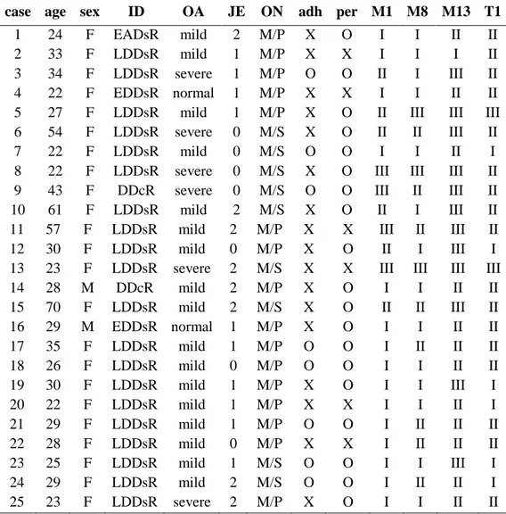
Table 1. Radiologic, operative findings and immunohistochemical expression of MMP-1, 8, 13 & TIMP-1 in TMJ disorder patients
case age sex ID OA JE ON adh per M1 M8 M13 T1
1 24 F EADsR mild 2 M/P X O I I II II 2 33 F LDDsR mild 1 M/P X X I I I II 3 34 F LDDsR severe 1 M/P O O II I III II 4 22 F EDDsR normal 1 M/P X X I I II II 5 27 F LDDsR mild 1 M/P X O II III III III 6 54 F LDDsR severe 0 M/S X O II II III II 7 22 F LDDsR mild 0 M/S O O I I II I 8 22 F LDDsR severe 0 M/S X O III III III II 9 43 F DDcR severe 0 M/S O O III II III II 10 61 F LDDsR mild 2 M/S X O II I III II 11 57 F LDDsR mild 2 M/P X X III II III II 12 30 F LDDsR mild 0 M/P X O II I III I 13 23 F LDDsR severe 2 M/S X X III III III III 14 28 M DDcR mild 2 M/P X O I I II II 15 70 F LDDsR mild 2 M/S X O II II III II 16 29 M EDDsR normal 1 M/P X O I I II II 17 35 F LDDsR mild 1 M/P O O I II II II 18 26 F LDDsR mild 0 M/P O O I I II II 19 30 F LDDsR mild 1 M/P X O I I III I 20 22 F LDDsR mild 1 M/P X X I I II I 21 29 F LDDsR mild 1 M/P O O I II II II 22 28 F LDDsR mild 0 M/P X X I II II II 23 25 F LDDsR mild 1 M/S O O I I III I 24 29 F LDDsR mild 2 M/S O O I II II I 25 23 F LDDsR severe 2 M/P X O I I II II ID: internal derangement of disc, OA: osteoarthritis, JE: joint effusion, DDcR: disc displacement with reduction, EDDsR: early stage of disc displacement without reduction, LDDsR: late stage of disc displacement without reduction, ON: operation name, M/P: meniscoplasty, M/S: meniscectomy, adh: adhesion, per: perforation, M1: MMP-1, M8: MMP-8, M13: MMP-13, T1: TIMP-1
12
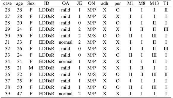
Table 1. Radiologic, operative findings and immunohistochemical expression of MMP-1, 8, 13 & TIMP-1 in TMJ disorder patients
case age Sex ID OA JE ON adh per M1 M8 M13 T1 26 36 F LDDsR mild 1 M/P X O I I I II 27 38 F LDDsR mild 1 M/P X X I I I I 28 20 F LDDsR mild 0 M/P X O I I II I 29 24 F LDDsR mild 2 M/P X X I II II III 30 56 F LDDsR mild 2 M/S O O II I III I 31 33 F EDDsR normal 2 M/P X X I I II I 32 26 F LDDsR mild 0 M/P X X I II II III 33 24 F LDDsR mild 0 M/P X O II I III I 34 34 F EDDsR normal 1 M/P X X I I II I 35 21 M EDDsR mild 1 M/P X X I II I I 36 32 F LDDsR mild 0 M/S X O II II III II 37 25 F LDDsR mild 1 M/P X O I I I I 38 50 F LDDsR mild 1 M/P O O II I III I 39 47 F EDDsR normal 2 M/P X X I I I I ID: internal derangement of disc, OA: osteoarthritis, JE: joint effusion, DDcR: disc displacement with reduction, EDDsR: early stage of disc displacement without reduction, LDDsR: late stage of disc displacement without reduction, ON: operation name, M/P: meniscoplasty, M/S: meniscectomy, adh: adhesion, per: perforation, M1: MMP-1, M8: MMP-8, M13: MMP-13, T1: TIMP-1
1. The Result of Immunohistochemistry
All the experimental cases showed MMP-1, 8, 13 and TIMP-1 expression, but the range or cell type of expression was different. And the expression of each collagenase appeared at different sites in many cases and expression levels were also different (Fig. 1).
13
Fig.1 Immunohistochemistry. A1(x200) and A2(x400) show grade I expression,
with extremely small portions of retrodiscal tissue removed after meniscoplasty colorized. B1(x200) and B2(x400) show grade II expression, with higher numbers of positive tissue cells and clearer expression than grade I. C1(x200) and C2(x400) show grade III expression, with even expression in cytoplasm and broad staining.
A1
A2
C1
C2
14
2. Comparison with MRI findings.
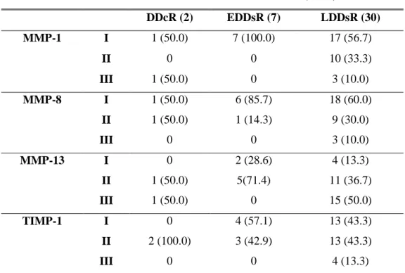
There was no case of normal position in research object. 2, 7, and 30 joints belonged to DDcR, EDDsR, and LDDsR, respectively, based on the status and status of joint disc. Regarding MMP-13 expression, 86.7 % of a group of LDDsR showed grade II or III, and the expression range of MMP-13 got wider as internal derangement became more degenerative. The concentration of MMPs expression was deeply tied to the status of diseases. Regarding MMP-13expression, the longer disc displacement lasted, the more enhanced expression was found (Table 2, 3).
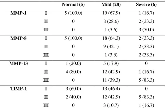
With regards to OA, 5, 28, and 6 joints belonged to normal, mild, and severe stage, respectively. In expression of MMP-13 of mild and severe stages, grade II and III had higher numbers. But other MMPs and TIMP-1 of mild stage dominantly showed grade I expression and MMP-1 expression showed that grade II and III were 5/6 in severe stage of OA. The severer OA became, the more expressions of MMP and TIMP appeared (Table 4, 5).
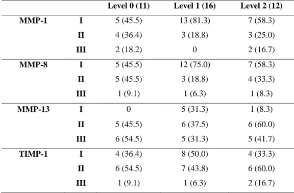
In joint effusion, 11 cases were level 0; 16, level 1; 12, level 2. There was no case of level 3 in research object. There was no significance in comparison of expression grade of MMPs or TIMP and joint effusion level (Table 6).
15
Table 2. Relation of TMJ internal derangement and MMPs & TIMP-1 expression ( ) % DDcR (2) EDDsR (7) LDDsR (30) MMP-1 I 1 (50.0) 7 (100.0) 17 (56.7) II 0 0 10 (33.3) III 1 (50.0) 0 3 (10.0) MMP-8 I 1 (50.0) 6 (85.7) 18 (60.0) II 1 (50.0) 1 (14.3) 9 (30.0) III 0 0 3 (10.0) MMP-13 I 0 2 (28.6) 4 (13.3) II 1 (50.0) 5(71.4) 11 (36.7) III 1 (50.0) 0 15 (50.0) TIMP-1 I 0 4 (57.1) 13 (43.3) II 2 (100.0) 3 (42.9) 13 (43.3) III 0 0 4 (13.3)
DDcR: disc displacement with reduction, EDDsR: early stage of disc displacement without reduction, LDDsR: late stage of disc displacement without reduction
Table 3. Statistical analysis of DDsR and MMP-13 expression
( ) %
EDDsR (7) LDDsR (30) p-value
MMP-13 I & II (22) 7 (100.0) 15 (50.0)
16
Table 4. Relation of osteoarthritis in TMJ and MMPs & TIMP-1 expression
( ) % Normal (5) Mild (28) Severe (6)
MMP-1 I 5 (100.0) 19 (67.9) 1 (16.7) II 0 8 (28.6) 2 (33.3) III 0 1 (3.6) 3 (50.0) MMP-8 I 5 (100.0) 18 (64.3) 2 (33.3) II 0 9 (32.1) 2 (33.3) III 0 1 (3.6) 2 (33.3) MMP-13 I 1 (20.0) 5 (17.9) 0 II 4 (80.0) 12 (42.9) 1 (16.7) III 0 11 (39.3) 5 (83.3) TIMP-1 I 3 (60.0) 13 (46.4) 0 II 2 (40.0) 12 (42.9) 5 (83.3) III 0 3 (10.7) 1 (16.7)
Table 5. Statistical analysis of osteoarthritis and MMP expression
( ) % normal & mild (33) severe (6) p-value
MMP-1 I & II 32 (97.0) 3 (50.0) III 1 (3.0) 3 (50.0) 0.008 MMP-8 I & II 32 (97.0) 4 (66.7) III) 1 (3.0) 2 (33.3) 0.056 MMP-13 I & II 22 (66.7) 1 (16.7) III 11 (33.3) 5 (83.3) 0.033
17
Table 6. Relation of joint effusion in TMJ and MMPs & TIMP-1 expression ( ) %
Level 0 (11) Level 1 (16) Level 2 (12)
MMP-1 I 5 (45.5) 13 (81.3) 7 (58.3) II 4 (36.4) 3 (18.8) 3 (25.0) III 2 (18.2) 0 2 (16.7) MMP-8 I 5 (45.5) 12 (75.0) 7 (58.3) II 5 (45.5) 3 (18.8) 4 (33.3) III 1 (9.1) 1 (6.3) 1 (8.3) MMP-13 I 0 5 (31.3) 1 (8.3) II 5 (45.5) 6 (37.5) 6 (60.0) III 6 (54.5) 5 (31.3) 5 (41.7) TIMP-1 I 4 (36.4) 8 (50.0) 4 (33.3) II 6 (54.5) 7 (43.8) 6 (60.0) III 1 (9.1) 1 (6.3) 2 (16.7)
3. Comparison with operative findings.
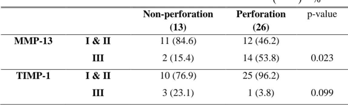
13 cases had no adhesion and perforation; 17, only perforation; 10, both adhesion and perforation. There was no case of only adhesion in research object. Groups of perforation or/and adhesion were grade II or III as for expression of MMP-13. MMP-13 expression increased when perforation occurred, which resulted in statistical significance (Table 7, 8).
18
Table 7. Relation of operation findings and MMPs & TIMP-1 expression ( ) %
Normal (13)
perforation (16)
Adhesion & perforation (10) MMP-1 I 11 (84.6) 8 (50.0) 6 (60.0) II 0 7 (43.8) 3 (30.0) III 2 (15.4) 1 (6.3) 1 (10.0) MMP-8 I 7 (53.8) 11 (68.8) 7 (70.0) II 5 (38.5) 3 (18.8) 3 (30.0) III 1 (7.7) 2 (12.5) 0 MMP-13 I 4 (30.8) 2 (12.5) 0 II 7 (53.8) 5 (31.3) 5 (50.0) III 2 (15.4) 9 (56.3) 5 (50.0) TIMP-1 I 6 (46.2) 5 (31.3) 5 (50.0) II 4 (30.8) 10 (62.5) 5 (50.0) III 3 (23.1) 1 (6.3) 0
Table 8. Statistical analysis of operation findings and MMP-13 & TIMP-1 expression ( ) % Non-perforation (13) Perforation (26) p-value MMP-13 I & II 11 (84.6) 12 (46.2) III 2 (15.4) 14 (53.8) 0.023 TIMP-1 I & II 10 (76.9) 25 (96.2) III 3 (23.1) 1 (3.8) 0.099
19
IV. Discussion
The combination of extracellular matrix, including collagen, relieves pressure and tensile force put on joints, however, when these forces start to hurt articular cells, various kinds of degenerative enzymes are secreted, degrading connective tissues. In this light, studies on the articular synovial fluid as a tool to detect TMJ disorders have been actively conducted on the assumption that damaged extracellular matrix and enzymes that control it are released into the synovial fluid. Some studies have suggested the involvement of MMPs in TMJ disease, as demonstrated by increased MMP-13 levels in synovial fluid from patients with ID (Srinivas et al., 2001). Since most human studies have been carried out in synovial fluid, the site of production of the enzyme has never been investigated in degenerated disc tissue. So this study aiming to examine more directly the retrodiscal tissue removed during the surgery observed collagenases and TIMP-1 immunoreactive patterns, which were classified into grade I, II, and III. This study also evaluated their association with ID, OA, JE, and operation findings-adhesion and perforation- in the retrodiscal tissue of TMJ.
MMP-1 (collagenase-1) is produced and immediately secreted from a variety of mesenchymal cells in response to specific inducers. MMP-8
20
(collagenase-2), in contrast, is primarily produced by neutrophils during their maturation in bone marrow, and it is then stored until the cells are stimulated to degranulate (Dioszegi et al., 1995; Jeffrey, 1998). MMP-13 (collagenase-3) is found in bone, normal and pathologic cartilage, and various epithelial cancers, and it is the predominant collagenase in mice and rat, with collagenase-1 being undetectable in these species (Jeffrey, 1998; Freije et al., 1994, Reboul et al., 1996; Mitchell et al., 1996). Although each mammalian collagenase can degrade all of the fibrillar collagens, the preferred substrates for MMP- 1, 8 and 13 are collagens type III, I and II, respectively (Knauper
et al., 1996; Hasty et al., 1987; Welgus et al., 1981). Gepstein and his
colleagues (2002) reported it has been established that although individual enzymes have similar substrate specificities, their patterns of expression are often distinct and the expression patterns are the characteristic of a certain tissue and cell type. In this study, all the experimental cases showed MMP-1, 8, 13 and TIMP-1 expression, but the range or cell type of expression was different. MMP-13 called ‘osteoblast collagenase’ showed high expression grade generally. The expression range of MMP-13 got wider as internal derangement or OA became more degenerative. This could verify an importance of collagen type II in the retrodiscal tissue as well as articular cartilage of TMJ. In the aspect of specificity of collagenases, it was observed
21
easily that MMP-1 called ‘fibroblast collagenase’ was expressed on fibroblast and MMP-8 called ‘neutrophil collagenase’ was expressed on neutrophil. And the expression of each collagenase appeared at different sites in many cases and expression levels were also different.
Some of the conflicting results could partially be explained by differences in the sensitivity of the assays used for determining MMPs and TIMPs such as immunohistochemistry, in situ hybridization, zymography, western blots and northern blots. Some of the studies examined MMPs in isolated chondrocytes, synovial fluids, cultured explants, or intact cartilages, which also can contribute to the different results (Gepstein et al., 2003). A study done by Huh and his colleagues (2003b) found that seven out of the eight joints with MMP-1 mRNA had OA, and the study also found that MMP-1 removed from the retrodiscal tissue can play a part in the destruction of cartilage and bone of mandibular condyle and fossa. Another study showed that MMP-1 is active in synovial fluid of patients with pain, in most cases, and that a high concentration of MMP-1 can increase joint inflammation (Ishimaru et al., 2000). As mentioned above, the role of MMP-1 is not yet clear. But it can be induced that MMP-1 is involved in disc displacement or other related diseases as collagen is a major component of the retrodiscal tissue. In this study, 5/6 cases out of the severe OA group expressed grade II
22
and III for MMP-1 expression. It indicates MMP-1 is associated with destruction of bone and cartilage around disc of TMJ even in the small-sized experimental group. But the association of MMP-8 with ID, OA, and surgical findings is not clearly shown.
Among the MMPs, MMP-13 appears to play a key role in joint connective tissue remodeling. MMP-13 can cleave type II collagen 10 times faster than MMP-1 (Otterness et al., 2000). MMP-13 tissue expression has also been demonstrated in articular cartilage during OA (Shlopov et al., 1997) and in the synovium of patients with rheumatoid arthritis (Lindy et al., 1997). Control disc cells were rarely immunopositive for MMP-13, although a few scattered fibroblast-like cells showed faint positive immunoreactions. And MMP-13 is upregulated in TMJ disc tissue from patients with IDs, MMP-13 immunolabelling increasing with degree of disc degeneration, and TMJ disc cells thus appear to play an active role in the synthesis of extracellular matrix-degrading proteinases (Leonardi et al., 2008). The expression range of MMP-13 in the retrodiscal tissue of TMJ got wider as internal derangement or OA became more degenerative in the present study. In particular, the severe OA group showed clear expression fitting into grade II or III, and this explains that MMP-13 as well as MMP-1 is related with OA of TMJ. In comparison of MMP-13 expression with the position and status
23
of articular disc, 86.7 % in the LDDsR group showed grade IIor III, which means that breakdown enzymes involved in articular disc degeneration become more activeas internal derangement progresses. Only an OA patient who went through meniscectomy due to severe adhesion and perforation of articular disc showed the case of grade IIIexpression with DDsR in MRI .
Marchetti and his colleagues’ study (1999) demonstrated that structural modifications of articular disc could be specific responses to changes in function of the TMJ and that variations in extrinsic stimuli may activate intrinsic factors such as MMPs that induce structural modifications in the discal tissue. The examination found that only the areas with myxoid change or hyalinization were stained or these areas resulted in much more considerable staining than others and that cell numbers increased in areas with myxoid change. Such differences appeared when the retrodiscal tissue deteriorated or responded to passed-on loading caused by aricular disc displacement, which suggests the association of MMPs.
In normal physiological processes, MMP activity is controlled at several levels. Loss of control of MMP activity appears to have serious consequences, and aberrations in MMP expression have been associated with several diseases. Though there is no general awareness of the expression of TIMP-1 in osteoarthrotic cartilage and synovium, it is thought that TIMP-1
24
correlates with the production of MMPs (Gomis-Ruth et al., 1997). It has been reported that there is a correlation between the concentration of TIMP-1 and MMP-1 (Naito et al., 1999), and the imbalance between them in the progression of OA results in the unrecoverable degradation of cartilage matrix. During OA, the production of TIMP-1 is correlated to the level of MMPs. Their imbalance in the progression of OA may lead to the destruction of cartilage (Wang et al., 2008). Whereas overexpression of collagenage-1, coupled with decreased production of TIMP-1, may impair healing, insufficient proteinase activity may lead to the accumulation of wound-associated tissue and delays in wound closure. Thus, the properly regulated, site-specific expression of collagenase, as well as other MMPs, may be needed to promote efficient wound repair (Parks WC et al., 1998). This study showed TIMP-1 expression was weaker than MMPs expression, which might be caused by the fact that experimental groups were patients with TMJ disorder. Four cases of grade III expression of TIMP-1 belonged to LDDsR and mild or severe stage of OA. TIMP-1 expression seemed to affect internal derangement when compared to osteoarthritis, but there is no significance. Nevertheless this study assumes that TIMP-1 can be active if MMP expression pattern fits into grade II or III without any change or destruction in joint structures.
25
The imbalance between MMP-1 and TIMP-1 also plays an important role in the degradation of extracellular matrix (Sawatsubashi et al., 1988). This imbalance and reduced expression of TIMP-1, which suppress the degradation of extracellular matrix, can explain destructive patterns against bone or cartilage. Follow up surveys on OA-related symptoms among TMJ disorder, as well as interaction of MMP-1, 13 and TIMP-1, would be required for clinical applications.
26
V. Conclusion
This study was aimed to observe how collagenases (MMP-1, 8, 13) and TIMP-1 were expressed in immunohistochemistry of retrodiscal tissue sections in order to understand how their expression pattern can be associated with internal derangement, osteoarthritis, joint effusion, and operation findings -adhesion and perforation- among TMJ disorder patients.
Among collagenases, MMP-13 plays a critical role in degrading of osteoarthritis as it is the predominant collagenase in collagen type II and it can also worsen internal derangement as breakdown enzyme of collagen of retrodiscal tissue in TMJ. MMP-1 is related to the degeneration of osteoarthritis. MMP-8 does not characterize specificity with regard to TMJ disorder. TIMP-1 expression seemed to affect internal derangement when compared to the expression in osteoarthritis, but there is no significance. This study could not explain the causal relation of imbalance between MMP and TIMP, but the activity of TIMP shows differences as degeneration of TMJ disorders indicates that TIMP can serve as the critieria of prevention of progressive disease because it is controlling collagenases.
27
References
Ali AM, Sharawy MM, O'dell NL, Al-Behery G (1993). Morphological alterations in the elastic fibers of the rabbit craniomandibular joint following experimentally induced anterior disk displacement. Acta Anat 147: 159-167. Breckon JJW, Hembry RM, Reynolds JJ, Meikle MC (1996). Identification of matrix metalloproteinases and their inhibitor in the articular disc of the craniotemporomandibular joint of the rabbbit. Archs oral Biol 4: 315-322. Dioszegi M, Cannon P, Van Wart HE (1995). Vertebraet collagenases. Methods Enzymol 248: 413-431.
Edwards JCW, Wilkinson LS, Soothill P, Hembry RM, Murphy G, Reynolds JJ (1996). Matrix metalloproteinases in the formation of human synovial joint cavities. J Anat 188: 355-360.
Freije JM, Diez-Itza I, Balbin M, Sanchez LM, Blasco R, Tolivia J, Lopez-Otin C (1994). Molecular cloning and expression of collagenase-3, a novel human matrix metalloproteinase produced by breast carcinoma. J Biol Chem 269:16766-16773.
Gaye C, Oliver F, Charlotte B, Tim EC, Barry B (2001). Early joint erosion and serum levels of matrix metalloproteinase-1, matrix metalloproteinase-3,
28
and tissue inhibitor of metalloproteinase-1 in rheumatoid arthritis. Arthritis Rheum 44: 2263-2274.
Gepstein A, Shapiro S, Arbel G, Lahat N, Livne E (2002). Expression of matrix metalloproteinases in articular cartilage of temporomandibular and knee joint of mice during growth, maturation, and aging. Arthritis&rheumatism 46: 3240-3250.
Gepstein A, Arbel G, Blumenfeld I, Peled M, Livne E (2003). Association of metalloproteinases, tissue inhibitors of matrix metalloproteinases, and
proteoglycans with development, aging, and osteoarthritis processes in
mouse temporomandibular joint. Histochem Cell Biol 120:23-32.
Gomis-Ruth FX, Maskos K, Betz M (1997). Mechanism of inhibition of the human matrix metalloproteinase stromelysin-1 by TIMP-1. Nature 389:77-81. Hasty KA, Jeffrey JJ, Hibbs MS, Welgus HG (1987). The collagen substrate specificity og human neutrophil collagenases. J Biol Chem 262:10048-10052. Helmy ES, Bays RA, Sharawy MM (1989). Histopathological study of the human TMJ perforated disk with emphasis on synovial membrane response. J Oral Maxillofac Surg 47: 1048-1052.
Huh JK, Kim HG, Ko JY (2003a). Magnetic resonance imaging of temporomandibular joint synovial fluid collection and disk morphology. Oral Surg Oral Med Oral Pathol Oral Radiol Endod 95:665-71.
29
Huh JK, Park KH, Choi MA, Kim HG (2003b). Expression of matrix metalloproteianase-1 and -2 in retrodiscal tissue of the temporomandibular joint. J Kor Oral Maxillofac Surg 29:212-218.
Iannaroon A, Wallon UM, Overall CM, Diewert VM (1996). Expression of 72-kDa gelatinase (Matrix metalloproteinase-2) in the developing mouse craniofacial complex. Arch Oral Biol 41: 1109-1119.
Ijima Y, Kobayashi M, Kubota E (2001). Role of interleukin-1 in induction of matrix metalloproteinases synthesized by rat temporomandibular joint chondrocytes and disc cells. Eur J Oral Sci 109(1):50-9.
Imai K, Ohta S, Matsumoto T (1997). Expression of membrane-type 1 matrix metalloproteinase and activation of progelatinase A in human osteoarthritic cartilage. Am J Pathol 151: 245-56.
Imai S, Konttinen YT, Jumppanen M, Lindy O, Ceponis A, Kemppinen P, Sorsa T, Santavirta S, Xu JW, Lopéz-Otín C (1998). High levels of expression of collagenase-3 (MMP-13) in pathological condition associated with a foreign-body reaction. J Bone Joint Surg Br 80: 701-10.
Ishibarshi H, Takenoshita Y, Ishibarshi K, Oka M (1996). Expression of extracellualr matrix in human mandibular condyle. Oral Surg Oral Med Oral Pathol Oral Radiol Endod 81:402-412.
30
inhibitor of metalloproteinase in serum and lavage synovial fluid of patients
with temporomandibular joint disorders. Br J Oral Maxillofac Surg 38:354-9.
Iwase T, Hasegawa Y, Ishiguro N, Ito T, Iwasada S, Kitamura S, Iwata H (1998). Synovial fluid cartilage metabolism marker concentrations in osteonecrosis of the femoral head compared with osteoarthrosis of the hip. J Rheumatol 25(3):527-31.
Jeffrey JJ (1998). Interstitial collagenases, Matrix Metalloproteinases. In: Parks WC and Mecham RP, eds. Academic Press, San Diego: pp. 15-42. Kanyama M, Kuboki T, Kojima S, Fujisawa T, Hattori T, Takigawa M, Yamashita A (2000). Matrix metalloproteinases and tissue inhibitors of metalloproteinases in synovial fluids of patients with temporomandibular joint osteoarthritis. J Orofac Pain 14: 20-30.
Kapila S, Lee C, Richards DW (1995). Characterization and identification of proteinases and proteinase inhibitors synthesized by temporomandibular joint disc cells. J Dent Res 74: 1328-1336.
Knauper V, Lopes-Otin C, Smith B, Knight G, Murphy G (1996). Biochemical characterization of human collagenase-3. J Biol Chem 271: 1544-1550.
Kubota E, Imamura H, Kubota T, Shibata T, Murakami K (1997). Interleukin 1 and stromelysin (MMP-3) activity of synovial fluid as possible markers of
31
osteoarthritis in the temporomandibular joint. J Oral Maxillofac Surg 55: 20-27.
Kubota E, Kubota K, Matsumoto J, Shibata T, Murakami K (1998a). Synovial fluid cytokines and proteinases as marker of temporomandibular joint disease. J Oral Maxillofac Surg 56: 192-8.
Kubota T, Kubota E, Matsumoto A, Kawai Y, Saito H, Mikuni-Takagaki Y, et al (1998b). Identification of matrix metalloproteinases (MMPs) in synovial fluid from patients with temporomandibular disorder. Eur J Oral Sci 106:992-998.
Leonardi R, Loretob C, Barbatoc E, Caltabianob R, Lombardoa C, Musumecib G, Muziod LL (2008). MMP-13 (collagenase-3) localization in human temporomandibular joint discs with internal derangement. Acta Histocem 110(4):314-8.
Lindy O, Konttinen YT, Sorsa T, Ding Y, Santavirta S, Ceponis A, López-Otín C (1997). Matrix metalloproteinase 13 (collagenase 3) in human rheumatoid synovium. Arthritis Rheum 40(8):1391-9.
Marchetti C, Piacentini C, Farina A, Bernasconi G, Calligaro A (1995). A microscopic and immunocytochemical study of structural changes in dysfunctional human temporomandibular joint discs. Arch Oral Biol 40: 549-557.
32
Marchetti C, Bernasconi G, Reguzzoni M, Farina A (1997). The articular disc surface in different functional conditions of the human temporomandibular joint. J Oral Pathol Med 26: 278-282.
Marchetti C, Cornaglia I, Casasco G, Bernasconi U, Baciliero U, Stetler-Stevenson WG (1999). Immunolocalization of gelatinase-A (matrix metalloproteinase-2) in damaged human temporomandibular joint discs. Arch Oral Biol 44: 297-304.
McDonnell S, Morgan M, Lynch C (1999). Role of matrix metalloproteinases in normal and disease process. Biochem Soc Trans 27: 234-40.
Mitchell PG, Magna HA, Reeves LM, Lopresti-Morrow LL, Yocum SA, R osner PJ, Geoghegan KF, Hambor JE (1996). Cloning, expression, and type II collagenolytic activity of matrix metalloproteinase-13 from human osteoarthritic cartilage. J Clin Inves 97:761-768.
Mohtai M, Smith RL, Schurman DJ, Tsuji Y, Torti FM, Hutchinson NI, Stetler-Stevenson WG, Goldberg GI (1993). Expression of 92-kD type IV collagenase/gelatinase (gelatinase B) in osteoarthritic cartilage and its induction in normal human articular cartilage by interleukin 1. J Clin Invest 92(1):179-85.
33
metalloproteinases (MMPs) and tissue inhibitor of metalloproteinase-1 (TIMP-1) in patients with knee osteoarthritis: comparison with generalized osteoarthritis. Rheumatology (Oxford) 38:510-5.
Otterness IG, Bliven ML, Eskra JD, te Koppele JM, Stukenbrok HA, Milici AJ (2000). Cartilage damage after intraarticular exposure to collagenase 3. Osteoarthritis Cartilage 8(5):366-73.
Parks WC, Sudbeck BD, Doyle GR, Saariahlo-Kere UK (1998). Matrix Metalloproteinases in Tissue Repair, Matrix Metalloproteinases. In: Parks WC and Mecham RP, eds. Academic Press, San Diego pp 263-290.
Reboul P, Pelletier JP, Tardif G, Cloutier JM, Martel-Pelletier J (1996). The new collagenase, collagenase-3, is expressed and synthesized by human chondrocytes but not by synoviocytes. A role in osteoarthritis. J Clin Inves 97:2011-2019.
Sawatsubashi M, Mizokami H, Tokunaga O, Shin T (1998). Expression of MMP-1, TIMP-1, and type Ⅰcollagen in laryngeal carcinoma. Mod Pathol 11:878-885.
Sharawy MM, Larke V, Helmy E (1987). Ultrastructural alterations in monkey TMJ following disc perforation. J Dent Res 66: 119.
34
Shlopov BV, Lie WR, Mainardi CL, Cole AA, Chubinskaya S, Hasty KA (1997). Osteoarthritic lesions: involvement of three different collagenases. Arthritis Rheum 40(11):2065-74.
Srinivas R, Sorsa T, Tjäderhane L, Niemi E, Raustia A, Pernu H, Teronen O, Salo T (2001). Matrix metalloproteinases in mild and severe temporomandibular joint internal derangement synovial fluid. Oral Surg Oral Med Oral Pathol Oral Radiol Endod 91(5):517-25.
Tanaka A, Kumagai S, Kawashiri S, Takatsuka S, Nakagawa K,Yamamoto E
(2001). Expression of matrix metalloproteinase-2 and -9 in synovial fluid of
the temporomandibular joint accompanied by anterior disk displacement. J
Oral Pathol Med 230:59-64.
Tiilikainen P, Pirttiniemi P, Kainulainen T, Pernu H, Raustia A (2005). MMP-3 and -8 expression is found in the condylar surface of temporomandibular joints with internal derangement. J Oral Pathol Med 34: 39-45.
Vaatainen U, Lohmander LS, Thoner E (1998). Marker of cartilage and synovial metabolism in joint fluid and serum of patients with chondromalacia of patella. Osteoarthritis Cartilage 6: 115-24.
Wang YL, Li XJ, Qin RF, Lei DL, Liu YP, Wu GY, Zhang YJ, Yan-Jin, Wang DZ, Hu KJ (2008). Matrix metalloproteinase and its inhibitor in
35
temporomandibular joint osteoarthrosis after indirect trauma in young goats. British Journal of Oral and Maxillofacial Surgery 46:192-197.
Welgus HG, Jeffrey JJ, Elisen AZ (1981). The collagen substrate specificity of himan skin fibroblast collagenase. J Biol Chem 256:9511-9515.
Wilkes C (1978). Arthrography of the temporomandibular joint in patients with the pain-dysfunction syndrome. Minn Med 61:645-652.
Yan C, Boyd DD (2007). Regulation of matrix metalloproteinase gene expression. J Cell Physiol 211(1):19-26.
36 국문요약
측두하악 관절 장애 환자에서 관절 원판 후조직의 collagenases (MMP-1, 8, 13) 및 tissue inhibitor of metalloproteinase-1 단백
발현 < 지도교수 김형곤 > 연세대학교 대학원 치의학과 고 원 경 세포외 기질과 세포 표면 분자를 퇴화시키는 기질 금속단백분해효소 (matrix metalloproteinase; MMP)는 금속단백분해효소의 조직 억제제 (tissue inhibitor of metalloproteinases; TIMP)와 함께 작용하여 태아 발육, 조직 형성, 창상 치유, 관절염, 종양 등의 생리적, 병리적 단계에서 중요한 역할을 한다. 측두하악 관절 장애는 주로 외상이나 저작계의 과도한 부하 등과 관련되며 그로 인한 관절 원판과 주변 조직의 변형에 의해 관절 잡음, 통증 및 개구장애 등의 병변으로 진행된다. 관절 원판 및 후조직은 주로 콜라겐(1 형과 2 형)으로 구성되는데 분해 효소의 활성화에 의해 결합조직의 구조적인 손상이 유발되면 관절 기능상의 장애를 초래하게 되는 것이다. 이에 본 연구는 기질 금속단백분해효소 중에서 관절 원판 후조직을 구성하는 콜라겐에 관여하는 분해효소인 collagenase 1, 2, 3 (MMP -1, 8, 13) 및 TIMP-1 의 발현 여부를 살펴보고, 발현 정도와
37 자기공명영상 사진, 수술 소견을 비교하여 측두하악 관절 장애에 있어서 이들 효소의 작용에 대해 알아보고자 하였다. 측두하악 관절 장애로 수술 받은 39 명 환자를 대상으로 수술시 제거된 관절 원판 후조직에 대해 면역조직화학 염색을 시행하여 MMP-1, 8, 13 과 TIMP-1 의 발현을 관찰하였다. MMP-1, 8, 13 과 TIMP-1 은 모든 실험군에서 발현을 보였으나 발색되는 세포나 부위에는 차이가 있었으며, MMP-13 (collagenase-3)은 다른 콜라겐분해효소와 비교하여 높은 발현 정도를 나타내었다. MMP-13 은 제 2 형 콜라겐을 분해에 우선적으로 작용하는 효소인 만큼 측두하악 관절의 관절 연골의 퇴화와 관련되는 골관절증의 악화에 큰 비중을 차지하며, 관절원판 후조직의 콜라겐 분해에도 중요한 역할을 함으로서 악관절 내장증의 심화에도 영향을 미친다. MMP-1 은 골관절증의 진행과 관련되며, MMP-8 은 측두하악 관절장애와 관련하여 특이성을 나타내지 않았다. TIMP-1 는 골관절증보다 악관절 내장증에서의 역할이 상승되어 보이나 통계학적인 유의성을 보이지는 않았다. 본 연구에서 MMP-TIMP 의 불균형에 대한 인과관계는 설명되지 못했지만, 측두하악 관절장애의 진행정도에 따라 차이를 보이는 TIMP 의 활성도는 콜라겐분해효소를 억제함으로서 질병의 진행을 예방할 수 있는 기준이 될 수 있을 것으로 보인다. 핵심되는 말: 콜라겐 분해효소, 기질 금속단백분해효소, 금속단백분해효소 조직억제제, 측두하악 관절 장애, 면역조직화학 염색