Feasibility of Sonography for
Intra-articular Injections in the Knee
Through a Medial Patellar Portal
Sang Hee Im, MD, Sang Chul Lee, MD, PhD, Yong Bum Park, MD, Sung-Rae Cho, MD, PhD, Jong Chan Kim, MD
Objective. The purpose of this study was to investigate the feasibility of using real-time high- resolution sonography to guide an injection needle into the intra-articular space within the knee. Methods. Eighty-nine patients with radiographically confirmed knee osteoarthritis (Kellgren-Lawrence grade 2 or 3) without an effusion were included. After sonographically guided or blind injection of hyaluronic acid (HA) and contrast dye through a medial patellar portal (MPP) into the knee joint, a radiographic image was made to ascertain whether the injected material had reached the intra-articular space. Result. Sonographically guided injections of HA into the knee joint had a significantly greater accuracy rate (95.6%) than blind injections (77.3%; P = .01). Conclusions. Intra-articular injections via an MPP using sonographic guidance may raise the accuracy rate in knee joint injections. Key words: injection; knee; sonography.
Received March 3, 2009, from the Department of Physical Medicine and Rehabilitation, Jeju National University Hospital, School of Medicine, Jeju National University, Jeju, Korea (S.H.I.); Department of Physical Medicine and Rehabilitation, Myongji Hospital, Kwandong University College of Medicine, Koyang, Kyunggi, Korea (S.C.L., J.C.K.); Department of Physical Medicine and Rehabilitation, Sanggye Paik Hospital, Inje University College of Medicine, Seoul, Korea (Y.B.P.); and Department of Rehabilitation Medicine and Research Institute of Rehabilitation Medicine, Yonsei University College of Medicine, Seoul, Korea (S.-R.C.). Revision requested March 30, 2009. Revised manuscript accepted for publication June 11, 2009.
Address correspondence to Sang Chul Lee, MD, PhD, Department of Physical Medicine and Rehabilitation, Myongji Hospital, Kwandong University College of Medicine, 697-24 Hwajung-dong, Dokyang-ku, Koyang, Kyunggi 412-270, Korea.
E-mail: [email protected] Abbreviations
HA, hyaluronic acid; MPP, medial patellar portal; MRI, magnetic resonance imaging; OA, osteoarthritis
he use of an intra-articular injection of hyaluron-ic acid (HA) has recently become widely accepted as a therapy for pain accompanying osteoarthritis (OA) of the knee.1In OA, there is a reduction in the elastoviscosity of the synovial fluid secondary to a decrease in the molecular weight and concentration of HA. Viscosupplementation is a therapeutic technique that addresses the decrease in synovial viscosity with the injec-tion of exogenous high-molecular-weight hyaluronan molecules.2 However, incorrect placement of an extra-articular HA injection causes discomfort to the patient and a reduced effect of HA.3
A number of factors emphasize the importance of accu-rate intra-articular injections of HA. First, a small volume (2–3 mL) of HA may not be expelled as easily as a larger volume of HA, which may dissipate into the joint through the soft tissues secondary to the injection pressure of the syringe.4Second, local corticosteroids may have an effect on nonspecific knee pain, even if administered periartic-ularly.5However, HA would not be expected to have any effect when applied to the tissue surrounding the joint.6 Third, the substantially higher cost of HA injections and the requirement for multiple injections increase the desirability that the preparation is delivered intra-articu-larly for a maximum effect.4Finally, incorrect placement of a soft tissue injection causes more discomfort to the patient during and after the procedure.
Needle placement is easily confirmed when an effusion is present. During knee joint aspiration for an effusion, the return of synovial fluid con-firms the intra-articular placement of the needle. On the other hand, accurate intra-articular placement of HA is difficult without guidance by real-time fluoroscopic imaging or a sonographic method for ‘‘dry’’ knee disease.7There are only a few studies that have evaluated the accuracy of needle placement into the intra-articular space of the knee joint in the absence of an effusion.6,8,9 The use of fluoroscopy for intra-articular injections in the knee has considerable impor-tance because this technique helps minimize the chance of injury associated with the injection and elevates the accuracy of the injection.6,9However, contrast media are costly and may not always be mixed with other substances for injection,10and repeated injections under fluoroscopy should be avoided because of the accumulated radiation.
Although a previous study showed the feasibili-ty of using sonography to guide intra-articular injections in the knee,10a need exists to further show its advantages, such as the accuracy rate and operability. Driven by a need for more accu-rate, safer, and less costly methods for articular injections, this study was undertaken to investigate the feasibility of using real-time high-resolution sonography to guide an injection nee-dle into the intra-articular space within the knee.
Materials and Methods
Eighty-nine patients with radiographically con-firmed knee OA (Kellgren-Lawrence grade 2 or 3) without an effusion who were symptomatic for at least 6 months and reported pain on most days for the previous 3 months were considered for enrollment in this study. To be eligible, the patients could not have inflammatory joint dis-ease, chondrocalcinosis (evidence from radio-graphs or synovial fluid analysis), or an infection in or around the study knee, and they could not be receiving anticoagulant therapy and could not have had viscosupplement treatment within the past 6 months. Only dry knees with no clinically detectable effusion and patients in whom the suprapatellar bursa was not discernible on sonography by the method reported in a previ-ous study10were included. Patients were
individ-ually randomized into sonographically guided and blind injection groups by a table of random numbers.
Approval from the Institutional Review Board was obtained at the outset of the study. The nature of the study was explained to the patients before the procedures, and informed consent was obtained in each case. The clinical efficacy of the intra-articular knee injection procedure was not the aim of this study; therefore, no attempt was made to correlate the treatment outcomes with any of the variables defined in the study. All of the patients underwent a clinical evaluation, a radiographic imaging study, and an intra-articular injection in the knee.
Preliminary Magnetic Resonance Imaging Study
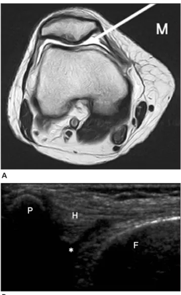
To estimate the optimal medial patellar portal (MPP) for injection, we evaluated the size of the fat pad and joint cavity using magnetic reso-nance imaging (MRI) of the medial part of the patellofemoral joints of 5 patients. On the basis of these preliminary data, we found potential MPPs for injection with small fat pads, which allow eas-ier access to the joint cavity during sonographi-cally guided injections (Figure 1).
Sonographic Examination
The procedure was performed by the sonogra-phy system operator, 1 sonogra-physiatrist, and 1 assis-tant. The patients in the sonographically guided injection group were placed in the supine posi-tion with a fully extended knee on the examina-tion table. We used an ultrasound machine (L12-5/38 mm, HDI 5000; Philips Healthcare, Bothell, WA) with a 7- to 12-MHz linear array probe. Before proceeding with an injection pro-cedure, an accurate sonographic examination of the medial side of the knee was repeated to iden-tify the most adequate MPP while shifting the probe up and down between the articular sur-faces of the patellofemoral joint near the mid-point of the patella. An MPP was selected in which the intra-articular space was visible with less of a fat pad.
For injections into the joint cavity, an adequate needle length is an essential requirement. The distance was measured from the surface to the target area by a built-in manual measurement of the sonographic image.
Injection Procedure
The treatment involved 3 intra-articular injections of high-molecular-weight HA (2 mL of 1% HA; molec-ular weight, 940–1020 kDa) into the affected knee at weekly intervals, according to the manufacturer’s recommended protocol (Hyalforte; Shin Poong, PhD, Kyunggi, Korea). Only the first injection of each case and its result were analyzed for this study.
A wide area of knee skin was prepared and draped. In the sonographically guided injection group, injections were performed with a 1.5-in (3.8-cm) or 2-in (5.1-cm) 21-gauge needle after estimation of the needle length by sonography. The probe was placed in close proximity to the puncture site, and the needle was advanced under direct sonographic guidance. The needle was aligned with the small side of the probe during insertion. Combined color Doppler imaging allowed a more precise assessment of the needle tip with detection of the flow of the solution as a bright color.
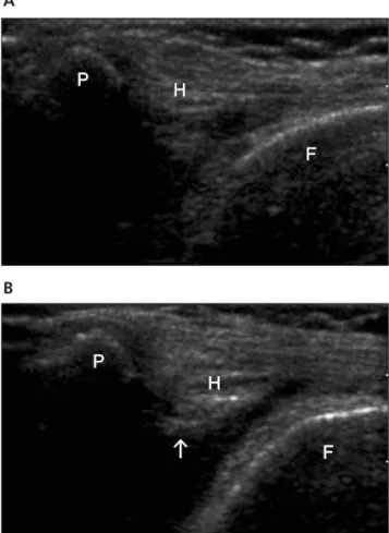
In the blind injection group, injections were performed with a 1.5-in (3.8-cm) 21-gauge nee-dle at the physician’s preference. The patellar border was outlined with a marking pen, and the injection was done at the point where the mid horizontal line of the patella met the medial bor-der of the patella (Figure 2).11
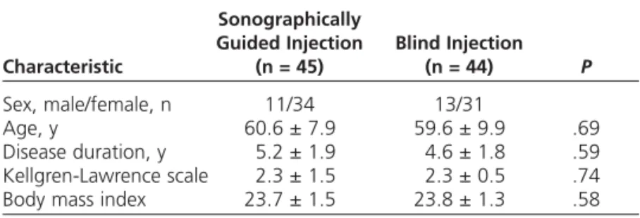
A 3-way stopcock was used for the study injec-tions, which allowed the passage of both the con-trast dye solution and the HA through the same needle with only a single needle placement. When the needle was positioned in the target area, 2 mL of HA was injected (Figure 3). After injection of HA, the stopcock was opened, and a small amount (0.5 mL) of contrast dye solution in the other syringe was injected to confirm the position of the needle tip by radiography.
Assessment
To assess the accuracy rate of sonographically guided intra-articular injections through the MPP, a radiographic imaging study was under-taken. A postinjection radiographic evaluation was performed 10 to 15 minutes after the injec-tion procedure. From the imaging results, we concluded whether the injected material had reached the intra-articular space. Any contrast material appearing in the extra-articular space was defined as a “failed” case.
A
B
Figure 1. Axial T2-weighted MRI of the knee joint (A) and cor-relative transverse sonogram (B). Asterisk indicates the targeted area; F, femur; H, Hoffa fat pad; M, medial side; and P, patella.
Figure 2. All landmarks were outlined with a marking pen, and blind MPP injections were performed with the lower limb extended on the examination table. The needle was advanced transversely between the articular surfaces of the patellofemoral joint at the midpoint of the patella.
Statistics
Statistical analysis was performed with SPSS version 13.0 software for Windows (SPSS Inc, Chicago, IL). An independent t test and Mann-Whitney U test were used to compare the demo-graphic and clinical characteristics of the patients. The χ2test was used for the comparison of success rates of the injection procedures.
P < .05 was considered statistically significant.
Results
Patient Characteristics
After being randomly assigned to 1 of 2 groups, 45 patients were included in the sonographical-ly guided injection group, and 44 were included in the blind injection group. The demographic and clinical characteristics of the patients in the 2 groups were comparable (Table 1).
Success Rate of the Injection Procedure
The sonographically guided injection group had a significantly higher success rate of injections compared with the blind injection group (P = .01). Forty-three of 45 knees (95.6%) in the sonographically guided injection group had successful injections into the joint cavity on radiography, which revealed the presence of con-trast dye in the joint cavity. The 2 failed cases had some contrast material in the extra-articular space. Thirty-four of 44 knees (77.3%) in the blind injection group had successful injections into the joint cavity.
Discussion
Jones et al8reported that 66% of knee joint blind injections were intra-articular, and almost one-third were extra-articular; the response to steroid therapy was similar regardless of whether the injection was intra- or extra-articular. However, to achieve a maximal potential therapeutic ben-efit, HA-based preparations should be delivered directly into the intra-articular space and not into the fat pad or the subsynovial tissue layers.3 Because HA has a high viscosity, it is difficult for the clinician to perceive whether the dose of HA is passing into the soft tissue or the joint space because of its flow resistance in the needle.12 Therefore, there might be a greater possibility of extra-articular injections of HA than other drugs with less viscosity.
A recent study suggested that sonography could be used as an adjuvant tool for articular injections in the knee instead of fluo-roscopy.10With sonographic guidance, HA was injected into the intra-articular space via the suprapatellar bursa. When the bursa was not dis-cernible on sonography, air was injected into the lateral point of the knee just under the patella, and the visible air on sonography was regarded as a successful injection into the suprapatellar bursa. However, we cannot conclude that this was an injection procedure under sonographic guidance but rather a blind injection with confir-mation by sonography.
Although a number of clinical trials are being performed regarding the efficacy of intra-articular HA injections, there is little consensus in the lit-erature on the appropriate technique.4,7,8Esenyel B
Figure 3. Before (A) and after (B) a sonographically guided injection. The fat pad is elevated upward because of the inject-ed fluid. F indicates femur; H and arrow, Hoffa fat pad; and P, patella.
et al11evaluated the accuracy rate of intra-articu-lar blind knee injections using anteromedial, anterolateral, lateral midpatellar, and medial midpatellar portals in cadavers and reported that the accuracy obtained with the use of a medial midpatellar portal was significantly lower (accuracy rate, 56%) than other portals. However, this study did not explain the basis of the lower accuracy rate with the medial midpatellar portal approach. Furthermore, the injection procedure was undertaken only by surface landmarks.
Involvement of the lateral patellofemoral joint in OA is more common than the medial patellofemoral joint,13and many cases show later-al tilting of the patellofemorlater-al joint.14This sup-ports the idea that there is sufficient intra-articular space for an injection approach through an MPP. In addition, extension of the knee joint induces external rotation of the hip joint, which results in an easier approach for the medial side of the knee. The medial approach to the knee also has an advantage for patients with strokes or spinal cord injuries who have difficulty with internal rotation of the hip due to severe spasticity or cooperation problems. As the results of our pre-liminary MRI study showed, not only the lateral side but also the medial side of the knee had enough space for the injection of HA (Figure 1), and there was no structure that could be injured during injection except the fat pad on the al side of the knee. Therefore, we chose the medi-al approach through an MPP, which was shown to have a lower accuracy rate in a previous study,8 and expected that the approach through an MPP might affirm the feasibility of sonography for intra-articular injections in the knee.
With a sonographically guided knee injection, we avoided injection into the fat pad between the skin and joint space. In the cases with an incorrectly placed needle, we could correct the direction of the needle under sonographic guid-ance and minimize the chguid-ance of injury associ-ated with the injection. When the needle became less evident or when the needle tip was not detectable with increasing obliquity,15 color Doppler imaging was useful to monitor the accu-rate placement of the needle tip. In the practical procedure of injection of a mixture of HA and a small amount of saline or lidocaine, it was possi-ble to detect the needle tip with color Doppler
imaging. In addition, all vessels could be visual-ized easily by color Doppler imaging and were distinguishable from anechoic cavities.
This study was designed to clarify whether an injection from the medial aspect of the patellofemoral joint under sonographic guid-ance could raise the accuracy rate of this proce-dure over that of the blind technique, which had a low accuracy rate in previous reports. We achieved good results with a higher accuracy rate than the blind method; however, we still do not know which approach is most accurate. Therefore, further studies comparing the accura-cy rates of different approaches under sono-graphic guidance are needed to find the optimal portal for intra-articular injections in the knee.
In conclusion, intra-articular injections via an MPP using sonographic guidance resulted in good intra-articular delivery with a 95.6% accu-racy rate and a lower incidence of soft tissue infil-tration. This approach has advantages, such as convenience and lack of radiation hazards, which makes sonography preferable to fluo-roscopy. Furthermore, intra-articular injections via an MPP using sonographic guidance might be more useful in cases in which the suprapatel-lar bursa is not discernable on sonography with-out a joint effusion or with a small joint effusion.
References
1. American College of Rheumatology Subcommittee on Osteoarthritis Guidelines. Recommendations for the medi-cal management of osteoarthritis of the hip and knee. Arthritis Rheum 2000; 43:1905–1915.
2. Balazs EA, Denlinger JL. Viscosupplementation: a new con-cept in the treatment of osteoarthritis. J Rheumatol Suppl 1993; 39:3–9.
Table 1. Patient Characteristics Sonographically
Guided Injection Blind Injection
Characteristic (n = 45) (n = 44) P
Sex, male/female, n 11/34 13/31
Age, y 60.6 ± 7.9 59.6 ± 9.9 .69
Disease duration, y 5.2 ± 1.9 4.6 ± 1.8 .59 Kellgren-Lawrence scale 2.3 ± 1.5 2.3 ± 0.5 .74 Body mass index 23.7 ± 1.5 23.8 ± 1.3 .58 Values are mean ± SD or as otherwise indicated.
3. Lussier A, Cividino AA, McFarlane CA, Olszynski WP, Potashner WJ, De Médicis R. Viscosupplementation with hylan for the treatment of osteoarthritis: findings from clin-ical practice in Canada. J Rheumatol 1996; 23:1579–1585. 4. Wind WM Jr, Smolinski RJ. Reliability of common knee injection sites with low-volume injections. J Arthroplasty 2004; 19:858–861.
5. Sambrook PN, Champion GD, Browne CD, et al. Corticosteroid injection for osteoarthritis of the knee: peri-patellar compared to intra-articular route. Clin Exp Rheumatol 1989; 7:609–613.
6. Bliddal H. Placement of intra-articular injections verified by mini-air arthrography. Ann Rheum Dis 1999; 58:641–643. 7. Luc M, Pham T, Chagnaud C, Lafforgue P, Legré V. Placement of intraarticular injection verified by the back-flow technique. Osteoarthritis Cartilage 2006; 14:714– 716.
8. Jones A, Regan M, Ledingham J, Pattrick M, Manhire A, Doherty M. Importance of placement of intra-articular steroid injections. BMJ 1993; 307:1329–1330.
9. Waddell D, Estey D, Bricker DC, Marsala A. Visco -supplementation under fluoroscopic control. Am J Med Sports 2001; 3:237–241.
10. Qvistgaard E, Kristoffersen H, Terslev L, Danneskiold-Samsøe B, Torp-Pedersen S, Bliddal H. Guidance by ultra-sound of intra-articular injections in the knee and hip joints. Osteoarthritis Cartilage 2001; 9:512–517.
11. Esenyel C, Demirhan M, Esenyel M, et al. Comparison of four different intra-articular injection sites in the knee: a cadaver study. Knee Surg Sports Traumatol Arthrosc 2007; 15:573–577.
12. Jackson DW, Evans NA, Thomas BM. Accuracy of needle placement into the intra-articualr space of the knee. J Bone Joint Surg Am 2002; 84:1522–1527.
13. Elahi S, Cahue S, Felson DT, Engelman L, Sharma L. The association between varus-valgus alignment and patellofemoral osteoarthritis. Arthritis Rheum 2000; 43: 1874–1880.
14. Berqquist TH. Knee. In: Berquist TH (ed). Musculoskeletal Imaging Companion. 1st ed. Philadelphia, PA: Lippincott Williams & Wilkins; 2002:210–305.
15. Bianchi S, Zamorani MP. US-guided interventional proce-dures. In: Bianchi S, Martinoli C (eds). Ultrasound of the Musculoskeletal System. 1st ed. Berlin, Germany: Springer; 2007:891–917.