The differences in immune cells
engaged in cell mediated immunity
around chemotherapy of far
advanced pancreatic cancer
Seungmin Bang
Department of Medicine
The differences in immune cells
engaged in cell mediated immunity
around chemotherapy of far
advanced pancreatic cancer
Seungmin Bang
Department of Medicine
The differences in immune cells
engaged in cell mediated immunity
around chemotherapy of far
advanced pancreatic cancer
Directed by Professor
Si Young Song
The Master's Thesis
submitted to the Department of Medicine,
the Graduate School of Yonsei University
in partial fulfillment of the requirements for the
degree of Master of Medicine
Seungmin Bang
This certifies that the Master's Thesis
of Seungmin Bang is approved.
The Graduate School
Yonsei University
Acknowledgements
I am pleased to acknowledge a number of persons who supported me
to go through and complete this thesis.
First of all, I have to thank Prof. Si Young Song, thesis director with my
best appreciation. He always encouraged and stimulated me to have the
mind of research and insights of medicine with generosity, critical advice
and personal intimacy.
I would like to appreciate to Prof. Han-Soo Kim. He always helped
me out of troubles in performing experiments in the laboratory. And I
am grateful to Prof. Jongsun Kim, who read my thesis proposal and
offered important suggestions about the precise methods of experiments.
I am own to my wife, son, daugheter and parents so many things. If
there were no supports and careful concerns of them, it would be hard
to me that this thesis have come to light. With all my heart, I show the
best appreciation to my family.
Finally, I give sincere thanks to the lord God for guiding me not to
go betray and giving the much bless than I expected.
Contents
Abstract ··· 1
I. INTRODUCTION ··· 3
II. MATERIALS AND METHODS ··· 6
1. Subjects ··· 6
2. Reagents ··· 7
3. Collection of peripheral blood and preparation of PBMCs ··· 7
4. Immunophenotypic analysis (Flow cytometric analysis) ··· 7
5. Isolation and preparation of NK cells ··· 8
6. Assay of NK cells cytotoxicity ··· 8
7. T cell proliferation assay ··· 9
8. Determination of VEGF and IL-10 concentration in serum ··· 9
9. Statistical Analysis ··· 10
III. RESUTLS ··· 11
1. The cell components of cell mediated immunity of far advanced pancreatic cancer are decreased ··· 10
2. The combination chemotherapy of gemcitabine and cisplatin leads to numerical restoration of cell mediated immunity ··· 14
3. The combination chemotherapy of gemcitabine and cisplatin restores impaired cytotoxicity of NK cells and proliferative activity of T cells ··· 17
4. The immune-suppressive cytokines tend to be decreased with the combination chemotherapy of gemcitabine and cisplatin ··· 20
IV. DISCUSSION ··· 24
V. CONCLUSION ··· 29
VI. REFERENCES ··· 30
LIST OF FIGURES
Figure 1. The schedule of gemcitabine and cisplatin combination chemotherapy and blood sampling ··· 6 Figure 2. Representative data of PBMCs from healthy volunteer and a patient of
far advanced pancreatic cancer ··· 12 Figure 3. The change of cell subpopulations of CMI with combination
chemotherapy of gemcitabine and cisplatin ··· 16 Figure 4. The influence of gemcitabine and cisplatin chemotherapy for cytotoxic
activity of NK cells ··· 18 Figure 5. The influence of gemcitabine and cisplatin chemotherapy on the T cell
proliferation ··· 19 Figure 6. The influence of chemotherapy on the serum concentration of VEGF ··· 22 Figure 7. The influence of chemotherapy on the serum concentration of IL-10 ··· 23
LIST OF TABLES
Table 1. Comparison of peripheral blood leukocytes and DC counts between the patients of far advanced pancreatic cancer and healthy control · 13 Table 2. The influence of gemcitabine and cisplatin combination
chemotherapy on the peripheral blood leukocytes ··· 15 Table 3. The changes of serum VEGF and IL-10 in patients of pancreatic
Abstract
The differences in immune cells engaged in cell
mediated immunity around chemotherapy of far
advanced pancreatic cancer
Seungmin Bang
Department of Medicine
The Graduate School, Yonsei University
(Directed by Professor Si Young Song)
Pancreatic cancer is one of the most dreadful malignancies and the 2 year survival rate is only less than 10%. The poor prognsis of pancreatic cancer is related to the poor response for systemic chemotherapy and/or radiotherapy. The poor outcomes of conventional treatments require some new therapeutic modalities, such as gene therapy or immunotherapy. Recently, the combination of chemotherapy and immunotherapy is suggested to augment the effect of immunotherapy. However, little is known about possible effects of chemotherapy on the immunity of cancer patients. So, we tried to evaluate the effect of the combined chemotherapy with gemcitabine and cisplatin on the cell mediated immunity of pancreatic cancer patients.
Thirteen patients of far advanced pancreatic cancer were enrolled and 7 sex and age matched healthy volunteers were included as control group.
The number of immune cell subpopulations engaged in cell mediated immunity of patient group was lower than that of control, and the cytotoxicity of natural killer cells was impaired in patients group. And the concentrations of serum vascular endothelial growth factor and interleukin-10 in patients group were higher than control.
immunologic impairments of pancreatic cancer patients were generally recovered. In numberic aspect, the decreased cell number of dendritic cells, T cells and natural killer cells were restored after completion of chemotherapy. Especially, the count of dendritic cells after chemotherapy returned to and exceeded the value of healthy control. With the numeric recovery, impaired cytotoxicity of natural killer cells showed to be restored and the elevated vascular endothelial growth factor, interleukin-10 concentration were decreased with chemotherapy.
Taken these data into accounts, when the patient of pancreatic cancer receives one cycle of chemotherapy is one of the ideal timing of dendritic cells collection for immunotherapy. And to maximize the effect of immunotherapy, it is important to evaluate when the immunosuppression of pancreatic cancer patients is improved maxi-mally with systemic chemotherapy.
Key Words: pancreatic cancer, immunotherapy, chemotherapy, dendritic cell, cell
The differences in immune cells engaged in cell mediated immunity around chemotherapy of far advanced pancreatic cancer
Seungmin Bang
Department of Medicine
The Graduate School, Yonsei University
(Directed by Professor Si Young Song)
I. INTRODUCTION
Pancreatic cancer is the fifth most common malignancy in western countries and ranks the position of the eighth common malignancy in Korea.1,2 This malignancy is so dreadful that the annual incidence of newly diagnosed patients is almost same with the annual mortality and the 2 year survival rate is only less than 10%.3 In most cases of pancreatic cancer, it is so hard to diagnose in early stage with lack of specific clinical symptoms and signs. And a lot of cases that are thought to be detected in early stage tend to be proven to have microscopic or macroscopic foci of metastasis in liver, peritoneum or distant lymph nodes at the time of initial diagnosis.4
Furthermore, the conventional therapeutic modalities including systemic chemotherapy have been reported to be ineffective for the control pancreatic cancer. Even though the beneficial effect on survival is reported recently in cases of pancreatic cancers that were treated with postoperative adjuvant chemotherapy, the resectable cases are less than 10% of all cases of pancreatic cancer and the role of chemotherapy and radiotherapy is limited in the treatment of locally or far advanced pancreatic cancer.3,5-8
With the poor therapeutic outcomes of the conventional treatments, including chemo-therapy or chemoradiation chemo-therapy, the need for new therapeutic paradigm is growing.
Immunotherapy with dendritic cells (DC) or adoptive cytotoxic T lymphocytes (CTL) seems to be an attractive alternative.
After Coley's experimental treatment with erysipelas against cancer,9 immunotherapy was popularized over a century. Recently, with advance of understanding in the immune system, it has been clear that cell mediated immunity (CMI), composed of DC, CTL, and natural killer (NK) cell, plays a key role in tumor immunotherapy.10-13 The major concern for cancer immunotherapy is how can an effective anti-tumor CTL response be elicited? A number of studies have been revealed that an effective CTL response requires that T cells should be stimulated by specific antigen presenting cells (APCs) that are called DCs.9,14,15 DCs were first described as the morphologically distinct Langerhans cells in the skin,16 and have since been shown to be the most efficient APCs for the activation of naive T cells.9,16 With the discoveries of diverse tumor associated or specific antigens and the developments of methods for stimulating naive DCs or CTLs, current attempts such as cancer vaccine or adoptive transfer of T cells have been tested in clinical field.17-19
However, the initial optimism of the trials showed limited value on the regression of established tumors and lead to some pessimism. Even if the effective CTLs are transferred adoptively to the patients or tumor specific CTLs are generated by DCs, there are several mechanisms by which tumor cells can escape from tumor-specific T cell surveillance in the tumor microenvironment. The immunosuppressive mechanisms induced by tumors include diminished responses to recall antigens, decreased prolife-rative T cell responses and loss of cytokine production, defective signal transduction in T cells and NK cells, and increased apoptosis among the CD8+ T cell in peripheral blood lymphocytes from cancer patients and mice with experimental tumors.20-22 To overcome these obstacles, a few recent trials in combination of chemotherapy, radio-therapy or chemoradioradio-therapy with immunoradio-therapy have been attempted.25-29 These combined trials are based on the ideas that apoptosis of tumor cell induced by the chemotherapeutic agents or radiation, is a good target for the DC. DCs, pulsed with the apoptotic tumor cell, can make naive T cells to be activated. With this mechanism, anti-tumor effect can be more potentiated. In other words, it is apparent that neither
of these two types of treatment by itself has been sufficient to eradicate the established cancers, despite of the potency of cytotoxic anti-cancer agents or radiation, and the great specificity that can be achieved with immunotherapy.
Although the combination of these two different modalities holds enormous potential for possibility of success, the knowledge to the effect on the host immunity, especially on the CMI, of the chemotherapy or radiation therapy seems to be very limited. Just empiricism has dominated in the choice and scheduling of combinations.
With the shortage of information on the change of host immunity during the conventional treatments, we analyzed the influences of systemic chemotherapy on the immunity of the patients of far advanced pancreatic cancer. First, we evaluated whe-ther the numeric and functional defects in immunity are present or not in patients of pancreatic cancer. Second, we analyzed the effect of chemotherapy, composed of gemcitabine and cisplatin, on the immunity of the patients of pancreatic cancer the numeric change of cellular components, including DCs, CTLs, NK cells and the change of tumor associated immunosuppressive cytokines such as interleukin-10 (IL-10), vas-cular endothelial growth factor (VEGF).
With this study, we will be more informative of the ideal timing of immunotherapy when combined with conventional therapy.
II. MATERIALS AND METHODS
1. Subjects
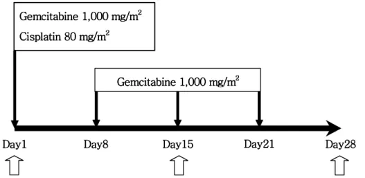
The study included 13 patients diagnosed as far advanced pancreatic cancer and planned to receive combination chemotherapy of gemcitabine and cisplatin. The patients who had a history of prior chemotherapy or radiotherapy were excluded. The chemo-therapeutic regimen consisted of an administration of gemcitabine 1,000 mg/m2 weekly (days 1, 8, and 15) followed by a 1-week rest and cisplatin 80 mg/m2 as a 2-hour infusion in 500 ml normal saline on day 1 of each 28-day cycle with adequate prehy-dration and mannitol infusion accompanied by adequate urinary output. The administ-ration of gemcitabine was 4-6 hours prior to cisplatin. With informed consents, the peripheral venous blood was obtained serially on the day 1, 15, 28 before the start of infusion of the drugs (Figure 1).
The peripheral blood of 7 volunteers who were matched age and sex and no history of internal malignancy or severe morbidity were sampled for health control.
Figure 1. The schedule of gemcitabine and cisplatin combination chemotherapy
and blood sampling. Blank arrows represent the day of blood sampling for study.
Gemcitabine Gemcitabine Gemcitabine Gemcitabine 1,000 mg/m1,000 mg/m1,000 mg/m1,000 mg/m2222 Cisplatin 80 mg/m Cisplatin 80 mg/m Cisplatin 80 mg/m Cisplatin 80 mg/m22 22 Gemcitabine 1,0 Gemcitabine 1,0 Gemcitabine 1,0 Gemcitabine 1,000 mg/m00 mg/m00 mg/m00 mg/m222 2 Day1 Day1Day1
Day1 Day8Day8Day8 Day8 Day15Day15 Day15Day15 Day21Day21Day21Day21 Day28Day28 Day28Day28
2. Reagents
Culture media used were RPMI-1640, supplemented with 10% fetal bovine serum (FBS, HyClone, USA), penicillin, streptomycin (1,000 unit/ml; Gibco BRL), 2 mM L-glutamine, 20 nM HEPES (sigma, St. Louis, MO, USA).
Fluorochrome-labelled monoclonal antibodies was used to analyze phenotypes of cells in peripheral blood mononuclear cells (PBMCs). They were: Lineage-FITC/CD11c-PE, CD4-FITC/CD8-PE, CD3-FITC/CD16+CD 56-PE, CD4-FITC/CD25-PE, CD34-FITC/CD14-PE, CD3-FITC/CD19-PE (BD pharmingen, San Jose, CA, USA).
For magnetic NK cell sorting, a commercial kit, NK cell isolation kit II (Militenyi Biotec Inc., Auburn, CA, USA) was used. This kit has two components. The first one is the hapten-antibody cocktail (Cocktail of hapten-conjugated monoclonal antibodies against CD3, CD14, CD19, CD36, and anti-IgE antibodies) and the other is anti-hapten MicroBeads (colloidal super-paramagnetic MACS MicroBeads conjugated to a monoclonal anti-hapten antibody).
3. Collection of peripheral blood and preparation of PBMCs
Peripheral blood samples were collected from 7 normal healthy, age and sex matched volunteers, and 13 patients with far advanced pancreatic cancer. Peripheral blood samples of the patients were collected on the day 1, 15, 28 of the first cycle of combination chemotherapy of gemcitabine and cisplatin (Figure 1). Blood samples were collected into EDTA tube. PBMCs and sera were isolated by centrifugation over Ficoll-Hypaque (Amersham Biosciences, NJ, USA) gradients at 20 , 900℃ g for 30min. After careful collection of sera, PBMCs were collected and washed 2 times with phosphate buffer solution (PBS).
4. Immunophenotypic analysis by flow cytometry
PBMCs were stained with the panel of monoclonal antibodies (20μl/106 cells) listed in reagents. And the cells were incubated for 15 min at 4 . After incubation, the℃
samples were centrifuged at 4 , 1,500℃ rpm for 5 min and the supernant fractions were removed. For fixing, 4% paraformaldehyde 100μl were added in each sample. Two-color flow cytometric analyses were performed, using a FACScan flow cytometer (Becton Dickinson). Data were analyzed with WINMdi program (ver. 2.8, USA). Each analysis included at least 10,000 events.
5. Isolation and preservation of NK cells
The isolation of NK cells from PBMCs was performed by depletion of non-NK cells with magnetic activating cell sorting (MACS) system according to the manufacturer's instruction. In brief, the prepared PBMCs were centrifuged at 300 g for 10 minutes and pipetted off supernant completely. The cell pellet was resuspended in 80μl of buffer solution (PBS, supplemented with 0.5% bovine serum albumin and 2 mM EDTA) per 107 total cells and 20μl of hapten-antibody cocktail mentioned in reagents per 107 total cells. After incubation for 10 minutes at 6℃ and washing two times with buffer solution, add 20μl of anti-hapten microbeads and 80μl of buffer solution per 107 total cells. After additional incubation for 15 minutes at 4℃ and washing with buffer, the cell pellet was resuspended with 500μl of buffer solution. The cell suspension was applied onto Midi MACS column for depletion of non-NK subpopulation. The purity of the enriched NK cells was evaluated by flow cytometry routinely and exceeded 90%.
6. Assay of NK cell cytotoxixity
Cytotoxic activity of NK cells was measured by JAM assay.28 NK cells were washed with PBS, resuspended in RPMI-1640 medium and combined with [3H]thymidine labeled K562 leukemia cells (105 cells/ml) at the desired effector/target (E/T) ratios in triplicate wells of V-bottomed microtiter plates. After overnight incubation at 37℃ in a humidified atmosphere containing 5% CO2, the contents of each well were harvested onto glass fiber mats using a Skatron multiple sample harvester and [3H]thymidine recovery was determined by liquid scintillation counting. Percent specific killing of
K562 target cells was calculated using the following formula: %specific killing = (S-E/S) × 100
where E is the experimentally retained DNA in the presence of NK cells (in cpm) and S is the retained DNA in the absence of NK cells (spontaneous). Data are presented as the mean percent specific killing of triplicate samples (±SD) from a representative experiment.
7. Assay of proliferative activity of T cells
For assay of the activity of T cells, phytohemagglutinin (PHA, Sigma, USA) was used.29 The final concentration of PHA was 10μg/ml, which are optimal for maximum proliferative responses. The cells were cultured at a final concentration of 105 cells/ml in 200μl of medium in 96 well microtiter plates. During 72 h of incubation at 37 ,℃ 5% CO2 and 100% humidity, cells were labelled with [3H]thymidine (6.7 Ci/mmol, 2 gCi/well) for 18 h at 48 h incubation. And after 72 h incubation, cells were harvested on glass fiber filters. The dried filters were placed in scintillation cocktail and label incorporation was measured as counts per minute (cpm).
8. Determination of VEGF and IL-10 concentrations in serum
Serum VEGF and IL-10 concentrations were determined using a commercially available enzyme-linked immunoassay (ELISA) designed to measure VEGF165 levels (Pierce endo-gen, Rockford, USA) and IL-10 levels (BD biosciences, San Diego, CA).
The assay for determining the concentration of VEGF employs the quantitative sand-wich enzyme immunoassay technique using recombinant human VEGF165 and antibodies raised against the recombinant protein. The assay exhibits no significant cross-reacti-vity with other angiogenesis factors and has a sensiticross-reacti-vity of 8.0 pg/ml. And the assay for IL-10 uses a monoclonal antibody specific for IL-10. The lower threshold of sensitivity of the IL-10 assay was 2 pg/ml. All samples were assayed in duplicate and the mean value was used for VEGF determination.
9. Statistical analysis
The differences in cell subpopulation and serum concentration of VEGF and IL-10 bwtween the healthy control and patients of pancreatic cancer were analyzed with Mann-Whitney U test. And the difference of those parameters on the day 1, 15, 28 in pancreatic cancer patients were analyzed with Kruskal-Wallis test.
III. RESULTS
Thirteen patient of far advanced pancreatic cancer were enrolled in the study and 7 volunteer who matched sex and age were enrolled as control group. The mean age of patients and controls were 58±10.2 and 59±6.1 years old, respectively. All the patients of pancreatic cancer had at least one site of distant metastasis.
1. The cell components of cell mediated immunity in patients with far advanced pancreatic cancer are decreased
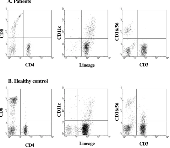
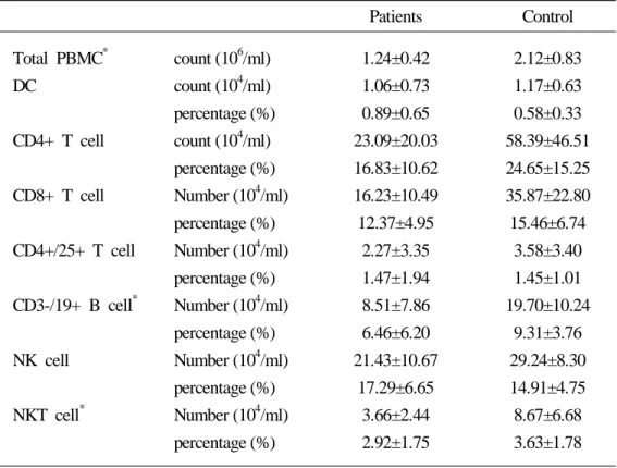
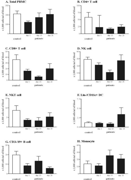
To evaluate the quantitative defect of CMI of the patients of far advanced pancreatic cancer, PBMCs of patients and control who matched sex and age were stained with specific monoclonal antibodies against cell-type specific markers; CD11c for detection of DCs, CD16, CD56 for NK cells, CD4, 8, 25 for T lymphocytes, CD19 for B lymphocytes, and CD14 for monocytes (Figure 2). The total PBMCs were significantly decreased in the patients of pancreatic cancer, compared with the healthy control group (p<0.05). CD3-/CD19+ B cell and CD3+/CD16+/CD56+ NKT cell subpopulation was significantly decreased. Although there was no statistical significance, Lineage-/ CD11c+ DCs, CD4+ T cell, CD8+ T cell and CD3-/CD16+/CD56+ NK cells were decreased in the patients of pancreatic cancer (Table 1).
Lineage Lineage CD3 CD3 A. Patients B. Healthy control CD4 C D 1 1 c C D 11 c C D 1 6 /5 6 C D 1 6 /5 6 CD4 C D 8 C D 8
Figure 2. Representative data of PBMCs from healthy volunteer and a patient of far
advanced pancreatic cancer. Isolated PBMCs were stained with antibodies against cell-type specific markers to measure immunological parameters. FITC conjugated antibodies were CD4, Lineage cocktail (Lin-), CD3 and PE conjugated antibodies were CD8, CD11c, CD16, CD56.
Table 1. Comparison of peripheral blood leukocytes and DC counts between the patients of far advanced pancreatic cancer and healthy control
Patients Control Total PBMC* DC CD4+ T cell CD8+ T cell CD4+/25+ T cell CD3-/19+ B cell* NK cell NKT cell* count (106/ml) count (104/ml) percentage (%) count (104/ml) percentage (%) Number (104/ml) percentage (%) Number (104/ml) percentage (%) Number (104/ml) percentage (%) Number (104/ml) percentage (%) Number (104/ml) percentage (%) 1.24±0.42 1.06±0.73 0.89±0.65 23.09±20.03 16.83±10.62 16.23±10.49 12.37±4.95 2.27±3.35 1.47±1.94 8.51±7.86 6.46±6.20 21.43±10.67 17.29±6.65 3.66±2.44 2.92±1.75 2.12±0.83 1.17±0.63 0.58±0.33 58.39±46.51 24.65±15.25 35.87±22.80 15.46±6.74 3.58±3.40 1.45±1.01 19.70±10.24 9.31±3.76 29.24±8.30 14.91±4.75 8.67±6.68 3.63±1.78 *p-value<0.05, Mann-Whitney U test
2. The combination chemotherapy of gemcitabine and cisplatin leads to numerical restoration of cell mediated immunity
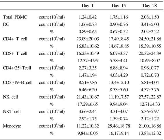
For analyzing the changes of cell subpopulations along to the gemciatbine and cisplatin combination chemotherapy, we performed serial immuno-phenotyping of PBMCs obtained on the day 1, day 15, and day 28. The total PBMCs count restored to the level of healthy control without temporal cytopenia during the course of chemotherapy. Other-wise, the counts of CD4+ T cells, CD8+ T cells, NK cells and DCs showed temporal decrement on the day 15 and exceeded the pretreatment value on the day 28. Even though the patterns of these populations were similar, the value of NK cells on the day 28 was recovered to the level of healthy control. Furthermore, the absolute DC counts restored and exceeded the healthy control value on the day 28 (Figure 3). On the contrary, the absolute count of CD4+/25+ regulatory T cell, CD3-/19+ B cell and monocytes sustained on the day 15 and eventually decreased below the pretreatment value on the day 28 (Table 2).
Table 2. The influence of gemcitabine and cisplatin combination chemotherapy on the peripheral blood leucocytes
Day 1 Day 15 Day 28 Total PBMC DC CD4+ T cell CD8+ T cell CD4+/25+Tcell CD3-/19+B cell NK cell NKT cell Monocyte count (106/ml) count (104/ml) % count (104/ml) % count (104/ml) % count (104/ml) % count (104/ml) % count (104/ml) % count (104/ml) % count (104/ml) % 1.24±0.42 1.06±0.73 0.89±0.65 23.09±20.03 16.83±10.62 16.23±10.49 12.37±4.95 2.27±3.35 1.47±1.94 8.51±7.86 6.46±6.20 21.43±10.67 17.29±6.65 3.66±2.44 2.92±1.75 11.22±10.32 9.84±10.05 1.75±1.16 0.90±0.76 0.67±0.52 17.49±8.45 14.67±8.85 6.07±3.37 5.58±4.41 6.88±8.94 4.03±4.29 13.4±12.10 8.33±5.60 11.19±7.57 9.94±9.04 3.31±4.07 1.59±0.74 25.46±18.78 16.17±9.14 2.08±1.50 3.41±5.00 2.02±2.22 24.50±21.86 15.39±10.55 20.32±24.39 10.65±8.07 0.96±0.77 0.72±0.70 5.81±4.04 4.37±3.76 27.57±22.87 12.71±4.33 5.36±5.97 2.12±1.22 21.00±16.86 13.88±12.32
Figure 3. The change of cell subpopulations of CMI with gemcitabine and cisplatin
combination chemotherapy.
3. The combination chemotherapy of gemcitabine and cisplatin restores impaired cytotoxicity of NK cells and proliferative activity of T cells 1) JAM assay for NK cell cytotoxicity
To evaluate the cytotoxicity of NK cells of the patients of far advanced pancreatic cancer, we performed JAM test with K562 cells. The NK cell subpopulations isolated from the PBMCs of 4 patients of far advanced pancreatic cancer and 4 healthy controls were used.
The mean percent of DNA loss by pretreatment NK cells was 6.76±7.87% and 9.95±11.48% in healthy control (Figure 4). However, this functional impairment of NK cells was recovered with gemcitabine and cisplatin combination chemotherapy and eventually restored to the level of healthy control (6.76±7.87 on day 1, 10.36±14.49 on day 15, 12.28±6.41 on day 28).
2) T cell proliferation activity with PHA stimulation
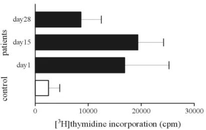
To evaluate the proliferative activity of T cell, 10μg/ml of PHA was used. The absolute value of [3H]thymidine incorporation was measured. The T cells subpopulation isolated from 7 patients of far advanced pancreatic cancer and 4 from healthy controls were used.
The proliferative activity of T cells without PHA was significantly lower than that stimulated with PHA. On the contrary to the decreased cytotoxic activity of NK cells, the proliferative activity of T cell in the absence of PHA in pancreatic cancer patients was more activated than that of healthy control at the time of pretreatment (723.0± 964.0 vs 86.9±56.9). And the cpm values with stimulation of PHA showed similar patterns (2,506.7±4,246.6 vs 16,879.1±22,064.6). Interestingly, the value of [3H]thymidine incorporation in pancreatic cancer patients showed temporal increment on day 15 and reciprocal decrement on day 28 (16,879.1±22,064.6, 19,366.3±10,824.1, 8,628.9± 10,174.9). However, the activity on day 28 of pancreatic cancer patients was higher than that of control group (Figure 5).
Figure 4. The influence of gemcitabine and cisplatin chemotherapy for cytotoxic
activity of NK cells. Cytotoxic activity of NK cells was measured by JAM assay. NK cells were cocultured with [3H]thymidine labeled K562 leukemia cells (105cells/ml) in triplicate. Thymidine recovery was determined by liquid scintillation counting. Percent specific killing of K562 target cells was calculated.
Figure 5. The influence of gemcitabine and cisplatin chemotherapy on the T cell
proliferation activity. Peripheral blood T cells (105cells/ml) were cultured with PHA (10 ug/ml). [3H]thymidine incorporation was measured on 72 hours of culture. Results represent the mean ±SE of the values.
4. The immune-suppressive cytokines tend to be decreased with the combination chemotherapy of gemcitabine and cisplatin
1) The influence of chemotherapy on serum VEGF
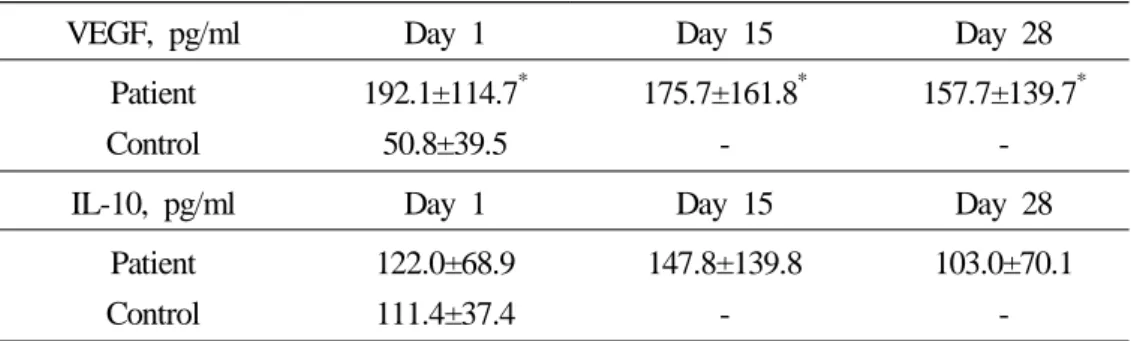
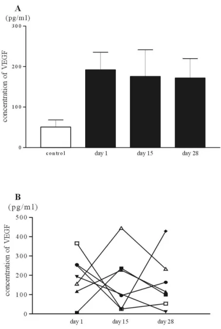
The pretreatment serum VEGF levels in patients (192.1±114.7 pg/ml) with pancreatic cancer were significantly higher than those in controls (50.8±39.5 pg/ml). The serum VEFG concentrations of pancreatic cancer patients were decreased with one cycle of gemcitabine and cisplatin combination chemotherapy (Table 3), even though the values of day 28 in patient group was higher than those of control group (Figure 6). Though the mean values of VEGF of pancreatic cancer patients decreased with chemotherapy, the change of VEGF in individual patients showed variable responses. The serum con-centration of VEGF in some patients decreased with chemotherapy, but in other patients steadily increased with chemotherapy and in the remaining, the value of serum VEGF decreased on the day 15 and slightly increased again on the day 28.
2) The influence of chemotherapy on serum IL-10
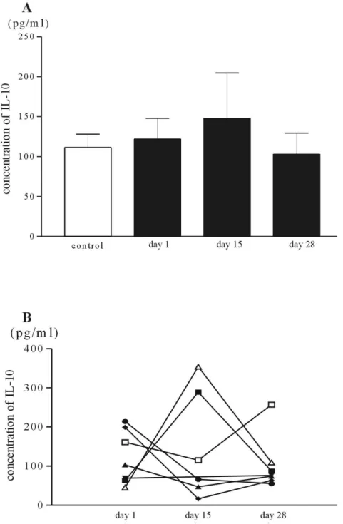
The pretreatment serum IL-10 values in patients with pancreatic cancer (122.0±68.9 pg/ml) were higher than those of control (111.4±37.4 pg/ml), however the difference did not show statistical significance. And the serum IL-10 levels on the day 15 in patients of pancreatic cancer were more elevated than those of pretreatment (Table 3). However, the values of IL-10 become lower than those of pretreatment after comple-tion of one cycle of gemcitabine and cisplatin combinacomple-tion chemotherapy (Figure 7). In the aspect of individual patients, the changes of IL-10 were similar to that of VEGF.
Table 3. The changes of serum VEGF and IL-10 in patients of pancreatic cancer with gemcitabine and cisplatin combination chemotherapy
VEGF, pg/ml Day 1 Day 15 Day 28 Patient Control 192.1±114.7* 50.8±39.5 175.7±161.8* -157.7±139.7* -IL-10, pg/ml Day 1 Day 15 Day 28
Patient Control 122.0±68.9 111.4±37.4 147.8±139.8 -103.0±70.1 -*p< 0.05, Mann-Whitney U test with control
Figure 6. The influence of chemotherapy on the serum concentration of VEGF. The
bars are the mean value of serum concentration of VEGF of pancreatic cancer patients and controls, A. The serial changes of the serum VEGF in individual patients are plotted, B.
Figure 7. The influence of chemotherapy on the serum concentration of IL-10. The
bars are the mean value of serum IL-10 of pancreatic cancer patients and controls, A. The serial changes of the serum IL-10 in individual patients are plotted, B.
IV. DISCUSSION
It is well known that the cancer patients or experimental animals with large tumors may have a defective immune system which is secondary to their disease, and immune dysfunctions associated with the presence of progressively growing tumors.19-22 Espe-cially, in contrast to the antigen-specific immune dysfunctions, so called 'tumor escape phenomenon' including the inability of tumor-specific CD4+ T cells and CD8+ T cells to respond to tumor antigens, as observed mainly in mouse experimental systems,23 advanced cancer is often associated with a more global change in the immune system that is not specific for the antigens expressed by the tumor. The generalized immuno-deficiency in patients of advanced cancer includes decreased reactivity of delayed type hypersensitivity test, defects in the production of Th1 cytokines like IFN-γ, IL-2 and TNF-α, defects in cytotoxic and proliferative functions of T cells and NK cells and secretion of immunosuppressive molecules.20,22,24
In the current study, the cell mediated immunity of pancreatic cancer patients showed defects in the aspect of cell quantity as well as of cellular functions. First, the total number of PBMC was significantly lower than healthy control. Furthermore, the cell subpopulations involving cell mediated immunity such as DCs, CD8+ CTLs, NK cells were decreased compared with sex and age matched healthy controls. In addition to these quantitative defects, the cytotoxic activity of NK cells seems to be impaired in the patients of pancreatic cancer, while the immunosuppressive cytokines-VEGF, IL-10 showed higher level than healthy control. These results may represent that the immu-nity, especially cell mediated immunity in the patients of far advanced pancreatic cancer is impaired numerically as well as functionally.
Despite the great specificity that can be achieved with immunotherapy, this treat-ment by itself has been insufficient to eradicate the malignancy. And this limit of immunotherapy attributes partially to the immunodeficient condition of the host with malignancy, revealed in our study.
Recently, many trials have been done to overcome this limit of immunotherapy. Just as cancer chemotherapy began with the use of single agents and evolved into
combination therapy, immunotherapy has been combined with chemotherapy or radio-therapy. Rosenberg et al.28 reported in their sequential treatements of metastatic melanoma that the administration of antitumor autologous lymphocyte populations can be far more active in mediating tumor regression in vivo when administered after nonmyeloablative chemotherapy than when administered without prior chemotherapy. And Takahashi et al.29 reported a good response with nonmyeloablative stem cell transplantation in unresectable pancreatic cancer patients. This trial used the concepts 'graft versus tumor (GVT) response' of hematologic malignancies. In allogenic bone marrow transplantation of hematologic malignancies, delayed GVT response is thought to be more important than early GVT response which are mediated by direct donor T cell allorecognition. In other words, dying tumor cells may provide antigen presenting cells including DCs with a plentiful source of tumor antigens in combination with danger signals that may enable DCs to initiate antitumor T cell responses and thus maintain tumor immune surveillance. This suggestion supported by the experiment of Rad et al.30 They reported that coculture of immature DCs with tumor cells treated with the alkylating agents-melphalan and chlorambucil leads to enhanced autologous and allogenic T cell activation, up-regulation of surface expression of MHC and costimulatory molecules, and increased IL-12 secretion. And exposure of DCs to DNA purified from tumor cells treated with alkylating agents also increased their T cell stimulating capacity, expression of CD86, and IL-12 secretion. Similar to this experi-ment, Nikata and Gabrilovich26 showed that the combination of γ-irradation leaded to accumulate DCs in transplanted tumor area of mice remarkably and resulted in a significant T cell response to the specific antigens.
In our study, we also evaluated the effect of chemotherapy in the immunity of patients of far advanced pancreatic cancer. Gemcitabine, a deoxycytidine analog, acts by intracellular activation into phosphorylated metabolites, such as gemcitabine triphos-phate, which competes with endogenous deoxycystidine triphosphate for incorporation into DNA, and gemcitabine diphosphate, which inhibits ribonucleotide reductase, and inhibit the DNA synthesis.33,34 Cisplatin, a platinum agent intercalates with double stranded DNA and forms platinum-DNA adduct. Experimental evidence of a synergistic
interaction between gemcitabine and cisplatin, probably related to an increase in platinum-DNA adduct formation and apoptosis of tumor cells.35,36
In the aspect of cell counts, one cycle of the combination chemotherapy of gem-citabine and cisplatin leads to variable results according to the cell subpopulation. Total PBMCs restored to the value of healthy control in relatively early stage of chemotherapy and more increased after completion of one cycle of chemotherapy. Otherwise, the cell subpopulations engaged in cell mediated immunity - CD8+ T cells, CD4+ T cells, NK cells, NKT cells, and DCs - showed temporal drop in early phase of the chemotherapeutic schedule and finally restored at least the value of pretreat-ment. Interestingly, DCs showed minimal drop in the mid of chemotherapy and resto-ration after completion of one cycle of chemotherapy. And the post chemotherapeutic value of DCs exceeded that of healthy control although it did not show statistical significance. This early restoration of DCs to the level of normal value was reported in the study of Markowicz et al.37 Their study with the patients of variable hema-tologic and solid tumors showed that DCs of peripheral blood were depleted and restored rapidly after chemotherapy and the early renewal of the DC population was followed by the recovery of DCdependent primary responsiveness to the neoantigen -keyhole limpet hemocyanin. Taken into consideration that DCs position the key role in currently available immunotherapy, this early restoration of DCs is an important clue for considering the timing of DCs collection for immunotherapy.
In accordance to the numeric recovery of immune cells, the cytotoxicity of NK cells and the proliferative activity of T cell were restored with chemotherapy. The cytotoxic activity of NK cells to K562 leukemia cells, which was decreased in pret-reatement phase, was restored relatively early phase of chemotherapy. Even though the NK cell subpopulation showed depletion on the day 15 of chemotherapy, it revealed that the cytotoxic activity already went into the recovery and exceeded the level of healthy control. Otherwise, the proliferation activity of T cells for PHA showed a different pattern of recovery. On the day 15, the mid time of the chemotherapy schedule, the value of [3H]Thymidine incorporation was increased and decreased below that of pretreatment after completion of one cycle of chemotherapy. However, the
proliferative activity of T cell in the patients of pancreatic cancer at the time of diag-nosis was superior to that of normal healthy control. This results are some different from others that have reported that the T cell proliferation activity was impaired in patients of malignancies. Even though this data should be reevaluated with repeated experiments, it is possible that T cells of patients of malignant diseases may be normal or more activated condition. However, the immunologic environments surrouding the T cells may be harzadous and immunosuppressive. For example, the immunosuppressive cytokines - TGF-β, IL-10 and VEGF- are proven to play a role for unresponsiveness of lymphocytes to antigens.38,39 TGF-β is well known immu- nosuppressive cytokines. In pancreatic cancer, Teraoka et al.40 showed that TGF-1 inhibit the attachment of lymphocytes to cancer cells and decreased cell cytotoxicity. And Bellone et al.41 reported that aberrant production fo TGF-β and IL-10 in pancreatic cancer patients skews T cell cytokine production patterns in favor of a Th2 immunophenotype.
Whether the immunosuppressive cytokines are increased or not in the patients of far advanced pancreatic cancer was evaluated with the sera of patients and controls. For this, we evaluated the serum concentration of IL-10 and VEGF. As shown above, the value of VEGF in patients group was markedly increased than healthy control. And the concentration of serum VEGF seemed to be decreased with one cycle of gem-citabien and cisplatin combination chemotherapy. However, when plotting the serial values of serum VEGF in individual patients, variable patterns of changes appeared. Some of the patients showed consecutive decrement of the VEGF with chemotherapy, and the inverse pattern occurred in others. It may be related with the difference of the sensitivity to chemotherapeutic agents. So, the VEGF value may be correlated with the prognosis of pancreatic cancer patients. Although it is some apart from our study, Karayiannakis et al.42 showed the higher level of serum VEGF in pancreatic cancer patients at the time of diagnosis correlate with advanced and metastatic disease and poor prognosis.
The serum concentration of IL-10 in patients group was also higher than control group. And the IL-10 after one cycle of chemotherapy became lower than pretreat-ment with transient elevation on the day 15. The serial changes in individual patients
also showed variable patterns similar to VEGF.
In summary, we found that the immunity of pancreatic cancer patients was defected in numerical and functional aspects. And with gemcitabine and cisplatin combination chemotherapy, these immunologic defects seemed to be restored. Especially, the numeric recovery of DCs population and the functional restoration of cytotoxicity of NK cells were rapidly recovered and exceeded the level of healthy control after one cycle of chemotherapy. In addition, the serum concentration of VEGF, IL-10 also decreased with chemotherapy.
V. CONCLUSION
Although our study has some limitations, because most chemotherapy are adminis-tered consecutively with 3 to 4 weeks interval rather than a single cycle, we found some suggestive data for considering ideal timing of immunotherapy combined with systemic chemotherapy and for the change of immunity of pancreatic cancer patients with systemic chemotherapy.
Taken into account that DCs population recovered rapidly and exceeded the value of healthy control with one cycle of gemcitabine and cisplatin combination chemo-therapy, when the patients of pancreatic cancer receive one cycle of chemotherapy may be one of the ideal timing of DCs collection for immunotherapy.
And the immunosuppressive environments, including VEGF, IL-10 and suppressed cytotoxic functions of NK cells, T cells seem to be improved with chemotherapy. So, cellular adoptive immunotherapy should be installed at the time of maximizing these improvements.
REFERENCES
1. Greelee RT, Murray T, Bolden S, Wingo PA. Cancer statistics, 2000. CA, Cancer, J Clin 2000;50:7-33.
2. Ministry of Health and Welfare Republic of Korea. 2002 Annual Report of the Korean Central Cancer Registry (Published in 2003).
3. Jemal AT, Murray J, Samuels A, Ghafoor A, Ward E, Thun M. Cancer statistics, 2003. CA Cancer J Clin 2003;53(1):5-26.
4. Hiraoka T. Extended radical resection of cancer of the pancreas with intraopera-tive radiotherapy. Clin Gastroenterol 1990;4:985-993.
5. Neoptolemos JP, Stocken DD, Friess H, Bassi C, Dunn JA, Hickey H, et al. A Randomized Trial of Chemoradiotherapy and Chemotherapy after Resection of Pancreatic Cancer. N Engl J Med 2004;350:1200-1210.
6. Geer RJ, Brennan MF. Prognostic indicators for survival after resection of pancreatic adenocarcinoma. Am J Surg 1993;165:68-72.
7. Sener SF, Fremgen A, Menck HR, Winchester DP. Pancreatic cancer: A report of treatment and survival trends for 100,313 patients diagnosed from 1985-1995, using the National Cancer Database. J Am Coll Surg 1999;189:1-7.
8. Sohn TA, Yeo CJ, Cameron JL, Koniaris L, Kaushal S, Abrams RA, et al. Resected adenocarcinoma of the pancreas - 616 patients: Results, outcomes, and prognostic indicators. J Gastrointest Surg 2000;4:567-79.
9. Coley WB. The treatment of malignant tumors by repeated inoculation of erysipelas: with a report of ten original cases. Am J Sci 1893;105:487-511.
10. Lanzavecchia A. Identifying strategies for immune intervention. Science 1993;260: 937-944.
11. Pardoll DM. Cancer vaccines. Nat Med 1998;4:525-531.
12. Colaco C. Why are dendritic cells central to cancer immunology? Mol Med Today 1999;1:14-17.
13. Smyth MJ, Godfrey DI, Trapani JA. A fresh look at tumor immunosurveillance and immunotherapy. Immunolo nature 2001;2:293-299.
14. Young JW and Inaba K. Dendritic cells as adjunvant for class I major histocom-patability complex-restricted anti-tumor immunity. J Exp Med 1996;183:7-11.
15. Hart I and Colaco C. Fusion induces tumor rejection. Nature 1997;388:627-628. 16. Banchereau J and Steinman RM. Dendritic cell and the control of immunity. Nature
1998;392:245-252.
17. Rosenberg SA. A new era for cancer immunology based on the genes that encode camcer antigens. Immunity 1999;10:281-287.
18. Kono K, Takahashi A, Ichihara F, Amemiya H, Iizuka H, Fujii H, et al. Prognostic significance of adoptive immunotherapy with tumor-associated lymphocytes in patients with advanced gastric cancer: A randomized trial. Clin Cancer Res 2002; 8:1767-1777.
19. Fumiko I, Koji K, Akihiro T, Hiromichi K, Hidemitsu S, et al. Increased popula-tions of regulatory T cells in peripheral blood and tumor-infiltrating lymphocytes in patients with gastric and esophageal cancers. Clin Cancer Res 2003;9:4404-4408. 20. Young RC, Corder MP, Haynes HA, DeVita VT. Delayed hypersensitivity in
Hodgkin's disease. A study of 103 untreated patients. Am J Med 1972;52:63-68. 21. Alexander JP, Kudoh S, Melsop KA, Hamilton TA, Edinger MGR, Tubbs R, et
al. T cells infiltrating renal cell carcinoma display a poor proliferative response even though they can produce interleukin 2 and express interleukin 2 receptors. Cancer Res 1999;59:2950-2956.
22. Kiessling R, Wasserman K, Horiguchi S, Kono K, Sjoberg J, Pisa P, et al. Tumor-induced immune dysfunction. Cancer Immunolo Immunother 1999;48:353-362. 23. Marindola FM, Jaffe EM, Hicklin DJ, Ferrone S. Escape of human solid tumors
from T-cell recognition: Molecular mechanisms and functional significance. Adv Immunol 2000;74:181-273.
24. Kiertscher SM, Luo J, Dubinett SM, Roth MD. Tumors promte altered maturation and early apoptosis of monocyte-derived dendritic cells. J Immunol 2000;164:1269-1279.
25. Mitchell MS. Combination of anticancer drugs and immunotherapy. Cancer Immunolo Immunother 2003;52:686-692.
26. Nikitina EY and Gabrilovich DI. Combination of -irradation and dendritic cell administration induces a potent antitumor response in tumor-bearing mice: Approach to treatment of advanced stage cancer. Int J Cancer 2001;94:825-833.
Microen-capsulated cell-mediated treatment of inoperable pancreatic carcinoma. Lancet 2001; 357:1591-1592.
28. Rosenberg SA, Yang JC, Robbins PF, Wunderlich JR, Hwu P, Sherry RM, et al. Cell transfer therapy for cancer: Lessons from sequential treatments of a patient with metastatic melanoma. J Immunology 2003;63:385-393.
29. Takahashi T, Omuro Y, Matsumoto G, Sakamaki H, Maeda Y, Hiruma K, et al. Nonmyeloablative Allogeneic Stem Cell Transplantation for Patients With Unresec-table Pancreatic Cancer. Pancreas 2004;28:e65e69.
30. Rad AN, Pollara G, Sohaib SM, Chiang C, Chain BM, Katz DR. The differential influence of allogenic tumor cell death via DNA damage on dendritic cell maturation and antigen presentation. Cancer Res 2003;63:5143-5150.
31. Matzinger P. The JAM test A simple assay for DNA fragmentation and cell death. J Immunol Methods 1991;145:185-192.
32. Ceuppens JL, Baroja ML, Katrien L, Damme JV, Billiau A. Human T cell acti-vation with phytohemagglutinin The function of IL-6 as an accessory signal. J Immunol 1988;141:3868-3874.
33. Hertel LW, Boder GB, Kroin JS, Rinzel SM, Poore GA, Todd GC, et al. Eval-uation of the antitumor activity of gemcitabine (2',2'-difluoro-2'-deoxycytidine). Cancer Res 1990;50:4417-4422.
34. Huang P, Chubb S, Hertel LW, Grindey GB, Plunkett W. Action of 2',2'-difluoro-deoxycytidine on DNA synthesis. Cancer Res 1991;51:6110-6117.
35. Bergman AM, Ruiz van Haperen VW, Veerman G, Kuiper CM, Peters GJ. Synergistic interaction between cisplatin and gemcitabine in vitro. Clin Cancer Res 1996;2:521-530.
36. van Moorsel CJ, Pinedo HM, Veerman G, Vermorken JB, Postmus PE, Peters GJ. Scheduling of gemcitabine and cisplatin in Lewis lung tumour bearing mice. Eur J Cancer 1999;35:808-814.
37. Markowicz S, Walewski J, Zajda K, Wiechno PJ, Skurzak HM, Giermek J, et al. Recovery of dendritic cell counts and function in peripheral blood of cancer patients after chemotherapy. Cytokines Cell Mol Ther 2002;7:15-24.
38. Sulitzeanu D. Immunosuppressive factors in human cancer. Adv Cancer Res 1993; 60:247-267.
39. Fortis C, Foppoli M, Gianotti L, Galli L, Citterio G, Consgno G, et al. Increased interleukin-10 serum levels in patients with solid tumors. Cancer Lett 1996;104: 1-5.
40. Teraoka H, Sawada T, Nishihara T, Yashiro M, Ishikawa T, Nishino H, et al. Enhanced VEGF production and decreased immunogenecity induced by TGF-1 promote liver metastasis of pancreatic cancer. Cancer Res 2001;85:612-617.
41. Bellone G, Anna T, Artusio E, Mareschi K, Carbone A, Tibaudi D, et al. Tumor-associated transforming growth factor-β and interleukin-10 contribute to a systemoc Th2 immune phenotype in pancreatic carcinoma patients. Am J Pathol 1999;155: 537-547.
42. Karayiannakis AJ, Bolanaki H, Syrigos KN, Asimakopoulos B, Polychronidis A, Anagnostoulis S, et al. Serum vascular endothelial growth factor levels in pancreatic cancer patients correlate with advanced and metastatic disease and poor prognosis. Cancer Lett 2003;194:119-124.
국 국 국 국문문문문요요요요약약약약
진
진
진
진행
행성
행
행
성
성
성 췌
췌
췌장
췌
장
장
장암
암의
암
암
의
의 항
의
항
항
항암
암
암
암약
약
약물
약
물치
물
물
치
치
치료
료
료
료 전
전
전
전후
후
후
후의
의 세
의
의
세
세
세포
포
포
포매
매
매개
매
개
개
개
면
면
면
면역
역반
역
역
반
반응
반
응
응에
응
에 관
에
에
관
관여
관
여
여
여하
하
하
하는
는 면
는
는
면
면
면역
역
역
역세
세
세포
세
포의
포
포
의
의
의 변
변
변
변화
화
화
화
<
지도교수
송
송
송
송
시
시
시
시
영
영
영
영
>
연세대학교 대학원 의학과
방
방
방
방
승
승
승
승
민
민
민
민
10% 2 . 췌장암은 이하의 년 생존률을 보이는 예후가 매우 불량한 악성 종양이다 이처럼 불량한 췌장암의 예후는 대부분의 환자가 진단 당시 근치적 수술이 불가 능한 상태로 발견된다는 것과 기존의 항암제 및 방사선 치료에 불량한 반응을 보 . 이는데 기인한다 이처럼 기존의 항암치료에 대한 불량한 반응은 새로운 치료 방 , 법의 개발에 대한 강력한 동기를 부여하고 있으며 현재 유전자 치료 또는 면역 . 치료 등의 새로운 치료법이 시도되고 있다 특히 수지상 세포를 중심으로하는 세 포면역치료는 기존의 항암치료에 반응하지 않는 악성종양에 있어 보다 종양세포 , 에 특이적인 세포독성을 유발할 수 있다는 점등으로 인해 주목 받고 있으며 최 근 기존의 항암약물치료 또는 방사선 치료가 세포 매개 면역반응을 강화시킬 수 , 있음이 밝혀지고 이러한 근거를 토대로 한 항암약물치료와 세포면역치료의 병행 . 이 보다 양호한 치료 성적을 보일 수 있음이 보고되고 있다 그러나 현재까지 항 암약물치료가 악성종양 환자의 실제 면역체계에 미치는 영향에 대한 연구 및 항 . 암약물치료와 병행하는 면역치료의 최적의 시기에 대한 연구는 부족한 실정이다 gemcitabine cisplatin 이에 본 연구는 및 병합요법으로 치료 받은 진행성 췌장암 , 환자들의 세포 매개 면역상태의 변화를 분석하여 약물치료와 면역세포치료를 병 . 용하기에 적합한 시기를 알아보고자 하였다 2004년 월부터2 2004년 월까지 진행성 췌장암을 진단받고8 , gemcitabine 및cispaltin(n=13) (n=7) 병합약물치료를 받은 환자들 과 악성종양의 병력이 없는 건강한 성인 을 대상으로 환자 -대조군 연구를 시행하였다 환자군은. 28일의 gemcitabine/cisplatin 1 , ( 15 ), 1 ( 병합요법 주기를 기준으로 약물치료전 치료중간 제 일 및 주기 치료 후 제 28 )일 에 걸쳐 회 말초혈액을 채취하였으며 대조군은 회 채취하였다3 , 1 . ( , T , 환자군의 약물치료 전과 대조군의 말초혈액내 면역세포들 수지상세포 림프구 NK세포 을 유세포 분석을 통한 비교에서 환자군의 말초 혈액 단핵구 및 세포매) , CD4+ T , CD8+ T NK 개 면역반응에 관여하는 수지상세포 림프구 림프구 및 세포 , NK K562 등이 정상 대조군에 비해 감소되어 있음을 규명하였으며 또한 세포의 . 세포주에 대한 세포독성능이 정상 대조군에 비해 억제되어 있음을 확인하였다 , VEGF, IL-10 뿐만 아니라 체내 면역반응을 억제하는 것으로 알려진 의 혈청 농도 . 도 정상 대조군에 비해 췌장암 환자군에서 유의하게 증가되어있음을 확인하였다 Gemcitabine 및 cisplatin 병합화학치료를 시행받은 췌장암 환자에게서 면역세포 , 28 군들의 치료전 수적인 감소는 회복되는 양상을 보였으며 특히 수지상세포는 . 일간의 항암약물 치료 후 정상 대조군보다 수적팽창이 유도됨을 확인하였다 또 NK T . 한 세포 및 림프구도 항암 치료 전 수치까지 회복하는 것을 확인하였다 이 러한 면역세포들의 수적인 회복과 함께 억제되어 있던 세포기능들도 회복되는 양 , K562 NK 상을 보여 세포주에 대한 세포의 세포독성능은 약물치료와 더불어 지속 . 적으로 향상되는 양상을 보였다 또한 치료전 정상 대조군에 비해 증가되어 있던 VEGF, IL-10 . 혈청 농도도 약물치료에 따라 감소하는 결과를 보였다 이상의 결과에서 전이성 췌장암 환자의 세포매개 면역반응은 정상 대조군에 비 , , 하여 양적 질적으로 억제되어 있으며 이러한 세포매개 면역반응의 억제는 약물 . 치료를 통해 회복되는 경향을 보임을 확인하였다 특히 수지상세포의 빠르고 증 폭된 수적 회복은 진행성췌장암에 있어 면역세포치료를 위한 수지상세포의 모집 , 에 있어 적절한 시기를 결정하는데 중요한 근거가 될 것이며 나아가 항암약물치 료와 더불어 췌장암 환자들의 면역저하가 점차 개선되는 양상을 보이는 것으로 , 고려할때 항암약물치료와 병용하게되는 세포면역치료의 효과를 극대화 하기 위 해서는 항암약물치료를 통하 면역저하의 회복이 최대화 된 시점에 시행하는 것이 . 보다 강력한 종양 면역반응을 유도할 수 있을 것으로 생각된다 : 핵 핵핵 핵심심심심되되되는되는 말는는 말말말 췌장암 면역치료 항암약물치료 수지상세포 세포매개면역반응, , , ,