저작자표시-비영리-변경금지 2.0 대한민국 이용자는 아래의 조건을 따르는 경우에 한하여 자유롭게 l 이 저작물을 복제, 배포, 전송, 전시, 공연 및 방송할 수 있습니다. 다음과 같은 조건을 따라야 합니다: l 귀하는, 이 저작물의 재이용이나 배포의 경우, 이 저작물에 적용된 이용허락조건 을 명확하게 나타내어야 합니다. l 저작권자로부터 별도의 허가를 받으면 이러한 조건들은 적용되지 않습니다. 저작권법에 따른 이용자의 권리는 위의 내용에 의하여 영향을 받지 않습니다. 이것은 이용허락규약(Legal Code)을 이해하기 쉽게 요약한 것입니다. Disclaimer 저작자표시. 귀하는 원저작자를 표시하여야 합니다. 비영리. 귀하는 이 저작물을 영리 목적으로 이용할 수 없습니다. 변경금지. 귀하는 이 저작물을 개작, 변형 또는 가공할 수 없습니다.
A Thesis for the Degree of Master of Science in Medicine
The Effects of Assisted Ergometer Training
with a Functional Electrical Stimulation
on Exercise Capacity and Functional Ability
in Subacute Stroke Patients
So Young Lee
Department of Medicine
GRADUATE SCHOOL
JEJU NATIONAL UNIVERSITY
아급성기 뇌졸중 환자에서
기능적 전기자극을 이용한 에르고미터의 적용이
운동량과 기능적 능력에 미치는 영향
지도교수 강 사 윤
이 소 영
이 논문을 의학 석사학위 논문으로 제출함
2014년 2월
이소영의 의학 석사학위 논문을 인준함
심사위원장
...위 원
...위 원
...제주대학교 일반대학원
2014년 2월
The Effects of Assisted Ergometer Training
with a Functional Electrical Stimulation
on Exercise Capacity and Functional Ability
in Subacute Stroke Patients
So Young Lee
(Supervised by Professor Sa-Yoon Kang)
A thesis submitted in partial fulfillment of the requirement
for the degree of Master of Science in Medicine
2014. 2.
This thesis has been examined and approved
……….……
………..……
………..…………
……….
Department of Medicine
GRADUATE SCHOOL
JEJU NATIONAL UNIVERSITY
I
ABSTRACT
The Effects of Assisted Ergometer Training
with a Functional Electrical Stimulation
on Exercise Capacity and Functional Ability
in Subacute Stroke Patients
So Young Lee
Department of Medicine
GRADUATE SCHOOL
JEJU NATIONAL UNIVERSITY
Supervised by Professor Kang, Sa-Yoon
Objective: To determine if assisted ergometer training can improve the functional ability and aerobic capacity of subacute stroke patients and if functional electrical stimulation (FES) of the paretic leg during ergometer cycling has additional effects.
Methods: Sixteen subacute stroke patents were randomly assigned to the FES (n=8) group or the control (n=8) group. All patients underwent assisted ergometer training for 30 minutes (five times per week for 4 weeks). The electrical stimulation group received FES of the paretic lower limb muscles during assisted ergometer training. Peak oxygen consumption
II
(Vo2peak), metabolic equivalent (MET), respiratory exchange ratio (RER), resting and
maximal heart rate (RHR, MHR), resting and maximal blood pressure (RBP, MBP), maximal rate pressure product (MRPP), estimated anaerobic threshold (AT), submaximal rate pressure product (SRPP), submaximal rate of perceived exertion (SRPE) and exercise duration (Ex Dur) were determined with the exercise tolerance treadmil test before and after treatment. The six minute walk test (6MWT), Berg balance scale (BBS), and the Korean version of modified Barthel index (K-MBI) were evaluated at the beginning and end of treatment. Results: At 4 weeks after treatment, the FES assisted ergometer training group showed significant improvements in Vo2peak (p=0.02), MET (p=0.02), estimated AT (p=0.02), 6MWT
(p=0.01), BBS (p=0.01) and K-MBI (p=0.01). The control group showed improvements in only BBS (p=0.01) and K-MBI (p=0.02). However, there was no significant difference in exercise capacity and functional ability between the two groups.
Conclusion: This study demonstrated that ergometer training for 4 weeks improved the functional ability of subacute stroke patients. In addition, aerobic capacity was improved after assisted ergometer training with a FES only.
III
CONTENTS
ABSTRACT... I
CONTENTS... III
LIST OF TABLES... V
LIST OF FIGURES... VI
LIST OF ABBREVIATIONS... VII
I. INTRODUCTION... 1
II. MATERIALS AND METHODS... 4
1. Subjects
2. Intervention
3. Exercise treadmill testing protocol
4. Outcome measures
IV
1) Assessment of aerobic capacity
2) Assessment of functional ability
5. Statistical analysis
III. RESULTS... 16
1. General characteristics of the participants
2. Changes of parameters before and after treatment within each group
3. Changes of parameters before and after treatment between the two groups
IV. DISCUSSION... 23
V. CONCLUSION... 27
VI. REFERENCES... 28
VII. ABSTRACT IN KOREAN... 34
V
LIST OF TABLES
Table 1. Baseline characteristics of the two groups... 17
Table 2. Exercise capacity, functional ability and motor function at baseline….
……….. 18
Table 3. Changes of parameters before and after treatment within each group
VI
LIST OF FIGURES
Figure 1. EMG-FES 2000
Ⓡ(Cyber Medic, Seoul, Korea)…….………….… 6
Figure 2. Placement on the paretic quadriceps, tibialis anterior, hamstring and
gluteus maximus
………... 7
VII
LIST OF ABBREVIATIONS
6MWT, six minute walk test
AT, anaerobic threshold
BBS, Berg balance scale
CO
2,carbon dioxide
ECG, electrocardiogram
Ex Dur, exercise duration
FES, functional electrical stimulation
K-MBI, Korean - modified Barthel index
MdBP, maximal diastolic blood pressure
MET, metabolic equivalent;
MHR, maximal heart rate
MI, motricity index
MRPP, maximal rate pressure product
MsBP, maximal systolic blood pressure
VIII
RdBP, resting diastolic blood pressure
RER, respiratory exchange ratio
RHR, resting heart rate
RsBP, resting systolic blood pressure
SCI, spinal cord injury
SRPE, submaximal rate of perceived exertion
SRPP, submaximal rate pressure product
VE, pulmonary ventilation
Vo
2 peak, peak oxygen consumption
1
I. INTRODUCTION
Stroke is a leading cause of disability that impairs ambulatory activity and daily
living functions (Bobath, 1990; Gresham et al., 1975). Post-stroke functional deficits such as
postural imbalance, muscle weakness, or spasticity may promote physical inactivity and a
deconditioning state, which can lead to reduced cardiovascular fitness (Gresham et al., 1975;
Brown et al., 1998; Mayo et al., 1999; Szecsi et al., 2008; Jorgensen et al., 1999). The low
aerobic capacity eventually results in low ambulatory function, social participation, and
quality of life (Mayo et al., 1999). Therefore, it is essential to encourage aerobic exercise in
post-stroke patients, but very few can participate in intensive gait training immediately after
their stroke event (Hwang et al., 2003) because of muscle weakness, fatigue, or general
deconditioning (Janssen et al., 2008).
Cycling exercise is a safe, functional treatment that is accessible by these patients for the relearning of motor skills required for locomotion (Hunt et al., 2004; Takahashi et al., 2004). Pedaling is an aerobic exercise and a repetitive, functional activity, which requires reciprocal contraction of agonist and antagonist muscles of the lower limbs in a repeating pattern similar to that required for walking (Raasch et al., 1999). The motorized cycling ergometer is an especially useful treatment for severe disabled stroke patients, as automatic pedaling facilitates phasic, coordinated muscle activity with continuous use of the affected-side
2
extremity (Hancock et al., 2012). This exercise improves cardiovascular fitness such as peak oxygen consumption, workload, and endurance (Potempa et al., 1995). Also, because cycling is practiced in the sitting position, it is safe, easy to perform, and can be widely applied, regardless of the severity of motor impairment (Brown et al., 1997). Therefore, cycling has been proposed to have potential patient benefit when used as an adjunct to therapy after stroke (Potempa et al., 1995; Holt et al., 2011; Fujiwara et al., 2006; Katz-Leurer et al., 2003; Katz-Leurer et al., 2006; Quaney et al., 2009).
In addition, functional electrical stimulation (FES) exercise can increase strength and prevent atrophy of paralyzed muscles by using stimulation patterns that induce repetitive contractions in selected muscle groups (Sheffler et al., 2007). FES is a technique that uses electrical currents to induce repetitive contraction in selected muscle group affected by paralysis resulting from spinal cord injury (SCI), head injury, stroke and other neurological disorders. FES is primarily used to restore function in people with disabilities. Previous study (SY Lee et al., 2012) reported improvement of cardiovascular function after the application of electrical stimulation in healthy adults. FES could be an alternative strategy to increase the muscle work performed in severely disabled patients (Kesar et al., 2010; Santos et al., 2006). Motorized cycle ergometer combined with FES became available in 1984, and are used primarily as a therapy for people with SCI (Petrofsky et al., 1984). Several studies have reported using the FES-evoked leg cycle ergometer as a rehabilitation therapy for lower limb paralysis following SCI (Petrofsky et al., 1992; Petrofsky et al., 2004; Hooker et al., 1995;
3
Skold et al., 2002; Nash et al., 1996; Raymond et al., 1999; Raymond et al., 2002). The therapy was found to increase the number of activated muscle fibers, muscle mass, strength, functional performance, and circulation.
However, no previous study has investigated the effectiveness of assisted ergometer training with a FES on hemiparetic legs in subacute stroke patients, including evaluation of the patient’s functional ability and aerobic capacity. Therefore, the aims of this study were to evaluate assisted ergometer training with a FES in subacute stroke patients for improving their functional ability and aerobic capacity.
4
II. MATERIALS AND METHODS
Subjects
This study was conducted in 16 stroke patients who had been treated in the rehabilitation department of our hospital. All subjects provided written informed consent before the selection procedure. The study protocol was reviewed and approved by the Institutional Review Board. The participants (1) suffered a primary ischemic or hemorrhagic stroke as revealed by computed tomography or magnetic resonance imaging scans, (2) had an onset of stroke within 3 months, and (3) presented with mild to moderate paresis of the lower extremities (manual muscle function test ≥ grade 3). Patients who had (1) severe deficits in communication, memory, or understanding, (2) any additional neurological or orthopedic disease causing motor deficits, such as fractures, joint degenerative changes, or clinical instability of the hip or knee joint, (3) psychiatric problems or unstable cardiorespiratory disease, or (4) inability to tolerate, or contraindications to electrical stimulation (e.g., epilepsy, cancer, skin ulcer, pacemaker, pregnancy) were excluded.
5
Intervention
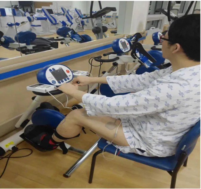
Eligible patients were randomly assigned to two groups. Eight patients (mean age 63.25±15.00 years) were assigned to the electrical stimulation group and another eight patients (mean age 63.25±14.12 years) to the control group. All patients underwent assisted ergometer training five times per week for 4 weeks to maintain a pedaling cadence of 30 rpm for 30 minutes. The electrical stimulation group received FES on the paretic quadriceps,
hamstring, gluteus maximus, and tibialis anterior muscles using two EMG-FES 2000Ⓡ
(Cyber Medic, Seoul, Korea) during assisted ergometer training. One was applied to paretic quadriceps and tibialis anterior, and the other was applied to hamstring and gluteus maximus (Fig. 1, 2). The stimulation protocol consisted of a symmetrical biphasic square pulse at 60 Hz, a constant pulse width of 300 µsec, and a duty cycle of 1.0 sec on and 1.0 sec off alternatively. We endeavored to synchronize FES stimulation timing to the cycling movement according to each muscle activation pattern, although we could not use an automatic synchronizing FES ergometer. The maximum power output of the stimulator was 100 mA. The stimulation intensity was increased within the range of motor threshold to the maximal comfortably tolerated intensity.
6
7
Figure 2. FES electrodes were applied to the paretic quadriceps, tibialis anterior, hamstring and gluteus maximus
8
Exercise treadmill testing protocol
Both groups underwent the symptom-limited low velocity graded treadmill test using a
modified Harbor protocol (Buchfuhrer et al., 1983; Macko et al., 1997). The exercise test was
performed using a CH2000, Integrated 12-lead Stress Testing ECG (Cambridge Heart,
Tewksbury, USA), Quark CPET Breath Gas Exchange data analysis (Cosmed Inc, Rome,
Italy), a T-2100 treadmill (GE, Fairfield, USA), and a BP (blood pressure) and HR (heart rate)
monitor. The initial round of treadmill walking without an incline was used to assess gait
safety and to select the target walking velocity for subsequent maximal-effort graded
treadmill testing. The zero-incline treadmill test was started at 0.5 mph and slowly advanced
by 0.1 mph increments according to the patient’s subjective tolerance and observer-rated gait
stability. Patients capable of performing ≥3 consecutive minutes of treadmill walking at ≥0.5
mph were allowed 15 min of seated rest, after which they underwent constant-velocity,
progressively graded treadmill exercise testing. The test was started with the participants
walking at zero-incline treadmill for the initial 2 min, followed by 2 min at 4% incline.
Then, the grade subsequently was increased 2% incline every 2 min until the test was
terminated at volitional exhaustion or in the presence of gait instability or cardiovascular
9
10
Outcome measures
Assessment of aerobic capacity
Peak oxygen consumption (Vo2 peak), metabolic equivalent (MET), resting and maximal
heart rate (RHR, MHR), resting and maximal blood pressure (RBP, MBP), maximal rate
pressure product (MRPP), exercise duration, respiratory exchange ratio (RER), estimated
anaerobic threshold (AT), submaximal rate pressure product (SRPP) and submaximal rate of
perceived exertion (SRPE) were determined before and after the exercise test.
Peak oxygen consumption (Vo2 peak) is the maximal oxygen uptake or the maximum
volume of oxygen that can be utilized in one minute during maximal or exhaustive
exercise. It is measured as milliliters of oxygen used in one minute per kilogram of body
weight, and it is one factor that can determine a capacity to perform sustained exercise.
Vo2 peak is also expressed in metabolic equivalents (MET), these are generally considered the
best indicator of cardiorespiratory endurance and aerobic fitness. Vo2 peak was reached by all
participants as defined by meeting the following criteria: (1) increase in Vo2 < 150 mL in the
final minute of exercise, i.e., a Vo2 plateau; (2) peak RER > 1.0; (3) peak SBP > 200 mmHg;
and (4) MHR within 15 beats per minute of the predicted maximal heart rate [220-
age] (Howley et al., 1995).
11
consumed in one breath. RER increase with the exercise intensity, and measured RER under
steady state conditions is commonly used to show the muscle’s oxidative capacity to get
energy (Simonson et al., 1990; Pendergast et al., 2000). The Vo2 peak and RER were
determined using a Quark CPET Breath Gas Exchange data analysis (Cosmed Inc, Rome,
Italy) as 15 sec averages during each stage of the test.
Resting heart rate (RHR) was recorded after a 15 min rest period, and maximal heart rate
(MHR) was analysed the average heart rate during the last 30 sec of exercise using an integrated 12-lead Stress Testing ECG (Cambridge Heart, Tewksbury, USA). Systolic (SBP) and diastolic blood pressure (DBP) were measured at rest, every 2 min during exercise. The rate pressure product (RPP) was defined as the workload or oxygen demand of the heart, and reflects hemodynamic stress. Maximal rate pressure product (MRPP) was multiplied of the heart rate and blood pressure (Thompson et al., 2009) with the maximal value defined as the average of the values recorded during the last 30 sec of the test.
And anaerobic threshold (AT) was estimated using gas exchange variables, including rate of pulmonary ventilation (VE), utilization of oxygen (O2) and production of carbon dioxide
(CO2). AT is the point at which lactate begins to accumulate in blood, causing an increase in
ventilation, and is important to document clinically. This is often termed the ventilator threshold (VT), provide better indices of aerobic fitness, especially for sustainable submaximal work and competitive endurance performance. The modified V-slope and the
12
ventilator equivalents methods have been reported to be sensitive and reliable for determining the AT (Gaskill, 2001). Ventilator equivalent method is the intensity of activity which causes the first rise in the ventilatory equivalent of oxygen (VE/O2) without a concurrent rise in the
ventilatory equivalent of carbon dioxide (VE/CO2). Modified V-slope method is the intensity
of exercise which, in a plot of the minute production of CO2 over the minute utilization of
oxygen (O2).
And, at point of estimated AT, submaximal rate pressure product (SRPP) and borg rating of perceived exertion (SRPE) (from 6 to 20) were recorded.
Exercise duration was defined as the time from the initiation to the termination of the exercise test protocol.
13
Assessment of functional ability
Functional abilities measured by the six minute walk test (6MWT), Berg balance
scale (BBS), and the Korean version of the modified Barthel index (K-MBI) were evaluated
at the beginning and the end of treatment.
Gait function was evaluated with the six minute walk test (6MWT). The test has been
widely used to measure of functional capacity targeted at people with at least moderately
severe impairment (Paul, 2003). It measures the distance that a patient can walk along a
flat 50 m track comfortably in a period of 6 minutes without support when possible.
Berg balance scale (BBS) was evaluated, which is a widely used clinical test of a person's
static and dynamic balance abilities (Blum, 2008). It is a valid instrument used for
evaluation of balance function by assessing the performance of 14 functional tasks.
Korean version of the BBS has been validated (Jung et al., 2006) (Appendix 1).
Korean-modified Barthel index (K-MBI) was used to evaluate activities of daily living
(ADL) functions. The modified Barthel index (MBI) is a measure of ADL, which shows the
degree of independence of a patient from any assistance. It covers 10 domains of functioning
(activities): bowel control, bladder control, as well as help with personal hygiene, bathing,
feeding, dressing, toilet use, transfers, walking, and climbing stairs. The MBI is a reliable and
14
15
Statistical analysis
Statistical analysis was performed using SPSS for Windows version 18.0 (SPSS Inc., Chicago, IL, USA). The Chi-square and Mann-Whitney tests were used to compare the baseline characteristics of the two groups. Wilcoxon’s rank sum test was also used to compare the exercise capacity and functional ability within each group at baseline and 4 weeks after testing. Repeated measures analysis of variance (ANOVA) was used for the inter- group comparison of all parameters of exercise capacity and functional ability between the two groups according to the time period until 4 weeks. A p-value < 0.05 was considered statistically significant.
16
III. RESULTS
General characteristics of the participants
The baseline characteristics of the participants are summarized in Table 1. Eight participants were assigned to each study group and each included four men. The mean age at enrolment was 63.25±15.00 years in the electrical stimulation group and 63.25±14.12 years in the control group. Demographic characteristics including gender, age, time since cardiovascular accident (CVA), height and weight were not significantly different.
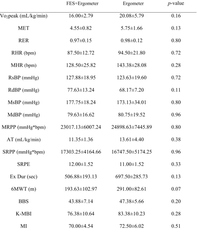
Data on the baseline exercise capacity, functional ability, and motor function (Mortricity Index- the hemiparetic leg score) (Appendix 3) of the two groups are presented in Table 2, and any significant differences were not found between the two groups.
17
Table 1. Baseline characteristics of the two groups
FES+Ergometer Ergometer p-value
Patients (numbers) 8 8
Gender (Male/Female) 4/4 4/4
Age (years) 63.25±15.00 63.25±14.12 0.96
Infarction/Hemorrhage 4/4 6/2
Side of lesion (Right/Left) 5/3 5/3
Time since CVA (days) 62.50±52.23 57.38±34.63 0.88
Height (cm) 158.13±8.80 162.13±9.37 0.38
Weight (kg) 60.76±7.26 55.00±22.96 0.96
Values are presented as mean±standard deviation or number of cases. FES, functional electrical stimulation., CVA, cerebrovascular accident.
18
Table 2. Exercise capacity, functional ability and motor function at baseline
FES+Ergometer Ergometer p-value
Vo2peak (mL/kg/min) 16.00±2.79 20.08±5.79 0.16 MET 4.55±0.82 5.75±1.66 0.13 RER 0.97±0.15 0.98±0.12 0.80 RHR (bpm) 87.50±12.72 94.50±21.80 0.72 MHR (bpm) 128.50±25.82 143.38±28.08 0.28 RsBP (mmHg) 127.88±18.95 123.63±19.60 0.72 RdBP (mmHg) 77.63±13.24 68.17±7.20 0.11 MsBP (mmHg) 177.75±18.24 173.13±34.01 0.80 MdBP (mmHg) 79.63±16.62 80.75±19.52 0.96 MRPP (mmHg*bpm) 23017.13±6007.24 24898.63±7445.89 0.80 AT (mL/kg/min) 11.35±1.36 13.61±4.40 0.38 SRPP (mmHg*bpm) 17303.25±4164.66 16747.50±5174.25 0.96 SRPE 12.00±1.52 11.00±1.52 0.33 Ex Dur (sec) 506.88±193.13 697.50±285.73 0.13 6MWT (m) 193.63±102.97 291.00±82.61 0.07 BBS 43.88±7.14 47.38±5.66 0.20 K-MBI 76.38±10.64 83.38±10.23 0.28 MI 70.00±4.54 72.50±6.02 0.51
Values are presented as mean±standard deviation.
FES, Functional electrical stimulation; Vo2 peak,peak oxygen consumption; MET, metabolic
equivalent; RER, respiratory exchange ratio; RHR, resting heart rate; MHR, maximal heart rate; RsBP, resting systolic blood pressure; RdBP, resting diastolic blood pressure; MsBP, maximal systolic blood pressure; MdBP, maximal diastolic blood pressure; MRPP, maximal
19
rate pressure product; AT, estimated anaerobic threshold; SRPP, submaximal rate pressure product; SRPE, submaximal rate of perceived exertion; Ex Dur, exercise duration; 6MWT, six minute walk test; BBS, Berg balance scale; K-MBI, modified Barthel index; MI, motricity index.
20
Changes of parameters before and after treatment within each group
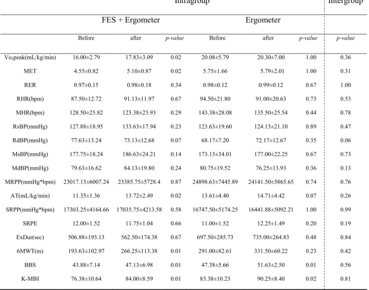
At 4 weeks after treatment, the FES assisted ergometer training group showed a significant improvement in both exercise capacity and functional ability (Table 3). Vo2 peak
increased from 16.00±2.79 mL/kg/min to 17.83±3.09 mL/kg/min (p=0.02), MET increased from 4.55±0.82 to 17.83±3.09 (p=0.02), and the estimated AT increased from 11.35±1.36 mL/kg/min to 13.72±2.49 mL/kg/min (p=0.02). In addition, 6MWT increased from 193.63±102.97 m to 266.25±113.38 m (p=0.01), BBS increased from 43.88±7.14 to 47.13±6.98 (p=0.01), and K-MBI increased from 76.38±10.64 to 84.00±8.59 (p=0.01). The control group showed no significant changes in exercise capacity, whereas statistically significant improvements occurred in the BBS (p=0.01) and the K-MBI (p=0.02).
21
Changes of parameters before and after treatment between the two groups
After 4 weeks, no significant intergroup differences were found in the exercise capacity and functional ability (Table 3).
22
Table 3. Changes of parameters before and after treatment within each group and between the two group
Intragroup Intergroup
FES + Ergometer Ergometer
Before after p-value Before after p-value p-value
Vo2peak(mL/kg/min) 16.00±2.79 17.83±3.09 0.02 20.08±5.79 20.30±7.00 1.00 0.36 MET 4.55±0.82 5.10±0.87 0.02 5.75±1.66 5.79±2.01 1.00 0.31 RER 0.97±0.15 0.98±0.18 0.34 0.98±0.12 0.99±0.12 0.67 1.00 RHR(bpm) 87.50±12.72 91.13±11.97 0.67 94.50±21.80 91.00±20.63 0.73 0.53 MHR(bpm) 128.50±25.82 123.38±23.93 0.29 143.38±28.08 135.50±25.54 0.44 0.78 RsBP(mmHg) 127.88±18.95 133.63±17.94 0.23 123.63±19.60 124.13±21.10 0.89 0.47 RdBP(mmHg) 77.63±13.24 73.13±12.68 0.07 68.17±7.20 72.17±12.67 0.35 0.06 MsBP(mmHg) 177.75±18.24 186.63±24.21 0.14 173.13±34.01 177.00±22.25 0.67 0.73 MdBP(mmHg) 79.63±16.62 84.13±19.80 0.24 80.75±19.52 76.25±13.93 0.36 0.13 MRPP(mmHg*bpm) 23017.13±6007.24 23385.75±5728.4 0.87 24898.63±7445.89 24141.50±5865.65 0.74 0.76 AT(mL/kg/min) 11.35±1.36 13.72±2.49 0.02 13.61±4.40 14.71±4.42 0.07 0.26 SRPP(mmHg*bpm) 17303.25±4164.66 17035.75±4213.58 0.58 16747.50±5174.25 16441.88±5092.21 1.00 0.99 SRPE 12.00±1.52 11.75±1.04 0.66 11.00±1.52 12.25±1.49 0.20 0.19 ExDur(sec) 506.88±193.13 562.50±174.38 0.67 697.50±285.73 735.00±264.83 0.48 0.84 6MWT(m) 193.63±102.97 266.25±113.38 0.01 291.00±82.61 331.50±60.22 0.23 0.42 BBS 43.88±7.14 47.13±6.98 0.01 47.38±5.66 51.63±2.50 0.01 0.56 K-MBI 76.38±10.64 84.00±8.59 0.01 83.38±10.23 90.25±8.40 0.02 0.81
Values are presented as mean±standard deviation.
FES, functional electrical stimulation; Vo2 peak,peak oxygen consumption; MET, metabolic
equivalent; RER, respiratory exchange ratio; RHR, resting heart rate; MHR, maximal heart rate; RsBP, resting systolic blood pressure; RdBP, resting diastolic blood pressure; MsBP, maximal systolic blood pressure; MdBP, maximal diastolic blood pressure; MRPP, maximal rate pressure product; AT, estimated anaerobic threshold; SRPP, submaximal rate pressure product; SRPE, submaximal rate of perceived exertion; Ex Dur, exercise duration; 6MWT, six minute walk test; BBS, Berg balance scale; K-MBI, modified Barthel index.
23
VI. DISCUSSION
The results of this study provide evidence that FES assisted ergometer training in the hemiparetic muscles of subacute stroke patients improved their aerobic capacities as well as functional abilities.
Several investigations (Szecsi et al., 2008; Janssen et al., 2008; Skold et al., 2002; Raymond et al., 2002; Ferrante et al., 2006; Ferrante et al., 2008; Ambrosini et al., 2011; Lo et al., 2012; Smart et al., 2013) havedemonstrated similar beneficial effects of FES assisted ergometer training in patients with SCI, heart failure, and stroke. Szecsi et al. (Szecsi et al., 2008) assessed whether FES-assisted cycling on the paretic leg in subjects with hemiparesis in subacute stroke could improve quantifiable biomechanical parameters such as smoothness, power, and symmetry of cycling. They showed that FES-assisted cycling was associated with strengthening the affected leg, reestablishing the side balance of forces, and improving pedaling control, particularly in patients with hemiparesis in the early phase of convalescence after stroke. Although they did not evaluate functional abilities and aerobic capacities, they noted that objective goals such as functional outcomes can be achieved with the clinical application of this therapy. Lo et al. (Lo et al., 2012) also reported that cycling exercise with FES on the hemiparetic leg improved postural control and spasticity of the extremities in subacute stroke patients. They suggested that the application of FES to the affected leg during cycling training has additional benefits,
24
especially for patients with high muscle tone. These findings emphasized the beneficial effects of FES on hemiparetic legs specifically in subacute stroke patients.
Additionally, FES induced cycling on the both legs (affected-side and intact-side) has demonstrated significant improvement in lower extremity strength and functional ability in subacute stroke patients. Ferrante et al. (Ferrante et al., 2008) and Ambrosini et al. (Ambrosini et al., 2011) evaluated the effectiveness of FES induced cycling on both legs in subacute stroke patients. Their studies revealed that FES cycling training significantly improved lower extremity strength, motor control, motor recovery, and walking abilities such as motricity index, trunk control test, upright motor control test, and maximum isometric voluntary contraction (MVC) compared to cycling only. These results confirmed that FES cycling training applied in the subacute stroke phase may play an important role in facilitating and accelerating motor recovery. The investigators concluded that the improvements they observed resulted from increased sensorial input provided to the brain by FES.
The improvements observed through the six minute walk test (6MWT), Berg balance scale (BBS), and the Korean version of modified Barthel index (K-MBI) in our study are in line with the results cited above. However, in our study, FES applied only to the muscles on the hemiparetic side was effective in improving functional abilities. This suggests that FES induced cycling may facilitate coordinated muscle activity, relearning of how to execute movements of paretic muscles voluntarily, and induction of continuous use of the affected-
25
side extremity.
Janssen et al. (Janssen et al., 2008) reported that their FES assisted cycling training program on paretic lower extremities in chronic stroke patients improved cycling performance, functional performance, and aerobic exercise such as power of output (PO max), Vo2 peak, 6MWT, and BBS performance. Except for muscle strength, it had no additional
effect over assisted ergometer only. Our study reached a similar conclusion in subacute stroke patients in that it showed improvements in aerobic capacities such as Vo2 peak, MET,
and estimated AT in the FES assisted ergometer training group within 4 weeks. These results could be explained by repetitive voluntary contraction of leg muscles as an adaptation to FES, which also promotes circulatory adaptations, hypertrophy, and histochemical changes in muscles, all of which are helpful in relearning how to execute movements. The changes in response to therapy increase metabolic capacity, prevent muscle atrophy, and induce functional recovery. Also, FES helps patients to focus on treatment during cycling, thus improving the efficiency of the exercise (Ambrosini et al., 2011). Moreover, improvement in estimated AT provides meaningful evidence that FES could be used to improve exercise endurance. Also, our study found that FES plus assisted motorized cycling was beneficial for aerobic capacity in subacute stroke patients.
There are several study limitations. The most serious limitation is the small number of subjects evaluated, making it insufficient to monitor the effects of FES training. Second, a therapeutic intervention period of 4 weeks might be too short to demonstrate the added
26
effects of FES training, compared to the control. Third, this study could not evaluate the changes in muscle strength, mass, and muscle spasticity, although they might have influenced the functional ability. Fourth, subjects could not perfectly synchronize FES stimulation timing with the angular movement of the ergometer motor. Finally, subjects did not use the cycling graded exercise stress test, which could be applied in the sitting position. Thus, symptom-limited exercise tests had to be performed by walking or running, and the participants might have already reached functional recovery at baseline exercise test. Accordingly, additional large scale, long term follow-up studies considering FES synchronization and other relevant factors should be conducted to evaluate functional ability and aerobic capacity after FES assisted ergometer training exercise.
27
V. CONCLUSION
This study demonstrated that ergometer training for 4 weeks improved the functional ability of subacute stroke patients. In addition, aerobic capacity was improved after assisted ergometer training with a FES only. Therefore, we suggest assisted ergometer training with a FES as a useful alternative therapy for increasing the aerobic capacity of subacute stroke patients.
28
VI. REFERENCES
Ambrosini E, Ferrante S, Pedrocchi A, Ferrigno G, Molteni F. Cycling induced by electrical stimulation improves motor recovery in postacute hemiparetic patients: a randomized controlled trial. Stroke 2011;42:1068-73.
Blum Lisa, Korner-Bitensky Nicol. Usefulness of the Berg balance scale in stroke rehabilitation: a systematic review. Physical therapy 2008;88:559-566.
Bobath B. Adult hemiplegia: evaluation and treatment. 3rd ed. London: Butterworth- Heinemann Oxford; 1990. p. 1-5.
Brown D, Kautz S, Dairaghi C. Muscle activity adapts to anti-gravity posture during pedalling in persons with post-stroke hemiplegia. Brain 1997;120:825-37.
Brown D, Kautz S. Increased workload enhances force output during pedaling exercise in persons with poststroke hemiplegia. Stroke 1998;29:598-606.
Buchfuhrer MJ, Hansen JE, Robinson TE, Sue DY, Wasserman K, Whipp BJ. Optimizing the exercise protocol for cardiopulmonary assessment. J Appl Physiol 1983;55:1558-64. Collin C Wade D. Assessing motor impairment after stroke: a pilot reliability study. J
Neurology Neurosurg Psychiatry. 1990;53:576-579.
Ferrante S, Pedrocchi A, Ferrigno G, Molteni F. FES cycling treatment on hemiplegic patients: preliminary results. 11th Annual Conference of the Functional Electrical Stimulation Society. Miyagi-Zao, Japan; 2006.
29
stimulation improves the muscular strength and the motor control of individuals with post-acute stroke. Eur J Phys Rehabil Med 2008;44:159-67.
Fujiwara T, Liu M, Tanuma A, Hase K, Tsuji T. Pedaling exercise for neuromuscular re- education: a review. Crit Rev Phys Rehabil Med 2005;17:163-78.
Gaskill, SE, Ruby AJ, Walker OA, Sanchez RC, Serfass, Leon AS. Validity and reliability of combining three methods to determine ventilatory threshold. Med. Sci. Sports Exerc 2001;33:1841-1848
Gresham GE, Fitzpatrick TE, Wolf PA, McNamara PM, Kannel WB, Dawber TR. Residual disability in survivors of stroke-the framingham study. N Engl J Med 1975;293:954-6. Hancock NJ, Shepstone L, Winterbotham W, Pomeroy V. Effects of lower limb reciprocal
pedalling exercise on motor function after stroke: a systematic review of randomized and nonrandomized studies. Int J Stroke 2012;7:47-60.
Holt R, Kendrick C, McGlashan K, Kirker S, Jenner J. Static bicycle training for functional mobility in chronic stroke: case report. Physiotherapy 2001;87:257-60.
Hooker SP, Scremin AM, Mutton DL, Kunkel CF, Cagle G. Peak and submaximal physiologic responses following electrical stimulation leg cycle ergometer training. 1995;32:361-6.
Howley ET, Bassett DR, Jr., Welch HG. Criteria for maximal oxygen uptake: review and commentary. Med Sci Sports Exerc 1995;27:1292-1301.
30
for integration of electric motor assist and functional electrical stimulation in paraplegic cycling: utility for exercise testing and mobile cycling. IEEE Trans Neural Syst Rehabil Eng 2004;12:89-101.
Hwang IS, Lee HM, Cherng RJ, Chen JJ. Electromyographic analysis of locomotion for healthy and hemiparetic subjects--study of performance variability and rail effect on treadmill.Gait Posture 2003;18:1-12.
Janssen TW, Beltman JM, Elich P, Koppe PA, Konijnenbelt H, de Haan A, et al. Effects of electric stimulation-assisted cycling training in people with chronic stroke. Arch Phys Med Rehabil 2008;89:463-9.
Jorgensen HS, Nakayama H, Raaschou HO, Olsen TS. Stroke. Neurologic and functional recovery the Copenhagen Stroke Study. Phys Med Rehabil Clin N Am 1999;10:887-906. Jung HY, Park, JH, Shim JJ, Kim MJ, Hwang MR, Kim SH. Reliability test of Korean
version of Berg balance scale. J Korean Acad Rehab Med 2006;30:611-618
Jung HY, Park BK, Shin HS, Kang YK, Pyun SB, Paik NJ, Kim SH, Kim TH, Han TR. Development of the Korean version of modified Barthel index (K-MBI): Multi-center study for subjects with stroke. J Korean Acad Rehabil Med 2007;31:283-297.
Katz-Leurer M, Carmeli E, Shochina M. The effect of early aerobic training on independence six months post stroke. Clin Rehabil 2003;17:735-41.
Katz-Leurer M, Sender I, Keren O, Dvir Z. The influence of early cycling training on balance in stroke patients at the subacute stage. Results of a preliminary trial. Clin Rehabil
31
2006;20:398-405.
Kesar TM, Perumal R, Jancosko A, Reisman DS, Rudolph KS, Higginson JS, et al. Novel patterns of functional electrical stimulation have an immediate effect on dorsiflexor muscle function during gait for people poststroke. Phys Ther 2010;90:55-66.
Lee SY, Im SH, Kim BR, Choi JH, Lee SJ, Han EY. The effects of neuromuscular electrical stimulation on cardiopulmonary function in healthy adults. Ann Rehabil Med 2012;36:849-56.
Lo HC, Hsu YC, Hsueh YH, Yeh CY. Cycling exercise with functional electrical stimulation improves postural control in stroke patients. Gait Posture 2012;35:506-10.
Macko RF, Katzel LI, Yataco A, Tretter LD, DeSouza CA, Dengel DR, et al. Low-velocity graded treadmill stress testing in hemiparetic stroke patients. Stroke 1997;28:988-92. Mayo NE, Wood-Dauphinee S, Ahmed S, Gordon C, Higgins J, Mcewen S, et al. Disabil
Rehabil 1999;21:258-68.
Nash MS, Montalvo BM, Applegate B. Lower extremity blood flow and responses to occlusion ischemia differ in exercise-trained and sedentary tetraplegic persons. Arch Phys Med Rehabil 1996;77:1260-5.
Enright PL. The six-minute walk test. Respir Care 2003;48:783-785.
Pendergast DR, Leddy JJ, Venkatraman JT. A perspective on fat intake in athletes. J. Am. Coll Nutr. 2000;19:345–50.
32
spinal cord injured patients. Cent Nerv Syst Trauma 1984;1:57-74.
Petrofsky JS, Stacy R. The effect of training on endurance and the cardiovascular responses of individuals with paraplegia during dynamic exercise induced by functional electrical stimulation. Eur J Appl Physiol Occup Physiol 1992;64:487-92.
Petrofsky JS, Laymon M. The effect of previous weight training and concurrent weight training on endurance for functional electrical stimulation cycle ergometry. Eur J Appl Physiol 2004;91:392-8.
Potempa K, Lopez M, Braun LT, Szidon JP, Fogg L, Tincknell T. Physiological outcomes of aerobic exercise training in hemiparetic stroke patients. Stroke 1995;26:101-5.
Quaney BM, Boyd LA, McDowd JM, Zahner LH, He J, Mayo MS, et al. Aerobic exercise improves cognition and motor function poststroke. Neurorehabil Neural Repair
2009;23:879-85.
Raasch CC, Zajac FE. Locomotor strategy for pedaling: muscle groups and biomechanical functions. J Neurophysiol 1999;82:515-25.
Raymond J, Davis GM, Climstein M, Sutton JR. Cardiorespiratory responses to arm cranking and electrical stimulation leg cycling in people with paraplegia. Med Sci Sports Exerc 1999;31:822-8.
Raymond J, Schoneveld K, Van Kemenade CH, Davis GM. Onset of electrical stimulation leg cycling in individuals with paraplegia. Med Sci Sports Exerc 2002;34:1557-62.
33
stimulation improves severe hand dysfunction for individuals with chronic stroke: a pilot study. 2006;30:175.
Sheffler LR, Chae J. Neuromuscular electrical stimulation in neurorehabilitation. Muscle Nerve 2007;35:562-90.
Simonson DC, DeFronzo RA. Indirect calorimetry: methodological and interpretative problems. Am. J. Physiol. 1990;258:399–12
Skold C, Lonn L, Harms-Ringdahl K, Hultling C, Levi R, Nash M, et al. Effects of functional electrical stimulation training for six months on body composition and spasticity in motor complete tetraplegic spinal cord-injured individuals. J Rehabil Med 2002;34:25-32. Smart NA, Dieberg G, Giallauria F. Functional electrical stimulation for chronic heart failure:
a meta-analysis. Int J Cardiol 2013;167:80-6
Szecsi J, Krewer C, Müller F, Straube A. Functional electrical stimulation assisted cycling of patients with subacute stroke: kinetic and kinematic analysis. Clin Biomech 2008;23:1086-94.
Takahashi T, Takazawa M, Nishiyama Y, Nakano E, Handa Y. FES cycling chair for the lower limbs disabled people with electric motor power assist. 9th Annual Conference of the International FES Society; 2004 Sep; UK. Bournemouth; 2004. 247-9.
Thompson WR, Gordon NF, Pescatello LS. ACSM's guidelines for exercise testing and prescription. 9thed. Baltimore:LippincottWilliams&Wilkins;HubstaLtd;2009.
34
VII. ABSTRACT IN KOREAN
아급성기 뇌졸중 환자에서
기능적 전기자극을 이용한 에르고미터의 적용이
운동량과 기능적 능력에 미치는 영향
이 소 영
제주대학교 일반대학원 의학과
지도교수 강 사 윤
목 적 : 본 연 구 는 아 급 성 기 뇌 졸 중 환 자 에 서 에 르 고 미 터 유 산 소 운 동 이 운동심폐기능과 보행, 기능적 능력에 미치는 영향과 기능적 전기자극을 이용하여 에르고미터 운동을 시행 하였을 때 미치는 부가적 효과에 대하여 알아보고자 한다. 방법: 총 16 명(실험군 8 명, 대조군 8 명)의 아급성기 뇌졸중 환자들이 연구에 모집되었다. 무작위로 두 군으로 나누어 실험군은 마비측 다리에 신경전기자극을 적용하도록 하고 대조군은 적용하지 않도록 하여 하루 30 분씩, 주 5 회, 총 4 주 동안 에르고미터 유산소 운동을 시행하였다. 모든 피험자들은 증상 제한적, 단 계 적 저 속 트 레 드 밀 운 동 부 하 검 사 를 시 행 하 였 으 며 운 동 부 하 검 사 의 평 가35 지표들로 최고 산소소비량, 단위 대사당량, 최고 호흡교환율, 휴식시와 최고 심박수, 휴식시와 최고 수축기 및 이완기 혈압, 최고 심근산소소비량, 예측성 무산소 역치, 최대하 심근산소소비량, 최대하 자각적 운동강도, 평가 수행 시간을 측정하였다. 그 외에 기능적 상태 평가 지표로서 보행기능의 평가 지표인 6 분 걷기 검사와 균형 능력의 평가 지표인 버그 균형지수, 일상생활동작 평가 지표인 한글판 수정바델지수를 평가하였다. 4 주의 재활치료 후에, 모든 평가지표를 반복적으로 측정하였다. 결과: 4 주의 재활치료 후, 실험군에서는 최고 산소소비량, 단위 대사당량, 예측성 무 산 소 역 치 , 6 분 걷 기 검 사 , 버 그 균형 지 수 , 한 글 판 수정 바 델지 수 에서 , 대 조 군 에 서 는 버 그 균 형 지 수 , 한 글 판 수 정 바 델 지 수 에 서 유 의 한 호 전 이 관찰되었다. 그러나 재활치료 전후의 모든 평가지표의 변화량은 두 군간에 유의한 차이점이 관찰되지 않았다. 결론: 아급성기 뇌졸중 환자에서 4 주간의 에르고미터 유산소 운동은 환자의 기능적 능력 호전에 유의한 영향을 끼친다. 또한 기능적 전기자극을 이용하여 에 르 고 미 터 유 산 소 운 동 을 했 을 때 에 르 고 미 터 운 동 만 하 는 경 우 보 다 운동심폐기능에 부가적인 호전을 가져올 수 있다는 것을 알 수 있었다.
36
VIII. APPENDICES
44
48