ISSN 2234-3806 • eISSN 2234-3814
http://dx.doi.org/10.3343/alm.2016.36.6.617 www.annlabmed.org
617
Ann Lab Med 2016;36:617-619
http://dx.doi.org/10.3343/alm.2016.36.6.617
Letter to the Editor
Clinical Microbiology
Identification of
Pasteurella canis in a Soft Tissue
Infection Caused by a Dog Bite: The First Report in
Korea
Bongyoung Kim, M.D.1, Hyunjoo Pai, M.D.1, Kwang-hyun Lee, M.D.2, and Yangsoon Lee, M.D.3
Departments of Internal Medicine1, Orthopediatrics2, and Laboratory Medicine3, Hanyang University of College of Medicine, Seoul, Korea
Dear Editor,
Pasteurella canis is a small, gram-negative coccobacillus that is a part of the normal flora of healthy domestic animals, especially dogs [1]. Although P. canis is a well-known major pathogen of infections caused by dog bites, only few cases of human infec-tions with P. canis have been reported [2-5]. We report a case of soft tissue infection caused by P. canis.
A 54-yr-old woman without underlying diseases visited the emergency department with multiple lacerations caused by a dog bite incurred two days prior. She had received immediate treatment with wound dressing, antibiotics (amoxicillin/clavu-lanic acid), and tetanus immunoglobulin at another hospital on the day she was bitten. However, the wound was worsening. The initial vital signs were as follows: temperature, 37.7°C; pulse rate, 88/min; respiratory rate, 20/min; blood pressure 108/73 mmHg. The wounds were located on the right distal arm (3 cm in the anteromedial area, 3 cm in the anterolateral area, and 4 cm in the posterior area with muscle exposure). Abnormalities such as fracture were absent on plain radiographs.
The initial blood counts were as follows: white blood cells (WBCs), 10.4×10⁹/L (neutrophils, 80.5%); hemoglobin, 12.4 g/
dL; and platelets 173 ×109/L. No prominent abnormalities in
blood chemistry were observed on routine tests except for mildly
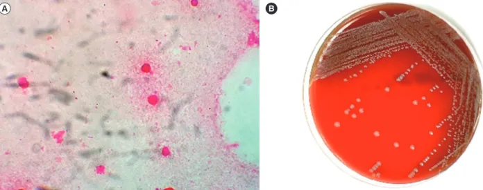
elevated acute phase reactants (C-reactive protein [CRP], 5.82 mg/dL; erythrocyte sedimentation rate [ESR], 52 mm/hr). The patient was admitted, and amoxicillin/clavulanic acid were ad-ministered immediately and maintained throughout the hospi-talization period. Tetanus and rabies vaccines were also given to prevent potential infections. Microbiological examination of the wounds was performed. A specimen was collected for Gram staining and culture. Gram-negative coccobacilli were observed on a direct gram-stained smear from the wound specimens (Fig. 1A). Pus specimens were inoculated on 5% sheep blood agar, chocolate agar, and MacConkey agar. After 24 hr, the growth of smooth, grayish-white colonies was observed on the blood agar and chocolate agar but not on the MacConkey agar (Fig. 1B). The colonies were positive for catalase and oxidase reactions. All isolates were identified with MicroScan Walkaway (Beckman Coulter, Brea, CA, USA) and Vitek automated bacterial identifi-cation systems 2 (bioMérieux, Marcy-l’Etoile, France) and a VI-TEK MS matrix-assisted laser desorption ionization mass spec-trometry (MALDI-TOF MS) system (bioMérieux).
For further identification of bacterial species, 16S ribosomal RNA (rRNA) gene sequencing was carried out. The universal primers 8F (5´-TTGGAGAGTTTGATCCTGGCTC-3´) and 801R (5´-GGCGTGGACTTCCAGGGTATCT-3´) were used to amplify
Received: March 7, 2016 Revision received: April 24, 2016 Accepted: June 17, 2016
Corresponding author: Yangsoon Lee
Department of Laboratory Medicine, Hanyang University Seoul Hospital, Hanyang University College of Medicine, 222-1 Wangsimni-ro, Seongdong-gu, Seoul 04763, Korea
Tel: +82-2-2290-9650, Fax: +82-2-2290-9193 E-mail: [email protected]
© The Korean Society for Laboratory Medicine.
This is an Open Access article distributed under the terms of the Creative Commons Attribution Non-Commercial License (http://creativecommons.org/licenses/by-nc/4.0) which permits unrestricted non-commercial use, distribution, and reproduction in any medium, provided the original work is properly cited.
Kim B, et al.
Pasteurella canis: first report in Korea
618
www.annlabmed.org http://dx.doi.org/10.3343/alm.2016.36.6.617parts of the 16S rRNA gene. The VITEK MS and Vitek 2 (bio-Mérieux) analyses identified P. canis, whereas the MicroScan Walkaway analysis identified Pasteurella multocida. The 16S rRNA gene sequences of the strain in this study were most closely related to those of the P. canis type strain. The identity was 99.1% with strain CCUG 12400 (GenBank accession num-ber AY362919).
An antimicrobial susceptibility test was performed by using the CLSI disk diffusion method [6]. The isolate was susceptible to ampicillin (zone diameter, 27 mm), penicillin (30 mm), tetra-cycline (30 mm), ceftriaxone (34 mm), ciprofloxacin (29 mm), and trimethoprim/sulfamethoxazole (24 mm). On hospital day 3, secondary wound closure with surgical abscess drainage and debridement was performed, and the patient was discharged on hospital day 12. One week later, the treatment was com-pleted with removal of the stitches at the outpatient clinic.
Approximately 15-20% of dog bite wounds and ≥50% of cat bite wounds contain a mixture of aerobic and anaerobic bacte-ria reflecting the oral flora of the biting animal [7]. Studying dog bites, Talan et al [1] reported that the most commonly identified aerobic and anaerobic bacteria are Pasteurella spp. (50%), Streptococcus spp. (46%), Staphylococcus spp. (46%), Fuso-bacterium spp. (32%), and Bacteroides spp. (30%). The same study identified P. canis in 26% of dog bites, which is a relatively high rate. However, this species has typically been overlooked in the clinical setting because gram-negative bacilli do not grow on MacConkey agar. In the present study, MicroScan Walkaway (Beckman Coulter) analysis identified the bacteria as P. multo-cida owing to the absence of a P. canis database.
Empirical antibiotics for dog and cat bites should cover Pas-teurella spp., Streptococcus spp., Staphylococcus spp., and an-aerobes. Therefore, β-lactam/β-lactamase inhibitors, a second-generation cephalosporin with anaerobic activity, or combination therapy with either penicillin and a first-generation cephalospo-rin or clindamycin and a fluoroquinolone are recommended [1]. Pasteurella spp. are generally susceptible to most antibiotics, including penicillin G, amoxicillin/clavulanic acid, piperacillin/ tazobactam, doxycycline, fluoroquinolones, advanced cephalo-sporins, and carbapenems [8-10]. Given this information, we chose amoxicillin/clavulanic acid for the initial treatment of this patient.
This report is the first of P. canis isolation from a dog bite wound in a human in Korea. The results of this study empha-size the importance of proper laboratory identification for the di-agnosis and treatment of infections caused by animal bites.
Authors’ Disclosures of Potential Conflicts of
Interest
No potential conflicts of interest relevant to this article were re-ported.
Acknowledgments
This study was supported by the research fund of Hanyang Uni-versity (HY-201400000002367).
Fig. 1. Microscopic and colonial morphology of Pasteurella canis. (A) Gram-negative coccobacilli on a direct gram-stained smear from the wound specimen (×1,000). (B) Smooth, grayish colonies on 5% sheep blood agar after 48-hr incubation.
Kim B, et al.
Pasteurella canis: first report in Korea
http://dx.doi.org/10.3343/alm.2016.36.6.617 www.annlabmed.org
619
REFERENCES
1. Talan DA, Citron DM, Abrahamian FM, Moran GJ, Goldstein EJ. Bacte-riologic analysis of infected dog and cat bites. Emergency Medicine Ani-mal Bite Infection Study Group. N Engl J Med 1999;340:85-92. 2. Albert TJ and Stevens DL. The first case of Pasteurella canis
bactere-mia: a cirrhotic patient with an open leg wound. Infection 2010;38:483-5.
3. Castellano I, Marín JP, Gallego S, Mora M, Rangel G, Suarez MA, et al. Pasteurella canis peritonitis in a peritoneal dialysis patient. Perit Dial Int 2011;31:503-4.
4. Hara H, Ochiai T, Morishima T, Arashima Y, Kumasaka K, Kawano KY. Pasteurella canis osteomyelitis and cutaneous abscess after a domestic dog bite. J Am Acad Dermatol 2002;46:S151-2.
5. Hazelton BJ, Axt MW, Jones CA. Pasteurella canis osteoarticular infec-tions in childhood: review of bone and joint infecinfec-tions due to pasteurella species over 10 years at a tertiary pediatric hospital and in the literature.
J Pediatr Orthop 2013;33:e34-8.
6. Clinical and Laboratory Standards Institute. Methods for antimicrobial dilution and disk susceptibility testing of infrequently isolated or fastidi-ous bacteria. 2nd ed. Approved guideline, M45-A2. Wayne, PA: Clinical and Laboratory Standards Institute, 2010.
7. Abrahamian FM and Goldstein EJ. Microbiology of animal bite wound infections. Clin Microbiol Rev 2011;24:231-46.
8. Lion C, Conroy MC, Carpentier AM, Lozniewski A. Antimicrobial suscep-tibilities of Pasteurella strains isolated from humans. Int J Antimicrob Agents 2006;27:290-3.
9. Stevens DL, Bisno AL, Chambers HF, Dellinger EP, Goldstein EJ, Gor-bach SL, et al. Practice guidelines for the diagnosis and management of skin and soft tissue infections: 2014 update by the Infectious Diseas-es Society of America. Clin Infect Dis 2014;59:147-59.
10. Citron DM, Warren YA, Fernandez HT, Goldstein MA, Tyrrell KL, Gold-stein EJ. Broth microdilution and disk diffusion tests for susceptibility testing of Pasteurella species isolated from human clinical specimens. J Clin Microbiol 2005;43:2485-8.