Kor J Oral Maxillofac Pathol 2013;37(4):133-140
염증성 사이토카인과 구강 편평세포암종
김도경
1), 배정윤
1), 박영진
1), 손화경
2), 이재훈
3), 김진
1)*
연세대학교 치과대학 구강병리학교실, 구강종양연구소1), 백석대학교 치위생학과2), 연세대학교 치과대학 보철학교실3)
<Abstract>
Inflammatory Cytokines and Oral Squamous Cell Carcinoma
Do Kyeong Kim
1), Jung Yoon Bae
1), Young Jin Park
1), Hwa Kyung Son
2), Jae Hoon Lee
3), Jin Kim
1)*
Department of Oral pathology, Oral Cancer Research Institute, Yonsei University College of Dentistry1), Department of Dental hygiene, Baekseok University2), Department of Prosthodontics, Yonsei University College of Dentistry3)
Inflammation functions as a double-edged sword against external stimulus. For instance, inflammation can have anti-cancer effect and simultaneously can play cancer-promoting factors. Recent studies have shown that cytokine plays an important role in tumor biology by influencing tumor growth, invasion and metastasis. We classify these cytokines by cancer type and review current knowledge of cytokines in terms of carcinogenesis. Here, we also focus on whether cytokines can act as biomarkers for early detection of oral squamous cell carcinoma (OSCC). This review will provide basis for further approach to study the role of cytokines in carcinogenesis and evaluating the possibilities of cytokines as biomarkers for cancer detection.
Key words:Cytokines, Biomarkers, Inflammation, Oral squamous cell carcinoma
* Correspondence: Jin Kim, Department of Oral Pathology, Oral Cancer Research Institute, Yonsei University College of Dentistry,134 Shinchon-Dong, Seoul 120-752, Republic of Korea. Tel: +82-2-2228-3030, Fax: +82-2-392-2959, E-mail: [email protected] * 본 논문은 교육부의 재원으로 한국연구재단의 지원을 받아 수행된 기초 연구사업 중 중점연구소 사업 (과제번호 : 2009-0094027)으로 이루어졌음.
Ⅰ. 서론
염증(Inflammation)은 외부의 여러 유해한 자극에 대한 방 어 기전으로서 조직 손상의 회복을 돕는 반면, 과도한 염증반응 은 질병을 유발할 수도 있기 때문에 양날의 칼과 같은 기능을 갖는다1). 이러한 염증은 주요 매개자인 염증성 사이토카인 (Inflammatory cytokine)을 분비하여 염증반응의 증폭과 지속 을 조절한다2). 특히, 암세포에서 발현되는 염증성 사이토카인 은 암세포의 성장과 전이, 신생 혈관 형성 등과 같은 암 발달 과정을 촉진시킨다3,4). 암에서 발현되는 사이토카인은 종류가 무척 다양하고, 조직 손상의 회복을 유도하는 사이토카인과 양성(Benign tumor) 및 악성 종양(Malignant tumor)에서 발현되는 사이토카인이 특별 히 구분되지 않기 때문에5), 암에서 사이토카인의 발현이 보이 더라도 암 특이적으로 나타내는 바이오마커(Biomarker) 라기 보다는 단순히 염증반응에서 나타나는 비특이적인 표지자 (non-specific indicator)로 여겨 왔다. 그러나, 최근에 암세포 에서 발현되는 일부 사이토카인은 암에 대한 단순한 숙주의 면역 반응 이외에 신생혈관 형성, 악성 종양의 전이 등 생물학 적 악성도와 관련이 있는 것으로 알려지면서 염증성 사이토카 인의 중요성이 대두되었다2,3,6). 현재까지 일부 사이토카인이 각 종 암에서 세포의 성장 및 진행에 관여한다고 보고되어 있으나 세포 및 조직에 따라 발현되는 사이토카인의 종류 및 발현 정도 가 다르기 때문에 임상적으로 암 진단 바이오마커 적용에 대해 논란의 여지가 있다. 더구나 외부 자극으로부터 숙주를 보호하 는 면역반응에서의 사이토카인과 대조적으로 암의 발생과 진행Fig. 1. The cytokine that links inflammation and cancer 과정에서 이들 인자들의 변화에 대한 연구는 잘 알려져 있지 않다. 그렇기 때문에 암에서 발현되는 사이토카인의 특이적인 특성을 규명한다면 암 진단을 위한 새로운 바이오마커의 독자 적인 확보가 가능할 것이다. 따라서 본 논문은 암에서 많이 발현되는 염증성 사이토카인을 검토하고, 구강 편평세포암종 관련 검토를 통하여 암 조기 진단 표지자로서 염증성 사이토카인의 가능성을 확인하기 위한 기초 자료로 삼고자 한다. 1. 암에서 발현되는 염증성 사이토카인(Inflammatory cytokine) 염증성 사이토카인은 암에서 발현되어 염증성 네트워크를 형성하면서 암 생성 및 발달을 매개하는 데 중요한 역할을 한다 고 알려져 있으며, 이러한 염증성 사이토카인 중 IL-1α, IL-1β, IL-6, IL-8, TNF-α는 여러 암에서 가장 많이 발현하는 사이토카 인으로 염증관련 전사인자인 NF-κB, STAT3에 의해 조절되는 중요한 암 촉진 매개자이다7-10)(Fig. 1).
IL-1은 IL-1α와 IL-1β로 나뉘며, 많은 암 및 암 미세 환경에서 생성되어 전이 및 혈관생성 유전자(MMP 등)와 성장 인자 (VEGF, IL-8, IL-6, TNF-α, TGF-β)의 발현을 유도하면서 암의
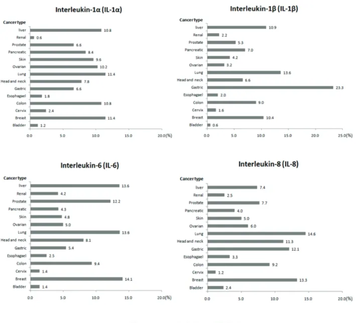
다단계 과정을 조절하는 주요 인자이다8). IL-6는 다양한 암에서 세포사멸(Apoptosis)을 억제하고 암의 성장을 촉진시키며, 최 근에는 상피세포의 간엽세포로의 변이(Epithelial-mesenchymal transition, EMT)를 매개하는 것으로 보고되어 암세포의 침윤 및 전이에 중요한 역할을 한다고 알려져 있다11,12). 또 다른 염증성 사이토카인인 IL-8은 비만세포, 중성구 등에서 생성되 어 염증반응에서 백혈구에 대한 화학주성인자로 작용하며 다 양한 암에서 암세포의 성장과 전이에 관여하는 것으로 알려져 있으며13), TNF-α는 병원성 자극에 의해 유도되는 염증반응에 관여하는 주요 인자이다. TNF-α는 세포를 변형, 증식하며 암을 촉진시킨다고 보고되어 있고, 또한 암 미세환경(Tumor microenvironment)에서 다른 염증성 사이토카인을 함께 유도 하며 염증성 반응을 조절한다14). 최근 10년 내에 등록된 논문을 대상으로 암에서 가장 많이 발현되는 다섯 가지 사이토카인, IL-1α, IL-1β, IL-6, IL-8, TNF-α 의 연구현황을 살펴보았다. 발생 부위별로 암을 분류하였으며
6)
, 각 암과 염증성 사이토카인을 함께(예를 들어, Bladder cancer and IL-1α) 검색어로 지정하였다. 그 결과 총 13,775편 의 논문이 검색되었으며, 이 중 IL-1α와 관련된 논문은 166편, IL-1β는 853편, IL-6는 3736편, IL-8는 1,910편, TNF-α는 7,110 편으로 분류되었다. IL-1α는 각각 19편씩 Breast cancer (11.4 %)와 Lung cancer (11.4 %)에서 가장 많은 연구 보고가 있음 을 확인하였고, IL-1β는 199편의 Gastric cancer (23.3 %), IL-6 는 526편의 Breast cancer (14.1 %), IL-8은 278편의 lung cancer (14.6 %), TNF-α는 1,102편의 Breast cancer (15.5 %) 에서 가장 많은 연구가 진행됨을 확인하였다(Fig. 2). 2. 자가 및 측 분비인자(Autocrine/Paracrine growth
effector)로서의 사이토카인
암 미세환경(Tumor microenvironment)은 섬유모세포 (Fibroblast), 내피세포(Endothelial cell), 염증세포(Inflammatory cell) 등으로 구성되어 있으며, 이러한 세포들은 사이토카인의 자가 및 측 분비(Autocrine / Paracrine manner)를 통해 자기 자신 또는 주변 세포들을 재구성(Reprogramming) 하면서 암
Table 1. Inflammatory cytokine as an autocrine and paracrine effector
Cytokine Forms of Secretion Cellular sources
IL-1α Autocrine Breast cancer cells, Cervix cancer cells, Skin cancer cells, Pancreatic cancer cells Paracrine Macrophage, Dendritic cells, B cells, Natural killer cells, Keratinocytes
IL-1β Autocrine Breast cancer cells, Skin cancer cells, Pancreatic cancer cells, Prostate cancer cells Paracrine Macrophage, Dendritic cells, B cells, Natural killer cells, Keratinocytes
IL- 6 Autocrine
Bladder cancer cells, Breast cancer cells, Cervix cancer cells, Esophageal cancer cells, Gastric cancer cells, Head and neck cancer cells, Lung cancer cells, Ovarian cancer cells, Skin cancer cells, Pancreatic cancer cells, Prostate cancer cells, Renal cancer cells, Liver cancer cells Paracrine Macrophages, T cells, B cells, Endothelial cells, Fibroblasts
IL- 8 Autocrine
Colon cancer cells, Gastric cancer cells, Head and neck cancer cells, Lung cancer cells, Skin cancer cells, Pancreatic cancer cells
Paracrine Macrophages, Mast cell, Neutrophil, T cells, Endothelial cells, Fibroblasts
TNF-α Autocrine Glioblastroma, Ovarian cancer cells, Prostate cancer cells, Renal cancer cells, Liver cancer cells Paracrine Macrophages, Natural killer cells, B cells, T cells, Neutrophils, Fibroblasts, Keratinocytes
즉, 동일한 사이토카인의 경우에도 세포에 따라 자가분비인 자 또는 측분비인자로 다른 기능을 수행한다. 따라서 암세포에 작용하는 대표적인 사이토카인을 자가 및 측 분비기능으로 나 누어 분류하였다(Table 1). IL-1α는 췌장암(Pancreatic cancer) 등의 암세포에서는 자가분비인자로서 작용하여 세포 증식에 관 여하고16), 측분비인자로서는 대식세포(Macrophage), 수지상세 포(Dendritic cell) 등에서 생성되어 암의 침윤을 촉진시키며, 암의 성장에 있어서 충분한 혈액공급을 유지하는 데에 기여한 다3). IL-6는 자가분비인자로서 암세포에서 분비되어 EMT와 같 은 암 미세환경의 변화를 통해 악성종양의 표현형을 유도하여 암의 악성도를 증가시키며 암세포의 성장을 촉진하며, 측분비 인자로서는 T림프구, B림프구, 대식세포, 섬유모세포 등 여러 세포에서 생성되며 류마티스 관절염(Rheumatoid arthritis), 건 선(Psoriasis), 암 등에서의 염증반응을 증가시킨다6,11,17). 그 밖에 사이토카인은 많은 암에서 자가분비(Autocrine manner)를 통해 암 세포의 증식(proliferation)을 촉진하여 직 접적으로 암 세포의 성장을 조절한다. 또한, 대식세포 등을 포 함한 면역세포와 섬유모세포 등에 생성된 사이토카인은 측분비 (Paracrine manner)를 통해서 면역 체계를 억제하여 암 미세 환경을 암이 촉진되는 환경으로 유도하고, 신생 혈관 생성, 전 이 등을 촉진하는 성장인자 및 사이토카인의 생성을 증가시켜 주변 세포가 암 세포의 성장을 간접적으로 조절하는 데 기여한 다18). 3. 암세포에서 발현되는 염증성 사이토카인의 수용체 (Receptor) 및 신호전달(Signaling pathway) 사이토카인의 자가 및 측분비(Autocrine / Paracrine manner) 에 의한 타깃 세포의 신호전달 체계(Signaling pathway)에서는 세포 표면에 존재하는 수용체(Receptor)가 외부로부터 오는 신 호를 감지하여 받아들이고 이러한 신호의 증폭을 통해 이후 대 사, 분비, 세포 성장과 같은 활동을 가능하게 한다. 많은 암 세포는 세포 표면의 사이토카인 수용체를 발현한다고 알려져 있으며, 수용체의 하향 신호전달체계가 암세포의 발달 및 생존을 조절하는 데에 중요한 역할을 한다(Table 2).
IL-1과 결합하는 수용체는 Immunoglobulin superfamily receptor의 종류로서, type1과 type2로 나뉘는데, 수용체와 결 합한 IL-1는 침윤 촉진인자 및 혈관 성장인자를 유도하며 발암
과정에 관여한다8,19). Type 1 cytokine receptor family 중 하나
인 IL-6 수용체는 IL-6와 그 수용체의 발현 정도에 따라서 암
환자의 예후에 영향을 미친다는 보고가 있으며20) 또한, IL-6
수용체의 하향 신호전달체계인 JAK-STAT 신호전달체계는 다 양한 신호전달체계(MAP kinase, EGFR, PI3K/AKT 등)와 상호
Receptor family Receptor- Cytokine (Ligand) Function Immunoglobulin Superfamily Receptors IL-1R1 IL-1R2 IL-1α
IL-1β Associated with cancer progression and a metastatic phenotype
,)
Type I Cytokine receptors IL-6R IL-6 Signals through JAK-STAT pathway
)
Associated with aggressiveness and a poor prognosis in cancer) Chemokine receptor
(G Protein-Coupled Receptors)
CXCR1
CXCR2 IL-8 Increases survival, proliferation and tumor cell migration
)
Tumor Necrosis Factor
Receptors (TNFR) TNFR TNF-α
Signals through NF-κB pathway)
Functions as co-stimulatory and co-inhibitory receptors,)
Table 2. Receptor family of cytokine and its function
Table 3. Cytokine in serum of patients with cancer
Cancer Secreted cytokine in serum
Bladder IL-6
Breast IL-1β, IL-6, IL-8, TNF-α
Cervix IL-6
Colon IL-6, IL-8
Esophageal IL-6
Gastric IL-6, TNF-α Head and neck IL-1β, IL-6, IL-8, TNF-α
Lung IL-6, TNF-α
Ovarian IL-6, IL-8 Skin IL-6, IL-8, TNF-α Pancreatic IL-6, IL-8
Prostate IL-6, TNF-α Renal IL-6, TNF-α Liver IL-6, TNF-α 작용하며 암 성장과 진행을 촉진하는 역할을 한다20,21). IL-8은 케모카인 수용체(G protein-coupled receptor)인 CXCR1과 CXCR2 수용체와 결합하며 이러한 수용체는 많은 암 조직에서 빈번히 발견된다.22) 암 미세환경에 의해 변형된 세포에서 발현 되는 케모카인 수용체는 암세포의 침윤 및 전이에 영향을 미친 다고 보고되고 있다15,23). TNF-α의 수용체와 TNF-α는 암 발달 과 관련해서 실험동물 모델 및 유전적 결실 등을 이용하여 연구 되고 있으며6), TNF receptor에 의해 매개되는 신호전달은 염증 관련 전사인자인 NF-κB와 MAP kinase 신호전달과 관련하여 암의 성장을 촉진한다고 보고되어 있다24,25). 4. 암 조기진단 표지자로서 염증성 사이토카인의 임상 적용 가능성 임상 영역의 암 진단에서 바이오마커(Biomarker)를 이용하 여 serum 내의 특정 단백질을 검출(detection)할 수 있는 방법 은 암의 조기 진단을 가능하게 하고 암 치료 및 예방효과를 높일 수 있다. 따라서 암 진단 바이오마커(Biomarker)로서 염 증성 사이토카인의 임상 적용 가능성을 확인하기 위해 암 환자 의 Serum내 사이토카인의 발현 확인이 필요가 있다. 각 암 환자의 serum 내에서 검출된 사이토카인을 나타내었다 (Table 3). 암에서 많이 발현되어 진다고 알려진 IL-1α, IL-β, IL-6, IL-8, TNF-α 의 사이토카인들을 확인한 결과 IL-6는 모든 암 환자의 serum 내에서 검출되었으나 IL-1α는 검출이 되지 않았다26-29). 즉, IL-6는 모든 암 환자의 serum내에서 검출됨에 따라 염증 반응에서 나타나는 비특이적인 표지자임을 확인하였고, 이와 같은 조사결과를 토대로 향후 암과 염증의 차이를 규명하는 연구에 후보 타깃으로서 IL-6의 단면적 가능성을 확인할 수 있 었다.
5. 구강 편평세포암종 조기진단 표지자로서 염증성 사이 토카인의 임상 적용 가능성
구강암의 약 90% 이상을 차지하는 구강 편평세포암종은 대
부분 stage III 와 IV의 진행된 단계에서 발견되고30), 따라서
병의 예후를 결정하는 5년 생존율이 약 60% 인 낮은 생존율의 암종이다31). 그러나 구강암은 조기 발견될 경우 기능 유지 등 의 예후가 우수하기 때문에 구강암의 조기 발견은 환자의 생존 율 향상에 매우 중요하다. 현재까지 구강암의 진단은 조직검사 를 통한 의사의 주관적 판단이 일반적이기 때문에 객관적인 표지자 및 조기진단에서의 사이토카인의 가능성을 확인하고자 한다.
구강암을 포함한 두경부 암(Head and neck squamous cell carcinoma, HNSCC) 환자의 serum을 대상으로 한 연구 보고에 서는26-29) IL-1β, IL-6, IL-8, TNF-α가 통계적으로 유의성이 있으 며 그 발현을 확인할 수 있는 반면, IL-1α 는 secretion level은 확인할 수 없었다(Table 3).
하지만 In vitro 실험을 진행한 IL-1α 관련 연구보고들에 따 르면32,33), 두경부 암에서 발현된 IL-1α 은 IL-6와 IL-8 과 같은 사이토카인을 유도하며 세포의 증식(proliferation)과 생존 (survival)을 촉진시키고, NF-κB와 AP-1과 같은 전사인자를 활 성화시켜, 악성 표현형(malignant phenotype)을 유지하는 데 에 주요 역할을 한다고 제시하고 있다. 또한, 본 실험실의 선행 연구에서는, 구강 편평세포암종(Oral squamous cell carcinoma, OSCC)에서 분비되는 IL-1α가 암 주위 섬유모세포 (Cancer-associated fibroblasts, CAF)와의 상호작용에 관여하는 주요 인자로서, 암 미세환경 내의 상호작용을 촉진하여 암 세포 의 침윤을 증가시켜 암 세포의 악성도를 증가시키는 것을 확인하 였다34).
즉, IL-1β, IL-6, IL-8, TNF-α 등의 사이토카인은 많은 암에서 도 높은 발현을 보이기 때문에 구강암 진단에서 특이적 표지자 로서의 가능성이 낮을 것으로 예상된다. 그에 반해 IL-1α 은 많은 암에서 높은 발현을 보이지는 않지만, 발현이 확인된다면 구강암에서 강한 악성 표현형을 나타내는 특이적인 표지자로서 가능성이 매우 높을 것으로 예상된다. 따라서 향후 추가적인 연구를 통해 구강암의 조기 진단 및 예후 결정 인자로서 IL-1α 의 검토가 필요하다고 사료된다.
Ⅱ. 결론
사이토카인은 종류가 매우 방대하고, 염증 반응에서 발현되 는 비특이적인 표지자로서 인식되어 암 진단 표지자로서의 가 능성이 배제되어 있었다. 하지만, 암에서 발현되는 염증성 사이 토카인은 암의 진행 과정에서 염증의 중요성을 제시하였으며 최근 활발히 진행되고 있는 염증과 암 관련성에 대한 연구에서 도 암에서 발현되는 사이토카인의 신호전달이 암세포와 암 미 세환경의 상호 작용을 촉진하여 암세포의 성장, 침윤, 이동에 영향을 줄 것이라고 제기되고 있다. 이를 토대로 본 연구에서는 객관적인 암 표지자로서의 염증 성 사이토카인의 가능성을 검토하였다. 이러한 조사 결과는 향 후 발암 과정에서 염증성 사이토카인 연구에 매우 도움이 될 것으로 생각한다. 또한, 본 연구는 향후 사이토카인을 이용한 구강 편평세포암종의 조기진단 및 예후 판정을 위해서 암 진단 표지자 연구에 유용한 자료로 활용될 것이다.Ⅲ. 참고문헌
1. Coussens LM, Werb Z: Inflammation and cancer. Nature 2002;420:860-867.
2. Multhoff G, Molls M, Radons J: Chronic inflammation in cancer development. Front Immunol 2011;2:98. 3. Dranoff G: Cytokines in cancer pathogenesis and
cancer therapy. Nat Rev Cancer 2004;4:11-22. 4. Grivennikov SI, Greten FR, Karin M: Immunity,
inflammation, and cancer. Cell 2010;140:883-899. 5. Punnonen R, Teisala K, Kuoppala T, Bennett B,
Punnonen J: Cytokine production profiles in the peritoneal fluids of patients with malignant or benign
gynecologic tumors. Cancer 1998;83:788-796.
6. Aggarwal BB, Shishodia S, Sandur SK, Pandey MK, Sethi G: Inflammation and cancer: how hot is the link? Biochem Pharmacol 2006;72:1605-1621.
7. Lin WW, Karin M: A cytokine-mediated link between innate immunity, inflammation, and cancer. J Clin Invest 2007;117:1175-1183.
8. Lewis AM, Varghese S, Xu H, Alexander HR: Interleukin-1 and cancer progression: the emerging role of interleukin-1 receptor antagonist as a novel therapeutic agent in cancer treatment. J Transl Med 2006;4:48.
9. Balkwill F: TNF-alpha in promotion and progression of cancer. Cancer Metastasis Rev 2006;25:409-416. 10. Yuan A, Chen JJ, Yao PL, Yang PC: The role of
interleukin-8 in cancer cells and microenvironment interaction. Front Biosci 2005;10:853-865.
11. Leu CM, Wong FH, Chang C, Huang SF, Hu CP: Interleukin-6 acts as an antiapoptotic factor in human esophageal carcinoma cells through the activation of both STAT3 and mitogen-activated protein kinase pathways. Oncogene 2003;22:7809-7818.
12. Sullivan NJ, Sasser AK, Axel AE et al: Interleukin-6 induces an epithelial-mesenchymal transition phenotype in human breast cancer cells. Oncogene 2009;28: 2940-2947.
13. Luca M, Huang S, Gershenwald JE, Singh RK, Reich R, Bar-Eli M: Expression of interleukin-8 by human melanoma cells up-regulates MMP-2 activity and increases tumor growth and metastasis. Am J Pathol 1997;151:1105-1113.
14. Kulbe H, Chakravarty P, Leinster DA et al: A dynamic inflammatory cytokine network in the human ovarian cancer microenvironment. Cancer Res 2012;72:66-75. 15. Mantovani A, Allavena P, Sica A, Balkwill F:
Cancer-related inflammation. Nature 2008;454:436-444. 16. Melisi D, Niu J, Chang Z et al: Secreted
interleukin-1alpha induces a metastatic phenotype in pancreatic cancer by sustaining a constitutive activation of nuclear factor-kappaB. Mol Cancer Res 2009;7:624-633. 17. Lederle W, Depner S, Schnur S et al: IL-6 promotes
malignant growth of skin SCCs by regulating a network of autocrine and paracrine cytokines. Int J Cancer 2011;128:2803-2814.
18. Lazar-Molnar E, Hegyesi H, Toth S, Falus A: Autocrine and paracrine regulation by cytokines and growth factors in melanoma. Cytokine 2000;12:547-554. 19. Apte RN, Voronov E: Interleukin-1--a major pleiotropic
cytokine in tumor-host interactions. Semin Cancer Biol 2002;12:277-290.
20. Azevedo A, Cunha V, Teixeira AL, Medeiros R: IL-6/IL-6R as a potential key signaling pathway in prostate cancer development. World J Clin Oncol 2011;2:384-396.
21. Colomiere M, Ward AC, Riley C et al: Cross talk of signals between EGFR and IL-6R through JAK2/STAT3 mediate epithelial-mesenchymal transition in ovarian carcinomas. Br J Cancer 2009;100:134-144.
22. Holmes WE, Lee J, Kuang WJ, Rice GC, Wood WI: Structure and functional expression of a human interleukin-8 receptor. Science 1991;253:1278-1280. 23. Balkwill F: Cancer and the chemokine network. Nat
Rev Cancer 2004;4:540-550.
24. Lind MH, Rozell B, Wallin RP et al: Tumor necrosis factor receptor 1-mediated signaling is required for skin cancer development induced by NF-kappaB inhibition. Proc Natl Acad Sci U S A 2004;101:4972-4977. 25. Rivas MA, Carnevale RP, Proietti CJ et al: TNF alpha
acting on TNFR1 promotes breast cancer growth via p42/P44 MAPK, JNK, Akt and NF-kappa B-dependent pathways. Exp Cell Res 2008;314:509-529.
26. Chen Z, Malhotra PS, Thomas GR et al: Expression of proinflammatory and proangiogenic cytokines in patients with head and neck cancer. Clin Cancer Res
1999;5:1369-1379.
27. Jablonska E, Piotrowski L, Grabowska Z: Serum Levels of IL-1b, IL-6, TNF-a, sTNF-RI and CRP in Patients with Oral Cavity Cancer. Pathol Oncol Res 1997;3:126-129. 28. Brailo V, Vucicevic-Boras V, Lukac J et al: Salivary and
serum interleukin 1 beta, interleukin 6 and tumor necrosis factor alpha in patients with leukoplakia and oral cancer. Med Oral Patol Oral Cir Bucal 2012;17:e10-15.
29. St John MA, Li Y, Zhou X et al: Interleukin 6 and interleukin 8 as potential biomarkers for oral cavity and oropharyngeal squamous cell carcinoma. Arch Otolaryngol Head Neck Surg 2004;130:929-935. 30. Mignogna MD, Fedele S, Lo Russo L: The World Cancer
Report and the burden of oral cancer. Eur J Cancer Prev 2004;13:139-142.
31. Elashoff D, Zhou H, Reiss J et al: Prevalidation of
salivary biomarkers for oral cancer detection. Cancer Epidemiol Biomarkers Prev 2012;21:664-672.
32. Wolf JS, Chen Z, Dong G et al: IL (interleukin)-1alpha promotes nuclear factor-kappaB and AP-1-induced IL-8 expression, cell survival, and proliferation in head and neck squamous cell carcinomas. Clin Cancer Res 2001;7:1812-1820.
33. Woodworth CD, McMullin E, Iglesias M, Plowman GD: Interleukin 1 alpha and tumor necrosis factor alpha stimulate autocrine amphiregulin expression and proliferation of human papillomavirus-immortalized and carcinoma-derived cervical epithelial cells. Proc Natl Acad Sci U S A 1995;92:2840-2844.
34. Jung DW, Che ZM, Kim J et al: Tumor-stromal crosstalk in invasion of oral squamous cell carcinoma: a pivotal role of CCL7. Int J Cancer 2010;127:332-344.