Feasibility of radiation dose reduction with
iterative reconstruction in abdominopelvic CT
for patients with inappropriate arm
positioning
Nieun Seo, Yong Eun ChungID*, Chansik An, Jin-Young Choi, Mi-Suk Park,
Myeong-Jin Kim
Department of Radiology, Yonsei University Health System, Yonsei University College of Medicine, Seodaemun-gu, Seoul, Korea
Abstract
Background
The arms-down position increases computed tomography (CT) radiation dose. Iterative reconstruction (IR) could enhance image quality without increasing radiation dose in patients with arms-down position.
Aim
To investigate the feasibility of reduced-dose CT with IR for patients with inappropriate arm positioning
Methods
Twenty patients who underwent two-phase abdominopelvic CT including standard-dose and reduced-dose CT (performed with 80% of the radiation dose of the standard protocol) with their arms positioned in the abdominal area were included in this study. Reduced-dose CT images were reconstructed using filtered back projection (FBP), hybrid IR, and iterative model reconstruction (IMR). These images were compared with standard-dose CT images reconstructed with FBP. Objective image noise in the liver and subcutaneous fat was mea-sured by standard deviation for the quantitative analysis. Then, two radiologists qualitatively assessed beam hardening artifacts, artificial texture, noise, sharpness, and overall image quality in consensus.
Results
Reduced-dose CT with all IR levels had lower objective image noise compared to standard-dose CT with FBP reconstruction (P<0.05). Quantitatively measured beam hardening arti-facts were similar in reduced-dose CT with iDose levels 5–6 and fewer with IMR compared to standard-dose CT. In the qualitative analysis, beam hardening artifacts and noise decreased as the IR levels increased. However, artificial texture was significantly aggra-vated with iDose 5–6 and IMR, and overall image quality significantly worsened with IMR.
a1111111111 a1111111111 a1111111111 a1111111111 a1111111111 OPEN ACCESS
Citation: Seo N, Chung YE, An C, Choi J-Y, Park
M-S, Kim M-J (2018) Feasibility of radiation dose reduction with iterative reconstruction in abdominopelvic CT for patients with inappropriate arm positioning. PLoS ONE 13(12): e0209754.
https://doi.org/10.1371/journal.pone.0209754
Editor: Amol Takalkar, BRF, UNITED STATES Received: June 22, 2018
Accepted: December 10, 2018 Published: December 31, 2018
Copyright:© 2018 Seo et al. This is an open access article distributed under the terms of theCreative Commons Attribution License, which permits unrestricted use, distribution, and reproduction in any medium, provided the original author and source are credited.
Data Availability Statement: All relevant data are
within the paper and its Supporting Information files.
Funding: This research was supported by the Basic
Science Research Program through the National Research Foundation of Korea funded by the Ministry of Education (NRF-2014R1A1A2057091) to YEC. The funder had no role in study design, data collection and analysis, decision to publish, or preparation of the manuscript.
Competing interests: The authors have declared
Conclusions
IR algorithms can reduce beam hardening artifacts in a reduced-dose CT setting in patients with arms-down position, and an intermediate level of hybrid IR allows radiologists to obtain the best image quality. Because the retrospective and single-center nature of our study lim-ited the number of patients, multicenter prospective clinical studies are required to validate our results.
Introduction
Abdominopelvic computed tomography (CT) is usually performed with arms positioned above shoulders because beam hardening artifacts and quantum mottle/photon starvation caused by arms in the scan range negatively affect image quality and increase radiation exposure. How-ever, some patients cannot raise their arms because of medical conditions such as trauma, drowsy mental status, or neurologic disease. The automatic exposure control (AEC) technology widely used in multi-detector CT systems automatically adjusts the tube current to the patient’s attenuation, guaranteeing good image quality at the lowest possible radiation dose independent of patient body volume [1–3]. Thus, AEC in CT scanners was expected to enable the scanning of patients with their arms positioned along the abdominal area while preserving good image quality. However, as AEC does not appropriately compensate for large body volume arising from arm-down positions, the degree of artifacts and radiation dose increases with AEC [4,5].
Several alternatives have been suggested to attain the best image quality in traumatized patients with incorrect arm positioning [5,6]. These methods have focused on modifying the arm position. For example, in patients who cannot elevate both arms, better image quality can be achieved by raising at least one arm [5]. In cases in which shoulder or arm injuries preclude arm raising, the positioning of both arms in front of the upper abdomen is better than posi-tioning both arms alongside the torso [5]. However, arms should be manipulated carefully because unexpected plexus injuries may occur, although the incidence of such injuries is low [7,8].
Iterative reconstruction (IR) algorithms are reconstruction methods extensively used to preserve image quality without increasing radiation doses during CT scans and have been proven to improve image quality compared to filtered back projection (FBP) reconstruction at the same radiation dose [9–13]. Considering that IR reduces image noise and artifacts com-pared to FBP reconstruction, we hypothesized that IR would help radiologists obtain images of acceptable quality in patients unable to raise their arms without a significant increase in radia-tion exposure. To our knowledge, there has been only one study that compared artifacts in CT images between FBP, full IR and hybrid IR without arm elevation [14]. However, the prior study did not focus on reduced-dose CT with IR in patients with arms-down position. There-fore, the purposes of this study were to investigate the feasibility of performing reduced-dose abdominopelvic CT with IR in patients with inappropriate arm positioning and to assess the optimal IR protocol for clinical use.
Methods and materials
Patients
This retrospective study was approved by institutional review board in the Severance Hospital, and informed consent was waived due to retrospective study design. In our hospital, CT tech-nicians asked patients if they could raise their arms above their shoulders before CT scanning.
In cases in which patients could not raise their arms or could not answer because of a drowsy mental status, their arms were positioned alongside the torso for CT scanning. From July 2015, a two-phase abdominopelvic CT protocol for patients with inappropriate arm position-ing consisted of reduced-dose scannposition-ing in the late arterial phase (LAP) and standard-dose scanning in the hepatic venous phase (HVP), to reduce radiation exposure during CT. Based on a previous phantom study result (S1 File), the radiation dose of the LAP was set to 80% of that of the standard protocol by decreasing the Dose Right Index (DRI). DRI is a discrete parameter designed to give consistent image quality for every patient size. By setting DRI, the default mAs is determined according to age group and patient size. After determining the default mAs using DRI, AEC is also applied. Using DRI, we attempted to adjust the radiation exposure of the LAP so that the patients would be exposed to similar amounts of radiation with arms positioned alongside the torso and arms located outside the scanning field. From July 2015 to February 2016, 20 consecutive patients (male:female ratio, 11:9; mean age± SD, 66.7± 12.8 years) who underwent two-phase abdominopelvic CT scans with unavailable stan-dard arms-up position were included in this study. The reasons for CT examinations in these patients were as follows: follow-up of underlying malignancy (n = 13; 4 cases of stomach can-cer, 4 cases of colon cancan-cer, 3 cases of lymphoma, and 2 cases of lung cancer), gastrointestinal bleeding (n = 2), fever (n = 3), and abdominal pain (n = 2). The mean body weight of these 20 patients was 57.4± 12.3 kg (range, 35–81), and the mean body mass index (BMI) was
21.8± 4.6 kg/m2
(range, 15.6–31.6). Among these patients, 16 patients could not raise both arms and four patients could not raise one arm.
CT acquisition and radiation dose measurements
CT scans were performed with a 128 slice CT with double z-sampling (Brilliance iCT, Philips Healthcare, Cleveland, OH, USA). CT was performed after intravenous injection of 2.0 mL/kg (up to a maximum 150 mL when patients weighed more than 75 kg) of iodinated contrast media (Omnipaque 300, Iohexol, GE Healthcare, Cork, Ireland) followed by a bolus injection of 20 mL of saline chaser. CT scans in the LAP and HVP were obtained 18 s and 55 s after the attenuation increased 100 HU compared to the baseline in the abdominal aorta. Identical CT parameters were used for the post-contrast scans in the LAP and HVP except for the DRI (radiation dose) and were as follows: applied automatic exposure control; reference kV, 120 kV; pitch, 0.899; rotation time, 0.5 s; beam collimation, 128× 0.625 mm, and section thickness, 3 mm. The DRI was set to 15 for the HVP and 13 for the LAP.
The CT dose index volume (CTDIvol) and dose length product (DLP) in the LAP and HVP were recorded. Then, the size-specific dose estimate (SSDE) was estimated from CTDIvol by using a conversion factor based on the water equivalent diameter, which was provided by the American Association of Physicists in Medicine [15].
Image reconstruction
Two IR methods, iDose and iterative model reconstruction (IMR), were used in this study. The iDose is a hybrid IR algorithm, and its levels (1 to 6 or 7 depending on the scan protocol) define the degree of noise removal. An increase in the iDose level indicates an increase in the strength of noise removal, with an approximately 11%–55% increase being reported relative to the corresponding FBP reconstruction [16]. IMR is an advanced model- and knowledge-based IR algorithm [17]. Image noise is reduced according to three IMR levels (1–3), and higher lev-els indicate larger reductions in noise.
Standard-dose CT scans (for the HVP) were reconstructed with FBP whereas reduced-dose CT scans (for the LAP) were reconstructed using FBP and the two previously mentioned IR
algorithms in the console. Therefore, nine imaging sets were generated for IR: iDose levels 1 to 6, and IMR levels 1 to 3. A total of 11 datasets, i.e., 1 FBP set each for the standard-dose and reduced-dose CT scans, and 9 IR sets for the reduced-dose CT scans, were analyzed for each patient. The reconstructed slice thickness was 3 mm without overlap, and was the same for all reconstruction image sets.
Quantitative image analysis
All quantitative measurements were made using software Image-J version 1.51 (http://imagej. nih.gov/ij). For all measurements, the size, shape, and location of the regions of interest (ROIs) were the same between the standard-dose CT images and the reconstructed reduced-dose CT image by using the copy-and-paste function of the software. One radiologist (with 4 years of experience in abdominal CT) measured objective image noise, which was defined as the stan-dard deviation (SD) of the mean CT number within a ROI. Objective image noises were mea-sured in subcutaneous tissue and liver. First, a circular ROI (21.0–582.2 mm2) was placed in the subcutaneous fat tissue of the abdominal wall to include an area with beam hardening arti-facts generated by the arm(s) in the abdominal area. Second, three circular ROIs (72.9–785.7 mm2) were positioned in the severely affected areas of the liver with beam hardening artifacts, and the mean value of these ROIs was used in the analysis. The areas of blood vessels, bile ducts, and focal hepatic lesions were avoided in the ROI measurements. In addition, the differ-ence between the maximum HU and minimum HU within each ROI, which represents the bright and dark streaks of the beam hardening artifacts, respectively, was calculated to assess changes in the beam hardening artifacts after IR was applied. Finally, the corrected CT number (HU), defined as [(maximum HU–minimum HU)/mean HU], was used in the analysis to adjust for the effect of different attenuation (or enhancement) of the liver between the LAP and HVP.
Qualitative image analysis
Two board-certified radiologists with 3 and 9 years of experience in abdominal CT reviewed the CT images in consensus. Two imaging sets, a standard-dose CT set and each reconstructed image set of reduced-dose CT, were displayed side by side for comparative analysis. The win-dow width and level was fixed at 350 and 50, respectively throughout the reconstructed image set. As clinical abdominal CT images from this CT scanner routinely include images recon-structed with IMR1 and iDose3, the raters were experienced with IMR1 and iDose3. Beam hardening artifacts, subjective image noise, artificial texture, margin sharpness, and overall image quality were graded using a five-point scale (S2 File). All reduced-dose CT images were scored compared to the standard-dose CT images. For beam hardening artifacts, artificial tex-ture and image noise, scores were graded as follows: 1, much more beam hardening artifacts (artificial texture or image noise) than the standard-dose CT; 2, slightly more than the stan-dard-dose CT; 3: similar to the stanstan-dard-dose CT; 4, slightly less than the stanstan-dard-dose CT; and 5: much less than the standard-dose CT. Margin sharpness and overall image quality were graded as follows: 1, much poorer than the standard-dose CT; 2, slightly poorer than the stan-dard-dose CT; 3, similar to the stanstan-dard-dose CT; 4, slightly better than the stanstan-dard-dose CT; and 5, much better than the standard-dose CT.
Statistical analysis
The differences in quantitative image noise between the standard-dose CT images and recon-structed image sets of the reduced-dose CT were compared using the pairedt-test. The Wil-coxon signed rank test was used to compare the qualitative scores of beam hardening artifacts,
artificial texture, subjective image noise, margin sharpness, and overall quality of the standard-dose CT and reduced-standard-dose CT images (S3 File). Statistical analyses were conducted using soft-ware SPSS version 23.0 (IBM Corp., New York, NY).P values smaller than 0.05 were consid-ered statistically significant.
Results
Radiation dose measurements
The mean CTDIvol of the standard-dose CT was 7.0± 1.8 mGy (range, 4.9–11.9) and the DLP was 439.9± 127.0 mGy�cm (276.8–796.3). The mean SSDE of the standard-dose CT was 9.8± 1.4 mGy. The mean CTDIvol of the reduced-dose CT was 5.7 ± 1.8 mGy (3.9–11.1) and the DLP was 358.8± 124.0 mGy�cm (220.3–742.8). The mean SSDE of the reduced-dose CT was 8.0± 1.4 mGy. The mean SSDE of the reduced-dose CT was 18.4% less than that of the standard-dose CT.
Quantitative analysis
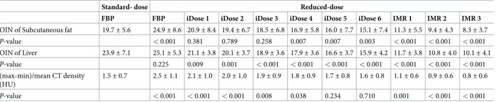
The results of quantitative measurement and analysis are summarized inTable 1. The objective image noise of the subcutaneous fat area with beam hardening artifacts in the reduced-dose CT with FBP was significantly higher than in the standard dose-CT with FBP (P < 0.001). The objective image noise of subcutaneous fat decreased as the iDose and IMR level increased. The objective image noises of subcutaneous fat in the reduced-dose CT with iDose levels between 4 and 6 and all IMR levels were significantly lower than those in the standard dose-CT with FBP (P < 0.05). In the liver with beam hardening artifacts, the objective image noises in the reduced-dose CT were significantly lower with all IR levels than that in the standard-dose CT with FBP (P < 0.05). The corrected CT numbers [(max–min)/mean value] for all IMR levels in the reduced-dose CT were significantly lower than those in the standard-dose CT with FBP
(P < 0.05).
Qualitative analysis
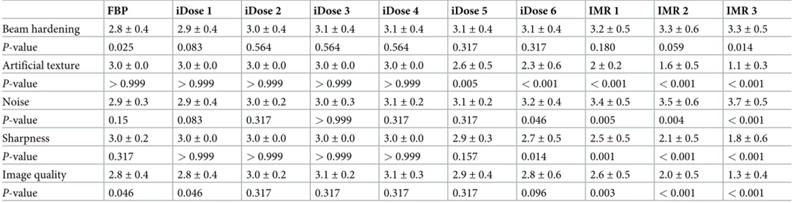
Fig 1andTable 2shows the results of qualitative analysis. The beam hardening artifacts in the reduced-dose scan with FBP reconstruction were significantly greater than those in the stan-dard-dose scan with FBP reconstruction (P = 0.025). When iDose or IMR was applied, the degree of beam hardening artifacts decreased as the IR level increased. Similarly, the subjective image noise decreased as the iDose and IMR levels increased. However, artificial texture and poor image sharpness increased as the iDose and IMR levels increased (Fig 2). The reviewers
Table 1. Objective image noise (OIN) of standard-dose CT and reduced-dose CT.
Standard- dose Reduced-dose
FBP FBP iDose 1 iDose 2 iDose 3 iDose 4 iDose 5 iDose 6 IMR 1 IMR 2 IMR 3
OIN of Subcutaneous fat 19.7± 5.6 24.9± 8.6 20.9 ± 8.4 19.4 ± 6.7 18.5 ± 6.8 16.9 ± 5.8 16.0 ± 7.7 15.1 ± 7.4 11.3 ± 5.5 9.4 ± 4.3 8.3± 3.7
P-value < 0.001 0.381 0.789 0.258 0.007 0.007 0.003 < 0.001 < 0.001 < 0.001 OIN of Liver 23.9± 7.1 25.1± 5.3 21.1 ± 3.8 20.1 ± 3.7 18.9 ± 3.6 17.9 ± 3.6 16.6 ± 3.7 15.9 ± 4.2 11.7 ± 3.8 10.8 ± 4.0 10.1 ± 4.1 P-value 0.225 0.009 0.001 < 0.001 < 0.001 < 0.001 < 0.001 < 0.001 < 0.001 < 0.001 (max-min)/mean CT density (HU) 1.5± 0.7 2.5± 1.1 2.1± 1.0 2.0± 1.0 1.9± 0.9 1.8± 0.9 1.7± 0.8 1.6± 0.8 1.1± 0.6 0.9± 0.6 0.8± 0.6 P-value < 0.001 < 0.001 < 0.001 0.008 0.038 0.234 0.710 0.001 < 0.001 < 0.001
Data are mean± standard deviation.
P-values are for differences between standard-dose FBP and each reconstruction of reduced-dose CT.
observed a strong artificial texture and degraded sharpness in the reduced-dose scans with iDose levels between 5 and 6, and all IMR levels (P < 0.05). The overall image quality was high-est with iDose levels of 3 and 4. IMR of all levels significantly decreased image quality com-pared to standard-dose CT scans with FBP reconstruction (P < 0.05).
Discussion
Our results showed that the image quality of reduced-dose scans with moderate levels of IR was similar to that of standard-dose scans with FBP reconstruction in patients who could not
Fig 1. Graphs illustrating qualitative scores of each reconstruction set of the reduced-dose CT. All reduced-dose CT images were scored compared to the
standard-dose CT images (e.g., 3: similar to the standard-standard-dose CT). Higher score indicates a better image quality. (A) Beam hardening artifacts and (B) subjective image noise decreased as iDose and IMR levels increased. However, iDose levels of 5 and 6, and all IMR levels negatively affect (C) artificial texture and (D) margin sharpness. (E) Overall image quality was highest with iDose levels of 3 and 4.
https://doi.org/10.1371/journal.pone.0209754.g001
Table 2. Subjective image quality scores of reduced-dose CT compared with standard-dose CT�.
FBP iDose 1 iDose 2 iDose 3 iDose 4 iDose 5 iDose 6 IMR 1 IMR 2 IMR 3
Beam hardening 2.8± 0.4 2.9± 0.4 3.0± 0.4 3.1± 0.4 3.1± 0.4 3.1± 0.4 3.1± 0.4 3.2± 0.5 3.3± 0.6 3.3± 0.5 P-value 0.025 0.083 0.564 0.564 0.564 0.317 0.317 0.180 0.059 0.014 Artificial texture 3.0± 0.0 3.0± 0.0 3.0± 0.0 3.0± 0.0 3.0± 0.0 2.6± 0.5 2.3± 0.6 2± 0.2 1.6± 0.5 1.1± 0.3 P-value > 0.999 > 0.999 > 0.999 > 0.999 > 0.999 0.005 < 0.001 < 0.001 < 0.001 < 0.001 Noise 2.9± 0.3 2.9± 0.4 3.0± 0.2 3.0± 0.3 3.1± 0.2 3.1± 0.2 3.2± 0.4 3.4± 0.5 3.5± 0.6 3.7± 0.5 P-value 0.15 0.083 0.317 > 0.999 0.317 0.317 0.046 0.005 0.004 < 0.001 Sharpness 3.0± 0.2 3.0± 0.0 3.0± 0.0 3.0± 0.0 3.0± 0.0 2.9± 0.3 2.7± 0.5 2.5± 0.5 2.1± 0.5 1.8± 0.6 P-value 0.317 > 0.999 > 0.999 > 0.999 > 0.999 0.157 0.014 0.001 < 0.001 < 0.001 Image quality 2.8± 0.4 2.8± 0.4 3.0± 0.2 3.1± 0.2 3.1± 0.3 2.9± 0.4 2.8± 0.6 2.6± 0.5 2.0± 0.5 1.3± 0.4 P-value 0.046 0.046 0.317 0.317 0.317 0.317 0.096 0.003 < 0.001 < 0.001
Data are mean± standard deviation.
�All reduced-dose CT images were scored compared with the standard-dose CT images (e.g., 3: similar to the standard-dose CT) P-values are for differences between standard-dose FBP and each reconstruction of reduced-dose CT.
raise their arms during CT scanning. This finding indicates that additional radiation exposure due to the arms-down position can be avoided while maintaining image quality using IR. This study was the first to investigate the feasibility of reduced-dose CT with IR in patients with inappropriate arm positioning. Objective and subjective image noise, including beam harden-ing artifacts, decreased as the IR levels increased. However, as artificial texture and poor image sharpness deteriorated image quality at iDose levels between 5 and 6 and all IMR levels, the overall image quality was highest at iDose levels between 3 and 4.
CT scans performed with arms along the body are not rare, especially in trauma patients or critically ill patients. Skeletal surveys using CT are also performed on multiple myeloma patients with the arms-down position on a routine basis [18]. In these cases, radiation expo-sure is known to increase compared to the standard arms-up position [4,6,19]. A previous study reported the corrected effective dose to be 18% higher in patients with one arm down and 45% higher in patients with two arms down [4]. We sought to keep similar radiation expo-sure to the standard arms-up position while maintaining image quality using IR in these
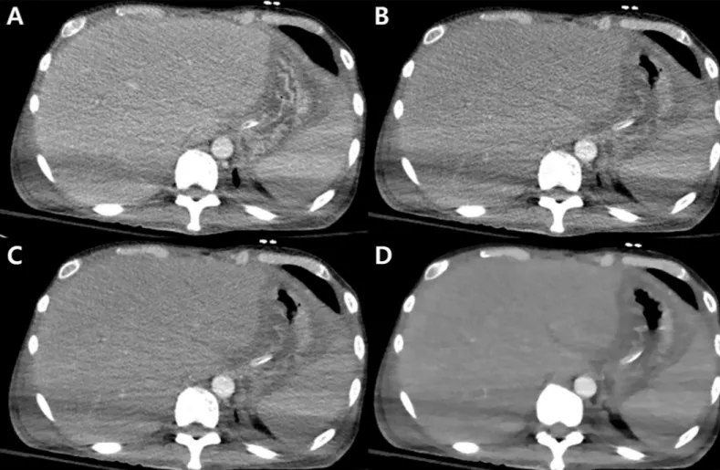
Fig 2. Multidetector CT images (A: standard-dose CT image; B-D: reduced-dose CT images) of a 45-year-old male patient with his two arms positioned on his sides. The section thickness was 3 mm. The window width and level was fixed at 350 and 50, respectively throughout the reconstructed image set. (A) The standard-dose
CT image with filtered back projection (FBP) reconstruction shows the presence of beam hardening artifacts associated with the arms-down position. (B) The reduced-dose CT image with FBP reconstruction shows more prominent hypodense and hyperdense artifacts in the liver and spleen compared with (A). (C) A reduced-reduced-dose CT image with hybrid iterative reconstruction (iDose level 4) had similar or slightly less beam hardening artifacts than (A) while image quality was preserved. (D) A reduced-dose CT image with iterative model reconstruction (IMR level 3) shows less beam hardening artifacts than (A-C). However, IMR negatively affected the overall image quality due to prominent artificial texture and poor margin sharpness.
patients. As we hypothesized, the quality of CT images obtained with IR at a lower radiation dose (18.4%) was similar to that of FBP images obtained with the standard radiation dose. The objective and subjective image noise of reduced-dose CT scans was even lower than the image noise of standard-dose CT scans when higher levels of iDose or IMR were applied to the reduced-dose CT scans.
One recent study using GE-CT and Toshiba-CT reported that full IR allowed more efficient streak artifact reduction compared with FBP and hybrid IR in abdominal CT without arm ele-vation [14]. However, the previous study only evaluated unenhanced CT images, and did not investigate the feasibility of reduced-dose CT with IR in patients without arm elevation. In our study, CT images with IMR also showed significant reduction of beam hardening artifacts. However, the severe blotch appearance of IMR significantly decreased the overall quality of the CT images. Other several studies on IR have evaluated beam hardening artifacts due to metallic materials [20–22]. A phantom study on prosthetic heart valve CT imaging demon-strated that IR significantly decreased hypodense and hyperdense artifact volumes by 53% and 20%, respectively, for iDose, and by 67% and 23%, respectively, for IMR, compared with FBP [20]. However, subjective image quality could not be assessed because this study was a phan-tom study. Although beam hardening artifacts due to metallic materials and those due to non-metallic materials might be somewhat different because non-metallic materials produce much higher attenuation, resulting in severe photon starvation and beam hardening, our study indi-cated that IR could be used to reduce beam hardening artifacts generated by the positioning of arms next to the torso.
The two IR algorithms investigated in this study, iDose and IMR, have distinct mechanisms for noise reduction. iDose is a hybrid IR algorithm that works in the projection and image domains [23]. IMR is a model-based algorithm that is fully iterative with forward and back-ward reconstructions [24–26]. The most significant difference between iDose and IMR is that IMR decreases quantum noise and non-random noise intrinsic to the geometry and optics of the imaging system [20]. Therefore, IMR was expected to have a greater impact on the reduc-tion of beam hardening artifacts, and this was demonstrated in this study. Despite significant improvements in objective and subjective image noise and beam hardening artifacts with IMR, the overall subjective image quality was significantly poorer with IMR than with stan-dard-dose FBP images. It seems that objective image quality does not exactly correlate with subjective rating, which could also be dependent on the reader’s experience with IR [27]. In a recent study on ultralow-dose chest CT, IMR reduced objective image noise compared to FBP and iDose, but the prominent blotchy images and poor demarcation of fine structures with IMR compromised its diagnostic confidence [28]. These previous results are in good agree-ment with our results. In addition, the effect of high iDose levels (5–6) was similar to that of IMR application. Therefore, an intermediate level of hybrid IR (iDose between 3 and 4) may be suitable to preserve image quality while lowering the radiation dose in patients in the arms-down position.
Our study has several limitations. First, the retrospective and single-center nature of our study limited the number of patients [29]. Notably, the BMI of our study population was quite low, because it was made up of Asian patients, and most of the patients had chronic diseases and/or were critically ill. Therefore, our study results cannot be readily generalized to over-weight patients. Multicenter prospective clinical studies including patients with various BMIs are required to validate our results. Second, standard-dose CT and reduced-dose CT images were scanned at different dynamic phases; therefore, the assessment of the objective and sub-jective image quality might be influenced by these different phases. However, repeated acquisi-tion of CT scans in the same dynamic phase is realistically unfeasible and unethical because of increased radiation exposure to the patient. Third, objective image noise was evaluated by
calculating the SD. Although several methods have attempted to evaluate image quality or noise such as grey-level co-occurrence matrices or histogram analysis, these methods were also unable to completely represent image quality. More accurate methods should be newly devel-oped in the future, but at this moment in time, SD measurement seems to be the only widely accepted method for the quantitative analysis of CT image quality. Also, other methods used in previous studies to reduce metallic artifacts, including measuring the volume of hyperdense and hypodense artifacts, were difficult to apply in our study, because the standard-dose and reduced-dose CT scans were obtained at different dynamic phases, and beam hardening arti-facts produced by the arms-down position were much weaker than those produced by metallic artifacts. Forth, subjective image quality was reviewed by two radiologists in consensus, which may decrease the generalizability of our results. Fifth, the results of this study cannot be gener-alized yet, because CT scanners from only one vendor were used. Further studies using IR techniques from other vendors are needed before we can generalize our study results. Finally, we did not investigate the effect of beam hardening artifacts on diagnostic efficacy, because this was a preliminary study and our main focus was investigating the feasibility of using IR at reduced-dose CT scans in this particular patient group.
In conclusion, IR algorithms can reduce beam hardening artifacts without increasing radiation dose in patients who cannot raise their arms, and an intermediate level of hybrid IR allow radiolo-gists to obtain images of the best quality without making patients change their arm positions.
Supporting information
S1 File. Phantom study results. (DOCX)
S2 File. Forms for qualitative assessment. (DOCX)
S3 File. Analysis plan. (DOCX)
Acknowledgments
This research was supported by the Basic Science Research Program through the National Research Foundation of Korea funded by the Ministry of Education
(NRF-2014R1A1A2057091).
Author Contributions
Conceptualization: Nieun Seo, Yong Eun Chung, Jin-Young Choi, Myeong-Jin Kim. Data curation: Nieun Seo, Yong Eun Chung.
Formal analysis: Nieun Seo, Yong Eun Chung, Chansik An. Funding acquisition: Yong Eun Chung.
Investigation: Nieun Seo, Yong Eun Chung, Chansik An, Mi-Suk Park.
Methodology: Nieun Seo, Yong Eun Chung, Chansik An, Jin-Young Choi, Mi-Suk Park, Myeong-Jin Kim.
Project administration: Yong Eun Chung. Resources: Yong Eun Chung.
Supervision: Yong Eun Chung, Jin-Young Choi, Mi-Suk Park, Myeong-Jin Kim. Validation: Nieun Seo, Yong Eun Chung, Chansik An, Jin-Young Choi, Mi-Suk Park,
Myeong-Jin Kim.
Visualization: Nieun Seo, Yong Eun Chung, Chansik An. Writing – original draft: Nieun Seo, Yong Eun Chung.
Writing – review & editing: Nieun Seo, Yong Eun Chung, Chansik An, Jin-Young Choi, Mi-Suk Park, Myeong-Jin Kim.
References
1. Kalra MK, Naz N, Rizzo SM, Blake MA. Computed tomography radiation dose optimization: scanning protocols and clinical applications of automatic exposure control. Curr Probl Diagn Radiol. 2005; 34: 171–181.https://doi.org/10.1067/j.cpradiol.2005.06.002PMID:16129235
2. Rizzo S, Kalra M, Schmidt B, Dalal T, Suess C, Flohr T, et al. Comparison of angular and combined automatic tube current modulation techniques with constant tube current CT of the abdomen and pelvis. AJR Am J Roentgenol. 2006; 186: 673–679.https://doi.org/10.2214/AJR.04.1513PMID:16498094 3. McCollough CH, Bruesewitz MR, Kofler JM Jr. CT dose reduction and dose management tools:
over-view of available options. Radiographics. 2006; 26: 503–512.https://doi.org/10.1148/rg.262055138 PMID:16549613
4. Brink M, de Lange F, Oostveen LJ, Dekker HM, Kool DR, Deunk J, et al. Arm raising at exposure-con-trolled multidetector trauma CT of thoracoabdominal region: higher image quality, lower radiation dose. Radiology. 2008; 249: 661–670.https://doi.org/10.1148/radiol.2492080169PMID:18936319 5. Kahn J, Grupp U, Maurer M. How does arm positioning of polytraumatized patients in the initial
com-puted tomography (CT) affect image quality and diagnostic accuracy? Eur J Radiol. 2014; 83: e67–71. https://doi.org/10.1016/j.ejrad.2013.10.002PMID:24189387
6. Karlo C, Gnannt R, Frauenfelder T, Leschka S, Bruesch M, Wanner GA, et al. Whole-body CT in poly-trauma patients: effect of arm positioning on thoracic and abdominal image quality. Emerg Radiol. 2011; 18: 285–293.https://doi.org/10.1007/s10140-011-0948-5PMID:21472460
7. Linsenmaier U, Krotz M, Hauser H, Rock C, Rieger J, Bohndorf K, et al. Whole-body computed tomog-raphy in polytrauma: techniques and management. Eur Radiol. 2002; 12: 1728–1740.https://doi.org/ 10.1007/s00330-001-1225-xPMID:12111064
8. Benneker LM, Bonel HM, Zumstein MA, Exadaktylos AK. A novel multiple-trauma CT-scanning protocol using patient repositioning may increase risks of iatrogenic injuries. Emerg Radiol. 2007; 13: 349–351; author reply 353.https://doi.org/10.1007/s10140-007-0577-1PMID:17342470
9. Hara AK, Paden RG, Silva AC, Kujak JL, Lawder HJ, Pavlicek W. Iterative reconstruction technique for reducing body radiation dose at CT: feasibility study. AJR Am J Roentgenol. 2009; 193: 764–771. https://doi.org/10.2214/AJR.09.2397PMID:19696291
10. Gervaise A, Osemont B, Lecocq S, Noel A, Micard E, Felblinger J, et al. CT image quality improvement using Adaptive Iterative Dose Reduction with wide-volume acquisition on 320-detector CT. Eur Radiol. 2012; 22: 295–301.https://doi.org/10.1007/s00330-011-2271-7PMID:21927791
11. Willemink MJ, Leiner T, de Jong PA, de Heer LM, Nievelstein RA, Schilham AM, et al. Iterative recon-struction techniques for computed tomography part 2: initial results in dose reduction and image quality. Eur Radiol. 2013; 23: 1632–1642.https://doi.org/10.1007/s00330-012-2764-zPMID:23322411 12. Park M, Chung YE, Lee HS, Choi JY, Park MS, Kim MJ, et al. Intraindividual comparison of diagnostic
performance in patients with hepatic metastasis of full-dose standard and half-dose iterative reconstruc-tions with dual-source abdominal computed tomography. Invest Radiol. 2014; 49: 195–200.https://doi. org/10.1097/RLI.0000000000000014PMID:24300843
13. Shin HJ, Chung YE, Lee YH, Choi JY, Park MS, Kim MJ, et al. Radiation dose reduction via sinogram affirmed iterative reconstruction and automatic tube voltage modulation (CARE kV) in abdominal CT. Korean J Radiol. 2013; 14: 886–893.https://doi.org/10.3348/kjr.2013.14.6.886PMID:24265563 14. Yasaka K, Furuta T, Kubo T, Maeda E, Katsura M, Sato J, et al. Full and hybrid iterative reconstruction
to reduce artifacts in abdominal CT for patients scanned without arm elevation. Acta Radiol. 2016. 2017/01/11.https://doi.org/10.1177/0284185116684675284185116684675. PMID:28068822 15. McCollough C, Bakalyar DM, Bostani M, Brady S, Boedeker K, Boone JM, et al. Use of Water
Equiva-lent Diameter for Calculating Patient Size and Size-Specific Dose Estimates (SSDE) in CT: The Report of AAPM Task Group 220. AAPM Rep. 2014; 2014: 6–23. PMID:27546949
16. Scibelli A. iDose4 iterative reconstruction technique. Philips Healthcare Websites.http://clinical. netforum.healthcare.philips.com/global/Explore/White-Papers/CT/iDose4-iterative-reconstruction-technique. 2011.
17. Mehta D, Thompson R, Morton T, Dhanantwari E, Shefer E. Iterative model reconstruction: simulta-neously lowered computed tomotraphy radiation dose and improve image quality. Med. Phys. Int. 2013; 1(2): 147–155.
18. Lambert L, Ourednicek P, Meckova Z, Gavelli G, Straub J, Spicka I. Whole-body low-dose computed tomography in multiple myeloma staging: Superior diagnostic performance in the detection of bone lesions, vertebral compression fractures, rib fractures and extraskeletal findings compared to radiogra-phy with similar radiation exposure. Oncol Lett. 2017; 13: 2490–2494.https://doi.org/10.3892/ol.2017. 5723PMID:28454425
19. Bayer J, Pache G, Strohm PC, Zwingmann J, Blanke P, Baumann T, et al. Influence of arm positioning on radiation dose for whole body computed tomography in trauma patients. J Trauma. 2011; 70: 900– 905.https://doi.org/10.1097/TA.0b013e3181edc80ePMID:20962679
20. Sucha D, Willemink MJ, de Jong PA, Schilham AM, Leiner T, Symersky P, et al. The impact of a new model-based iterative reconstruction algorithm on prosthetic heart valve related artifacts at reduced radiation dose MDCT. Int J Cardiovasc Imaging. 2014; 30: 785–793. https://doi.org/10.1007/s10554-014-0379-yPMID:24474347
21. Habets J, Symersky P, Leiner T, de Mol BA, Mali WP, Budde RP. Artifact reduction strategies for pros-thetic heart valve CT imaging. Int J Cardiovasc Imaging. 2012; 28: 2099–2108.https://doi.org/10.1007/ s10554-012-0041-5PMID:22476910
22. Jeong S, Kim SH, Hwang EJ, Shin CI, Han JK, Choi BI. Usefulness of a metal artifact reduction algo-rithm for orthopedic implants in abdominal CT: phantom and clinical study results. AJR Am J Roent-genol. 2015; 204: 307–317.https://doi.org/10.2214/AJR.14.12745PMID:25615752
23. Song JS, Lee JM, Sohn JY, Yoon JH, Han JK, Choi BI. Hybrid iterative reconstruction technique for liver CT scans for image noise reduction and image quality improvement: evaluation of the optimal iterative reconstruction strengths. Radiol Med. 2015; 120: 259–267.https://doi.org/10.1007/s11547-014-0441-9 PMID:25168773
24. Nelson RC, Feuerlein S, Boll DT. New iterative reconstruction techniques for cardiovascular computed tomography: how do they work, and what are the advantages and disadvantages? J Cardiovasc Com-put Tomogr. 2011; 5: 286–292.https://doi.org/10.1016/j.jcct.2011.07.001PMID:21875826
25. Yuki H, Oda S, Utsunomiya D, Funama Y, Kidoh M, Namimoto T, et al. Clinical impact of model-based type iterative reconstruction with fast reconstruction time on image quality of low-dose screening chest CT. Acta Radiol. 2016; 57: 295–302.https://doi.org/10.1177/0284185115575537PMID:25817455 26. Yoon JH, Lee JM, Yu MH, Baek JH, Jeon JH, Hur BY, et al. Comparison of iterative model-based
recon-struction versus conventional filtered back projection and hybrid iterative reconrecon-struction techniques: lesion conspicuity and influence of body size in anthropomorphic liver phantoms. J Comput Assist Tomogr. 2014; 38: 859–868.https://doi.org/10.1097/RCT.0000000000000145PMID:25321625 27. Pickhardt PJ, Lubner MG, Kim DH, Tang J, Ruma JA, del Rio AM, et al. Abdominal CT with
model-based iterative reconstruction (MBIR): initial results of a prospective trial comparing ultralow-dose with standard-dose imaging. AJR Am J Roentgenol. 2012; 199: 1266–1274.https://doi.org/10.2214/AJR.12. 9382PMID:23169718
28. Laqmani A, Avanesov M, Butscheidt S, Kurfurst M, Sehner S, Schmidt-Holtz J, et al. Comparison of image quality and visibility of normal and abnormal findings at submillisievert chest CT using filtered back projection, iterative model reconstruction (IMR) and iDose4. Eur J Radiol. 2016; 85: 1971–1979. https://doi.org/10.1016/j.ejrad.2016.09.001PMID:27776648
29. Taylor S, Van Muylem A, Howarth N, Gevenois PA, Tack D. CT dose survey in adults: what sample size for what precision? Eur Radiol. 2017; 27: 365–373.https://doi.org/10.1007/s00330-016-4333-3PMID: 27048530