Vol. 47, No. 2, pp. 237 - 242, 2006
The upper chest wall does not grow properly in children with spinal muscular atrophy (SMA) with paradoxical breathing. This suggests that long-term inability to take a deep breath in developing children may result in underdevelopment of the upper chest wall. In addition, a rapid and paradoxical breathing pattern is frequently observed in children with severe cerebral palsy (CP), which often corresponds to the under-development of the upper chest wall. The present study is designed to evaluate the ratio of the upper to lower chest wall in children with severe spastic quadriplegic CP, compared with normal children. We compared normal children with children that had spastic quadriplegic CP who did not have kyphosis or scoliosis. Test subjects were matched in terms of age, height, and weight. The diameters of upper chest (Dapex) and of lower chest (Dbase) were measured on the anteroposterior (AP) view of a chest X-ray and the Dapex to Dbase ratio was calculated. In selected cases the forced vital capacity (FVC) was measured using a Wright Respirometer. The Dapexto Dbase ratio was significantly lower in the CP group than in the control group (p<0.001). The ratio increased linearly with age (p<0.001) in both CP (R = 0.372) and control groups (R = 0.477). The FVC/preFVC showed significant correlation with the Dapex to Dbaseratio (R = 0.542, p < 0.01). The results of this study suggest a deviation of optimal chest wall structure in children with spastic quadriplegic CP.
Key Words: Chest wall, cerebral palsy, quadriplegia
INTRODUCTION
Cerebral palsy (CP) patients suffer from a high
incidence of respiratory dysfunction such as recurrent pneumonia, atelectasis, bronchiectasis, sleep apnea, chronic obstructive lung disease, and restrictive lung disease.1,2Respiratory dysfunction
is known to be a leading cause of death among individuals with CP.1 However, respiratory
dys-function in children with severe CP has not been well studied, possibly due to the difficulty of testing these uncooperative young children. Clini-cal symptoms such as poor coughing and airway clearance, respiratory muscle weakness and kyphoscoliosis are thought to be related to res-piratory dysfunction in these individuals.26
People of all ages take deep breaths or sigh regularly, and, of course, infants cry. These ac-tions stretch the respiratory structures. Children with CP, particularly quadriplegic CP, breathe in a poorly coordinated fashion, relying on the ab-dominal muscles instead of the chest muscles.7,8
Eventually, chest movement is restricted and the chest muscles weaken. As a result, the ability to take a large breath is impaired.3,9,10 The shallow
and low breathing volume in severe CP leads to the development of widespread microatelectasis and a decrease in lung distensibility.11 In
par-ticular, CP can lead to underdevelopment of the lung tissues and an alteration in chest wall devel-opment in children who are not fully grown.12-14
The development of pectus excavatum in children with spinal muscular atrophy (SMA) who did not receive early intervention,14 demonstrates the
impact of long term respiratory insufficiency on chest wall development.
An intercostal muscle weakness in patients with
Comparison of the Ratio of Upper to Lower Chest Wall in
Children with Spastic Quadriplegic Cerebral Palsy and
Normally Developed Children
Eun Sook Park,1,2 Jung Hyun Park,1 Dong-Wook Rha,1 Chang Il Park,1 and Chan Woo Park1
1Department and Research Institute of Rehabilitation Medicine, Yonsei University College of Medicine,2Brain Korea 21 Project for Medical Science, Yonsei University, Seoul, Korea.
Received June 15, 2005 Accepted November 8, 2005
Reprint address: requests to Dr. Eun Sook Park, Rehabilitation Hospital, Yonsei University of College of Medicine, 134 Shinchon-dong, Seodamun-gu, Seoul 120-752, Korea. Tel: 82-2-2228-3710, Fax: 82-2-363-2795, E-mail: [email protected]
spinal muscular atrophy results in a paradoxical ventilatory pattern.14-16 Kinematic analysis of
breathing revealed a chronic failure to expand the upper chest wall and lungs in these children.16It
is possible that the shallow respiration observed in severe CP leads to an inability to expand the upper chest wall which may ultimately affect upper chest development. This can result in a lower ratio of upper chest to lower chest diameter in children with severe CP compared with normally developed children. Although there are several methods for assessing upper chest volume or expansion, evaluation of these children can be difficult and would ideally involve a simple test that does not require the cooperation of the child. The ratio of the transverse diameters of the upper chest and lower chest measured on a chest radiograph is a simple method for the assessment of chronic hypoventilation of the upper chest compartment in these children. The present study compares the ratio of the transverse diameter of the upper chest to the lower chest in children with severe spastic quadriplegic CP and normally developed children.
MATERIALS AND METHODS
Spastic quadriplegic children of Gross Motor Function Classification System level 4 or 5 and normally developed children were recruited. Supine anteroposterior (AP) chest radiographs were taken and children with musculoskeletal problems such as scoliosis or kyphosis were ex-cluded from the study. A simple chest radio-graph was used to exclude those cases with a rotation of the facet joint or spinous process. Each group consisted of 112 children: 80 boys and 32 girls. The mean age was 42.7 ± 32.2 months in the
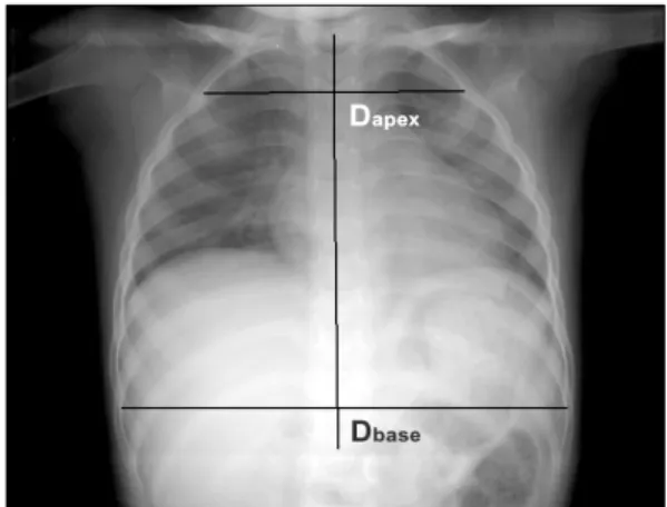
CP group and 43.1 ± 32.4 months in the control group (Table 1). Using the AP view of a chest radiograph horizontal lines were drawn from the inner margin of the rib on one side to the other side, perpendicular to the line connecting the spinous processes. The longest line of the 2nd rib (Dapex) and 9th rib (Dbase) were measured (Fig. 1)
and the percentage ratio of the upper to lower chest wall was calculated as Dapex/ Dbase× 100
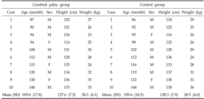
(%). In addition, 10 children over age 7 who were able to cooperate with the pulmonary function test were selected from each group. The mean age and height of children in the two groups were similar (p > 0.05; Table 2). The forced vital ca-pacity (FVC) was measured in a sitting position using a Wright Respirometer (Ferraris Develop-ment and Engineering Co Ltd., Edmonton, London, UK). Each patient was told to inhale as deeply as possible and to blow the entire volume through the spirometer. This process was repeated
Table 1. General Characteristics of Subjects
Subjects Cerebral palsy group Control group p value
Total (male/female) 112 (80/32) 112 (80/32) NS
Age (months) 42.7 ± 32.2 43.1 ± 32.4 NS
Growth grade (percentile) 50.7 ± 11.3 53.2 ± 13.3 NS
Values are mean ± standard deviation. NS, not significant.
Fig. 1. Measurement of the diameter of the chest apex (Dapex) and base (Dbase). Horizontal lines were drawn from
the inner margin of the rib on one side to the other side perpendicular to the line connecting the spinous pro-cesses. The longest line of the 2nd rib (Dapex) and the 9th
at least three times and the highest value was selected as the FVC. This study was approved by the local institutional Ethics Subcommittee for research involving human subjects.
An independent t-test was used to compare the ratio of upper to lower chest wall between the CP group and the control group. The relationship between the percentage ratio of chest wall and age or FVC was analyzed using coefficients of cor-relation. All data were analyzed with SPSS 11.0 for Windows.
RESULTS
The ratio of the upper to lower chest wall of the CP group was significantly lower than that of the
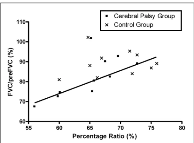
control group (p<0.05; Table 3). In addition, the ratio increased significantly with age in both the CP (R = 0.372, p<0.05) and control group (R = 0.477, p<0.05; Fig. 2). There was no gender dif-ference between the CP and control groups. Table 4 shows the results of the pulmonary function test for the 20 children who underwent the test. The percentage ratio of FVC to the predicted FVC (preFVC) was calculated. There was a significant correlation between the upper to lower chest wall ratio and the percentage ratio of FVC/preFVC (R= 0.542, p<0.05; Fig. 3).
DISCUSSION
The chest wall plays a dual role as a scaffold Table 2. Subjects Who Underwent the Pulmonary Function Test
Cerebral palsy group Control group
Case Age (month) Sex Height (cm) Weight (kg) Case Age (month) Sex Height (cm) Weight (kg)
1 87 M 120 27 1 86 M 118 29 2 90 M 121 26 2 92 M 122 25 3 94 M 124 23 3 95 F 116 24 4 94 F 114 25 4 98 M 125 26 5 108 M 131 30 5 102 M 128 29 6 112 M 128 28 6 112 M 136 24 7 115 F 135 24 7 116 M 133 28 8 120 M 134 32 8 119 M 137 31 9 130 F 136 35 9 132 F 138 33 10 140 M 133 33 10 144 M 130 36 Mean (SD) 109.0 (17.8) 127.6 (7.5) 28.3 (4.1) Mean (SD) 109.6 (18.5) 128.3 (7.9) 28.5 (4.0)
M, male; F, female; SD, standard deviation.
Table 3. Results of the Percentage Ratio of the Upper to Lower Chest Diameter
Cerebral palsy group Control group
Male (n = 80) 61.3 ± 4.9 65.4 ± 4.6*
Female (n = 32) 62.5 ± 5.7 66.0 ± 5.0*
Total (n = 112) 61.7 ± 4.3 65.7 ± 4.4*
Values are mean ± standard deviation. *p<0.001.
and a pump for the respiratory system. Normal respiratory function requires a scaffolding struc-ture that allows adequate mobility and girth for lung expansion. For optimal pump function, the scaffolding needs to be rigid enough to withstand the distorting force of the inward lung recoil but not so rigid as to impede ventilation. Deviation from the optimal chest wall structure can result in respiratory dysfunction17; however, there have
been few studies on the clinical implications of developmental abnormalities of the chest wall.
If left untreated, pectus excavatum can develop in patients with SMA type 1 or SMA type 2 with paradoxical breathing.14Kinematic analysis of the breathing pattern in these children reveals para-doxical decreases in the upper thoracic volume during autonomous inspiration.16 In the case of
CP, the adverse effects of a spinal deformity such as scoliosis, which commonly develops in non-ambulatory CP cases, on pulmonary function are well documented. However, there are very few reports on the effect of deviation from the optimal
chest configuration on respiratory function in children with CP. Many studies have described the rapid and paradoxical breathing pattern of subjects with severe CP.3,9-10 In developing chil-dren the long-term inability to take a deep breath may result in development of a bell shaped chest, similar to cases of SMA with paradoxical breath-ing. The present study shows that children with severe CP have a low upper to lower chest diameter ratio compared with normally devel-oped children, suggesting chronic hypoventilation of the upper thoracic compartment in children with CP.
The chest wall compliance of an infant is three times the normal compliance, resulting in para-doxical rib movement during inspiration. The highly compliant chest wall of infants predisposes them to deformation, necessitating that a portion of the respiratory muscle energy be expended in stabilizing the rib cage rather than in effecting ventilation.17 With increasing age the chest wall
stiffens17 and developmental changes in chest wall Fig. 2. Changes in the percentage ratio of the chest wall
with age. Percentage ratio was calculated as the Dapex/
Dbase diameter × 100. Filled squares and the solid line
represent the cerebral palsy group, whereas the cross marks and dotted line represent the control group.
Table 4. Results of the Pulmonary Function Test
Group FVC (mL) preFVC (mL) 100 × FVC/preFVC (%)
Control (n = 10) 1795 ± 399 1941 ± 370 92.5 ± 6.1
Cerebral palsy (n = 10) 1530 ± 348 1908 ± 351 79.9 ± 7.8
Values are mean ± standard deviation.
FVC, forced vital capacity; preFCV, predicted value of forced vital capacity.
Fig. 3.Relationship between the ratio of upper to lower chest wall diameter and the forced vital capacity. Percent-age ratio was represented as Dapex/Dbase diameter × 100.
The squares represent the cerebral palsy group, and the cross marks represent the control group.
compliance increase the respiratory efficiency. Therefore, the tendency for respiratory muscle fatigue, atelectasis, and respiratory failure d-ecreases with age.18-21 In the present study the
ratio of the upper to lower thoracic diameter increased with age in both CP and control groups. This can be explained in part by normal devel-opmental changes in chest wall compliance.
A deviation from the optimal chest wall struc-ture can result in mechanical insufficiency, respi-ratory muscle fatigue and failure, abnormal ventilation and abnormal resting lung volume.17 The correlation observed in the present study between the upper to lower chest wall ratio and the forced vital capacity demonstrates the rela-tionship between chest wall structure and respira-tory efficiency. Assessment of pulmonary me-chanics in children with severe CP is challenging due to the difficulty of testing subjects who are unable to cooperate. Therefore, a simple index representing lung function might be a useful screening test for respiratory risk. It is important to note that, although the ratio of the upper to lower chest wall was significantly related to the percentage ratio of FVC/preFVC in this study, this index may not directly represent lung func-tion. In the present study forced vital capacity was determined in a limited number of children who could cooperate with the testing procedure. Moreover, the ratio of the upper to lower chest wall appears to be related to a restrictive lung dysfunction rather than an obstructive lung dys-function. Various causes of respiratory dysfunc-tion in CP have been reported including: a restrictive pattern,5 abnormal compliance of the
chest wall and lung, mismatched ventilation and perfusion,3,4 upper airway obstruction,2 and
ob-structive sleep apnea.22 Therefore, the low ratio of
the upper to lower thoracic compartment can be considered to be one of several factors affecting lung function in these children.
The pectus excavatum of SMA type 1 can resolve, and the lungs and chest walls can grow more normally, if there is intervention with a high-span positive inspiratory pressure plus a positive end-expiratory pressure.14Therefore, it is
possible that the deviation from the optimal chest wall observed in children with CP can resolved if there is proper intervention in the early stages of
development.
In conclusion, this study revealed a deviation from optimal chest wall structure in children with spastic quadriplegic CP. The correlation between the ratio of the upper to lower chest wall diameter and the forced vital capacity indicates the impor-tance of the chest wall in respiration, and may provide a simple index for the assessment of respiratory function in children with CP.
REFERENCES
1. Strauss D, Cable W, Shavelle R. Causes of excess mortality in cerebral palsy. Dev Med Child Neurol 1999;41:580-5.
2. Seddon PC, Khan Y. Respiratory problems in children with neurological impairment. Arch Dis Child 2003;88: 75-8.
3. Blumberg ML. Respiration and speech in the cerebral palsied child. AMA Am J Dis Child 1955;89:48-53. 4. Hardy JC. Lung function of athetoid and spastic
quadriplegic children. Dev Med Child Neurol 1964;89: 378-88.
5. Bjure J, Berg K. Dynamic and static lung volumes of school children with cerebral palsy. Acta Paediatr Scand Suppl 1970;204:35-41.
6. Keesee PD. Abnormal postural reflex activity and voice usage deviations in cerebral palsy. Phys Ther 1976;56: 1358-60.
7. Benditt JO. Management of pulmonary complications in neuromuscular disease. Phys Med Rehabil Clin North Am 1998;9:167-85.
8. Redstone F. The effects of seating position on the res-piratory patterns of preschoolers with cerebral palsy. Int J Rehabil Res 2004;27:283-8.
9. Muller H. Speech. In: Finnie NR, editor. Handling the young child with cerebral palsy at home. 3rd ed. Oxford: Butterworth-Heinemann; 1997. p.112-7. 10. Stamer MH. Posture and movement of the child with
cerebral palsy. San Antonio, Texas: Therapy Skill Builders;2000. p.79.
11. Leopando MT, Moussavi Z, Holbrow J, Chernick V, Pasterkamp H, Rempel G. Effect of a soft Boston orthosis on pulmonary mechanics in severe cerebral palsy. Pediatr Pulmonol 1999;28:53-8.
12. De Troyer A, Deisser P. The effects of intermittent positive pressure breathing on patients with respiratory muscle weakness. Am Rev Respir Dis 1981;124:132-7. 13. Estenne M, De Troyer A. The effects of tetraplegia on chest wall statics. Am Rev Respir dis 1986;134:121-4. 14. Bach JR, Bianchi C. Prevention of pectus excavatum for
children with spinal muscular atrophy type 1. Am J Phys Med Rehabil 2003;82:815-9.
muscular atrophy: kinematic breathing analysis. Am J Phys Med Rehabil 1996;75:332-9.
16. Lissoni A, Aliverti A, Tzeng A, Bach JR. Kinematic analysis of patients with spinal muscular atrophy during spontaneous breathing and mechanical ventila-tion. Am J Phys Med Rehabil 1998;77:188-92. 17. Papastamelos C, Panitch HB, England SE, Allen JL.
Developmental changes in chest wall compliance in infancy and early childhood. J Appl Physiol 1995;78: 179-84.
18. Goldman MD, Grimby G, Mead J. Mechanical work of breathing derived from rib cage and abdominal V-P partitioning. J Appl Physiol 1976;41:752-63.
19. Mortola JP, Saetta M, Fox G, Smith B, Weeks S. Mechanical aspects of chest wall distortion. J Appl Physiol 1985;59:295-304.
20. Guslits BG, Gaston SE, Bryan MH, England SJ, Bryan AC. Diaphragmatic work of breathing in premature human infants. J Appl Physiol 1987;62:1410-5. 21. Heldt GP, Mcllroy MB. Distortion of chest wall and
work of diaphragm in preterm infants. J Appl Physiol 1987;62:164-9.
22. Kotagal S, Gibbons VP, Stith JA. Sleep abnormalities in patients with severe cerebral palsy. Dev Med Child Neurol 1994;36:304-11.