80 -접수일 : 2009.5.14. 통과일 : 2009.7.15. *교신저자 : 권 자 영 120-752, 서울특별시 서대문구 신촌동 134 연세대학교 의과대학 산부인과학교실 Tel: 02-2228-2230, Fax: 02-313-8357 E-mail: [email protected] 09-012
증 례
The Korean Journal of Ultrasound in Obstetrics and Gynecology Vol. 11, No. 2, June 2009선천성 장 협착증 태아에서의 탯줄 궤양에 의한 태아 사망
연세대학교 의과대학 산부인과학교실, 병리학교실*임경진⋅곽동욱⋅박용원⋅김영한⋅조남훈*⋅최준정*⋅권자영
Sudden Fetal Death Resulting from Intrauterine Hemorrhage Due to Umbilical
Cord Ulceration Associated with Congenital Intestinal Atresia: A Case Report
Kyung-Jin Lim, MD, Dong-Wook Kwak, MD, Yong-Won Park, MD, Young-Han Kim, MD, Nam-Hoon Cho, MD*, Jun-Chung Choi MD*, Ja-Young Kwon, MD
Division of Maternal-Fetal Medicine, Department of Obstetrics and Gynecology, Department of Pathology*, Yonsei University College of Medicine, Yonsei University Health System, Seoul, Korea
Congenital intestinal atresia is usually detected on prenatal ultrasound in the late second to third trimester, depicted as proximal bowel dilatation accompanied by polyhydramnios. Since the initial report in 1991 pertaining to the association between umbilical cord ulceration (UCU) and congenital intestinal obstruction, many studies have followed to investigate fetal cord hemorrhage and fetal death associated with congenital intestinal atresia. Nonetheless, UCU has not received much attention from many obstetricians. In light of this, we report a case of intrauterine hemorrhage from an umbilical cord ulcer in a fetus with jejunal atresia that resulted in intrauterine death.
The Korean Journal of Ultrasound in Obstetrics and Gynecology 11(2):80-83, 2009
Key Words: Congenital intestinal obstruction, Umbilical cord ulceration, Intrauterine fetal death
Congenital intestinal obstruction is highly associated with increased perinatal morbidity and mortality ascribed to bowel perforation, meconium peritonitis, meconium ileus, growth restriction, preterm delivery, and neonatal sepsis.1 However, since the first report of the relationship between intestinal atresia and umbilical cord ulceration (UCU) by Bendon et al. in 1991,2 the possibility of UCU eroding into the cord vessels is being raised as a potential cause of death for the fetus with intestinal obstruction. Following their report, an increasing number of studies have revealed the association of UCU and fetal hemorrhage to congenital intestinal obstruction. Nonetheless, not much attention has
been paid to this life-threatening complication when counseling or managing the fetus complicated by jejunal or ileal obstruction. Thus, we report for the first time in Korea a case pertaining to the intrauterine death of a fetus with intestinal atresia attributable to umbilical cord ulcer bleeding.
Case Report
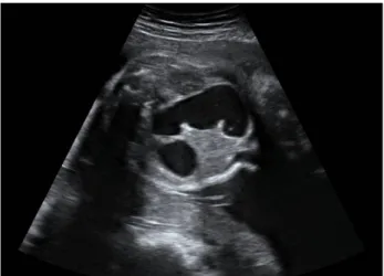
A 30-year-old Korean woman, gravida 2, para 1, was referred to our institute at 33 weeks of gestation presenting with fetal bowel dilatation. On ultrasound, markedly dilated intestines predominant on the left side of the abdomen, enlarged stomach, and polyhydramnios indicated by a high amniotic fluid index of 26 cm were observed (Fig. 1). Other structures were unremarkable. Based on these findings, fetal jejunal obstruction was suspected. She was instructed to
임경진 외 6인. 선천성 장 협착증 태아에서의 탯줄 궤양에 의한 태아 사망
81
-Fig. 1. Sonographic examination of the fetus at 34 weeks of
gestation. Marked jejunal dilatation up to 2 cm in diameter is noted.
(A) (B)
Fig. 2. Umbilical perforation site (indicated by an arrow) was
de-tected 1 cm from the fetal abdomen ((A) immediate postpartum. (B) after fixation).
Fig. 3. Microscopic examination of the umbilical cord depicts
cord ulceration with artery rupture indicated by an arrow. (H&E, ×40, ×100, original magnification).
perform daily fetal movement counting and fetal well-being testing, including non-stress test (NST) and biophysical profile scoring (BPS), which was done semiweekly. After 1 week of a normal NST and 2 days of a normal BPS, she visited the delivery room complaining of decreased fetal movement. Ultrasonographic finding demonstrated the absence of fetal heart activity and the amniotic fluid was echogenic, suggesting intra-amniotic hemorrhage. Due to unfavorable cervix, labor induction was planned. However, due to the uncontrolled emotional instability in regards to delivering a dead fetus, the family requested surgical delivery. Cesarean hysterotomy upon request was performed under spinal anesthesia to deliver a dead male fetus weighing 2760 gm. At the time of delivery, the amniotic fluid was
bloody and the general appearance of the fetus was unremarkable. The placenta was discoid, measured 25×22 cm2, and weighed 395 gm without any evidence of
retroplacental hematoma. The umbilical cord was 50 cm in length, containing 2 arteries and 1 vein. Closer examination of the umbilical cord demonstrated a pin-point surface ulceration 1 cm from the fetal abdominal insertion site (Fig. 2) and multiple longitudinal surface erosions. Microscopic examination of the ulceration site showed loss of Wharton’s jelly and amnion around the umbilical artery with fresh thrombus confirming umbilical artery rupture (Fig. 3).
Discussion
This is the first report in Korea about the intrauterine death of a fetus with jejunal atresia due to associated cord ulceration and umbilical artery perforation. In accord with previously reported cases, our case also presented with bloody amniotic fluid and ulceration on the proximal portion of the umbilical cord surface eroding into the artery on histopathologic examination.
Due to the lack of data on the association of UCU and fetal small intestinal obstruction, the precise incidence of UCU complicating the small intestinal obstruction has not been evaluated. Although Takuya et al. reported a tangible incidence of UCU associated with fetal small intestinal obstruction to be 5.6% based on their experience,3 the cases
were too small in number and not all umbilical cords were histopathologically examined, thus the actual incidence may have been underestimated.
대한산부인과초음파학회지 제11권 제2호, 2009
82
-T able 1. Reported cases of umbilical cord ulceration associated with intestinal atresia
Reference Case no. Anomalies Gestational week MOD Weight (g) Outcome
2 1 DA 31 CS 1830 Alive 2 2 JA 34 CS 2020 Alive 2 3 DA 30 V 1700 Stillbirth 4 4 DA 32 V 1630 Dead (10 months) 7 5 DA 35 CS 2500 Dead (6 days) 6 6 JA 32 CS 1998 Dead (0 day) 6 7 JA 33 CS 1580 Alive 6 8 DA 35 V 2366 Stillbirth 6 9 DA 36 V 2344 Stillbirth 6 10 DA 34 CS 1826 Alive 6 11 JA 37 CS 2366 Dead (0 day) 8 12 JA 35 CS 1934 Alive 8 13 JA 32 CS 2050 Alive 9 14 DA 34 NA 2484 Stillbirth 3 15 DA 35 CS 2675 Dead (0 day) 3 16 JA 31 CS 1500 Stillbirth
Present case 17 JA 34 CS 2760 Stillbirth
DA, duodenal atresia; JA, jejunal atresia; CS, cesarean section; V, vaginal; MOD, mode of delivery; NA, not available. In the literature, only 17 cases, including the present case,
are available to date that indicate a dismal perinatal outcome in pregnancies complicated by fetal intestinal obstruction and cord ulcer. Out of 17 cases, 10 (10/17, 58.8%) resulted in perinatal death, 3 (3/17, 17.6%) were alive but with hypoxic damage, and only 4 survived without any sequelae (Table 1). Such fatal outcome associated with UCU and fetal intestinal atresia is suggested to be due to cord vessel rupture resulting in massive intrauterine bleeding.2-9 The underlying mecha-nism of the development of UCU in the fetus complicated by intestinal obstruction has not yet been elucidated, however, the most plausible explanation for the initiating cause is tox-icity of gastric or intestinal juice, regurgitated from the fetal gastrointestinal tract into the amniotic fluid.6 In human em-bryology, pepsin and hydroxychloride are known to acidify the gastric content at near term, and zymogen granules that appear at 5 months have been reported to be quiescent until birth.10,11 Therefore, these enzymes are least likely to digest the umbilical cord in utero.11
On the other hand, bile acid, a powerful toxin, is produced from the fetal liver from 12 weeks of gestation, concentrated
in the gall bladder, and then secreted into the duodenum, causing the intestinal content to turn green. Thus, bile acid regurgitated and accumulated in the amniotic fluid due to obstruction distal to the ampulla of Vater seemed to the strongest candidate for the causative compound of UCU. To date, the sites of atresia associated with UCU are limited to the duodenum and jejunum but, not the esophagus or ileum, which would substantiate the proposed mechanism.
It is interesting to note that in all of the previously reported cases as well as our case, the perforation site of UCU was near the fetal abdomen. This may be attributed to the theoret-ically higher concentration of bile acid at the proximity of fe-tal mouth, but this theory needs verification.10 The onset of cord rupture superimposing on UCU in previous reports was later than 30 weeks of gestation (mean, 35 weeks of ges-tation). Ohyama et al. measured and compared total bile acid in the amniotic fluid of normal pregnancies to that of preg-nancy complicated by fetal intestinal obstruction beyond 30 weeks of gestation. They indicated that the incidence of UCU was high when amniotic total bile acid concentration exceeded 10.0 μmol/L at around 32 weeks, and that delivery
임경진 외 6인. 선천성 장 협착증 태아에서의 탯줄 궤양에 의한 태아 사망 83 -국문요약 선천성 장 협착증은 대부분 산전 초음파검사상 임신 2분기 말이나 3분기에 양수과다증을 동반한 장 확장으로 진단 할 수 있다. 1991년에 처음으로 선천성 장 협착증과 탯줄 궤양의 관계에 대해 보고가 된 이후로 선천성 장 협착증과 관련하여 탯줄 출혈로 인한 태아 사망에 대한 연구가 많이 이루어져왔 다. 그럼에도 불구하고, 이는 많은 산과 전문의들에게 관심을 받지 못했다. 이에 공장 협착이 있었던 태아에서 탯줄 궤양으로 인한 자궁 내 출혈로 자궁 내 태아 사망한 1예를 증례 보고하는 바이다. 중심 단어 : 선천성 장 협착증, 탯줄 궤양, 자궁 내 태아 사망
should be opted under such circumstances to prevent pend-ing cord perforation.6 However, due to the unavailability of a total bile acid assay kit in Korea, validating and integrating their suggestion into clinical practice is not possible. Although NST and fetal BPS is frequently performed as a fe-tal surveillance test in high risk pregnancy, it failed to predict fetal distress associated with cord perforation in the fetus with UCU.6 In our case, BPS performed 2 days before fetal death was assuring. Proper antenatal fetal monitoring to pre-dict adverse outcomes in the fetus with UCU has yet to be determined.
The present case suggests that we should alert to possible sudden nature of UCU and subsequent stillbirth in the fetus with proximal intestinal obstruction and perform thorough umbilical cord examination when encountered with such cases. Furthermore, investigation of the prevalence of UCU complicating intestinal atresia and establishment of manage-ment protocol to prevent UCU-related fetal death should be pursued based on a larger number of cases.
References
1. Rang S, Brice A. Prenatal bowel dilatation and the sub-sequent postnatal management. Early Human Development 2006; 82: 297-303.
2. Bendon RW, Tyson RW, Baldwin VJ, Cashner KA, Mimouni F, Miodovnik. 1991. Umbilical cord ulceration and
intestinal atresia: a new association? Am J Obstet Gynecol 1991; 164: 582-6.
3. Takuya K, Noriaki U, Shinkichi K, Sawai T, Hirano S, Wada K, et al. Umbilical Cord Ulcer Associated with Fetal Jejunal Atresia: Report of 2 Cases. Fetal Diagn Ther 2003; 18: 144-7. 4. Khong TY, Ford WDA, Haan EA. Umbilical cord ulceration in association with intestinal atresia in a child with detection 13q and Hirschsprung’s disease. Arch Dis Child Fetal Nenatal Ed 1994; 71: 212-3.
5. Yamanaka M, Ohyama M, Koresawa M, Kawataki M, Ohsaki I, Tanaka Y, et al. Umbilical cord ulceration and intestinal atresia. Eur J Obstet Gynecol Reprod Biol 1996; 70: 209-12.
6. Ohyama M, Itani Y, Yamanaka M, Imaizumi K, Nishi T, Ijiri R, et al. Umbilical cord ulcer: a serious in utero complication of intestinal atresia. Placenta 2000; 21: 432-5.
7. Khurana A, Huettner PC, Cole FS. Umbilical cord ulceration as a cause of hypoxic-ischemic encephalopathy: report of a case and review of the literature. J Perinatol 1995; 15: 423-5.
8. Shimizu S, Kawagishi R, Wada K, Arimato-Ishida E, Shimoya K, Murata Y. Fetal hemorrhage associated with congenital intestinal atresia. J Obstet Gynecol Res 2003; 29: 312-6.
9. Anami A, Morokuma S, Tsukimori K, Kondo H, Nozaki M, Sueishi K, et al. Sudden fetal death associated with both duodenal atresia and umbilical cord ulcer, a case and review. Am J Perinatol 2006; 23: 183-8.
10. O’Rahilly R. The timing and sequence of events in the development of the human digestive system and associated structures during the embryonic period proper. Anat Embryol 1978; 153: 123-36.
11. Park HW, Chae YM, Shin TS. Morphologic development of the pancreas in the staged human embryo. Yonsei Medical J 1992; 3: 104-8.