Cementless Metaphyseal Fitting Anatomic Total Hip
Arthroplasty with a Ceramic-on-Ceramic Bearing
in Patients Thirty Years of Age or Younger
Young-Hoo Kim, MD, Jang-Won Park, MD, and Jun-Shik Kim, MD
Investigation performed at The Joint Replacement Center, Ewha Womans University School of Medicine, Seoul, South Korea
Background: The number of midterm or long-term studies on the current generation of cementless total hip replace-ments with alumina-on-alumina ceramic bearings in patients younger than thirty years of age is limited. The purpose of this study was to evaluate the midterm results of the cementless metaphyseal fitting anatomic total hip prosthesis in patients younger than thirty years of age, with a particular emphasis on the prevalence of thigh pain, resorption of bone due to stress-shielding of the proximal part of the femur, aseptic loosening, and osteolysis.
Methods: We reviewed the cases of ninety-six patients (127 hips) who had a cementless total hip arthroplasty when they were thirty years or younger at the time of surgery. All surgical procedures were performed by a single surgeon. The most common diagnoses were osteonecrosis (54.3%) and developmental dysplasia of the hip (20.5%). Demographic data, the Harris hip score, Western Ontario and McMaster Universities Osteoarthritis Index (WOMAC), and University of California, Los Angeles (UCLA) activity scores were recorded. Radiographic evaluation was used to evaluate implant fixation and osteolysis. The minimum follow-up interval was ten years (mean, 14.6 years; range, ten to sixteen years).
Results: The mean preoperative Harris hip score, WOMAC score, and UCLA activity score were 41 points, 66 points, and 3 points, respectively. At the time of final follow-up, the mean Harris hip score, WOMAC score, and UCLA activity score were 95 points, 16 points, and 8 points, respectively. No patient had thigh pain after one year postoperatively. All of the femoral stems and all but one of the acetabular components were well-fixed at the time of final follow-up. No hip exhibited squeaking, ceramic fracture, loosening, or osteolysis at the time of the final follow-up.
Conclusions: These results in patients thirty years of age or younger suggest that the cementless metaphyseal fitting anatomic total hip prosthesis provides outstanding midterm fixation and substantial pain relief well into the second decade postoperatively. Moreover, the alumina-on-alumina ceramic bearing provides a high rate of survivorship without osteolysis. Level of Evidence: Therapeutic Level IV. See Instructions for Authors for a complete description of levels of evidence.
O
ne of the reasons for introducing the cementless total hip arthroplasty was to decrease the rate of aseptic loosening. The prevalence of aseptic loosening of the femoral component in patients with cementless total hip re-placements has been reported to be low, with revision-free survival of almost 100% at ten years1-5. These data are in agreement with the low prevalence of aseptic loosening of ce-mentless total hip replacements that has recently been de-scribed in many studies6-10
. Although mechanical fixation of cementless total hip arthroplasty components was improved
markedly, the prevalence of osteolysis was increased as poly-ethylene wear rates increase in young patients11,12
.
Strategies to reduce wear have focused on improvements in the bearing surface. In vitro testing has confirmed that ceramic-on-ceramic bearings have the lowest wear rates of all tested couples13
and would seem best suited to the highly active and young patients in whom long-term wear is a concern. Studies of newer ceramic bearings in patients with early to intermediate-term clinical follow-up have described low rates of revision and few reports of wear, periprosthetic bone loss, or osteolysis14,15
.
Disclosure: None of the authors received payments or services, either directly or indirectly (i.e., via his or her institution), from a third party in support of any aspect of this work. None of the authors, or their institution(s), have had any financial relationship, in the thirty-six months prior to submission of this work, with any entity in the biomedical arena that could be perceived to influence or have the potential to influence what is written in this work. Also, no author has had any other relationships, or has engaged in any other activities, that could be perceived to influence or have the potential to influence what is written in this work. The complete Disclosures of Potential Conflicts of Interest submitted by authors are always provided with the online version of the article.
One study of cementless total hip arthroplasty with ceramic-on-ceramic bearings in patients younger than forty-five years with an intermediate activity level demonstrated insignificant thigh pain together with an absence of osteolysis and aseptic loosening of the components on radiographs16.
The midterm and long-term published data on the cur-rent generation of cementless total hip arthroplasties with an alumina-on-alumina ceramic bearing in highly active patients younger than thirty years of age are limited. The purpose of the current study was to evaluate the midterm results of the use of the cementless anatomic metaphyseal fitting stem in highly active patients younger than thirty years of age.
Materials and Methods
Patients
B
etween March 1995 and April 2001, 137 total hip arthroplasties with alu-mina ceramic-on-alualu-mina ceramic bearings were performed in 106 pa-tients who were thirty years of age or younger; thirty-one papa-tients had a bilateral total hip arthroplasty. Patients were excluded if they were older than thirty years of age or had either a follow-up interval of less than ten years or other alter-native procedures, including osteotomy or bone graft. A total of ten patients (ten hips) were lost to follow-up prior to two years of follow-up. No patient died. Therefore, 127 hips in ninety-six patients were available for clinical and radiographic evaluation at a mean of 14.6 years (range, ten to sixteen years). The study protocol, including the consent forms, was approved by the insti-tutional review board at our institution. Three patients in this cohort had been previously reported in another publication16.The study group comprised sixty-two male and thirty-four female patients. The mean age (and standard deviation) at the time of surgery was 24 ± 5 years (range, nineteen to thirty years). The mean body weight was 64 ± 13 kg (range, 44 to 111 kg), and the mean height was 167 ± 9 cm (range, 150 to 183 cm). The mean body mass index was 26 ± 4 kg/m2(range, 22 to 36 kg/m2). The pre-operative diagnosis was femoral head osteonecrosis for sixty-nine hips (54.3%), developmental dysplasia of the hip for twenty-six (20.5%), traumatic arthritis for thirteen (10.2%), postinfectious ankylosis for twelve (9.5%), rheumatoid arthritis for three (2.4%), multiple epiphyseal dysplasia for two (1.6%), and childhood pyogenic arthritis for two (1.6%). The mean duration of follow-up was 14.8 years (range, ten to sixteen years). The presumed cause of the osteo-necrosis was alcohol abuse in forty-nine (71%) of the sixty-nine hips, idiopathic in eleven (16%), chronic corticosteroid treatment for bronchial asthma or skin disease in eight (12%), and posttraumatic in one hip (1%). No patient with alcohol or corticosteroid-induced osteonecrosis had an associated severe medical problem17-21.
Surgical Procedure
All procedures were performed by the senior author (Y.H.-K.) through a posterolateral surgical approach to the hip. A cementless Duraloc acetabular component (DePuy, Warsaw, Indiana) was used in all hips. These components were press-fit after the acetabulum had been underreamed by 1 mm. One or two screws were used for additional cup fixation in twenty-one hips (17%); the remainder did not require any screws. An alumina ceramic liner with a 28-mm internal diameter (BIOLOX-forte; CeramTec, Plochingen, Germany) was used in all hips regardless of the external diameter of the acetabular component, which ranged from 48 mm to 58 mm. The acetabular component was fixed with an inclination angle between 40° and 45° and anteversion between 20° and 30°. All patients received an Immediate Postoperative Stability cementless anatomic femoral component (IPS; DePuy, Leeds, United Kingdom) with a 28-mm alumina forte ceramic modular head (BIOLOX-forte; CeramTec). There was a satisfactory fit of this femoral stem in all patients.
The patients were allowed to stand on the second postoperative day, and then progressed to full weight-bearing with crutches as tolerated. They were
advised to use a pair of crutches for six weeks and walk with a cane thereafter as needed.
Clinical and Radiographic Evaluation
Clinical and radiographic follow-up examinations were done at three months, one year, and yearly thereafter. The Harris hip score22and the Western Ontario and McMaster Universities Osteoarthritis Index (WOMAC) score23were de-termined before surgery and at each follow-up examination. Patients scored thigh pain on a 10-point visual analog scale (with 0 indicating no pain and 10, severe pain). The level of activity of the patients after the total hip arthroplasty was assessed with use of the University of California, Los Angeles (UCLA) activity score24. We defined a limp as mild if patients moved their trunk and head <5 cm over the affected hip; moderate, when they moved 5 to 10 cm; and severe, when they moved >10 cm prior to the stance phase of gait.
The occurrence of any clicking or squeaking sound emanating from the ceramic-on-ceramic bearing was recorded.
The radiographs were analyzed by a research associate who had no knowledge of the patient’s identity. A supine anteroposterior radiograph of the pelvis with both hips in neutral rotation and no abduction was made for every patient (see Appendix). Anteversion of the acetabular component was mea-sured on the lateral radiograph of the hip as the angle between the horizontal line where the film cassette rested on the x-ray table and a second line marking the plane of the opening of the acetabular component.
Loosening of the femoral component was defined as a progressive axial subsidence of >3 mm, or a varus or valgus shift of >3°25. Definite loosening of the acetabular component was diagnosed when a change in the position of the component (>2 mm vertically and/or medially or laterally) or a continuous radiolucent line of >2 mm on both the anteroposterior and the lateral radio-graphs was seen26. Bone ingrowth into the acetabular component was con-sidered to have occurred when there was direct contact of the trabecular bone of the acetabulum and the acetabular component.
The sites of any osteolysis in the acetabulum were recorded according to the classification of DeLee and Charnley27and those in the femur, by the classification of Gruen et al.28. Osteolysis was defined as any discretely localized radiolucency that had been absent on radiographs made immediately after the total hip arthroplasty. Proximal femoral bone resorption was graded radio-graphically29. Measurement of the linear wear of the alumina ceramic liner was attempted but was below the level of detection by the method of Kim et al.30. Heterotopic ossification, if present, was graded according to the clas-sification of Brooker et al.31.
Statistical Analysis
Kaplan-Meier survival analysis32was used to estimate the probability of retention of the total hip replacement in relation to revision for any reason and included the entire cohort. Ninety-five percent confidence intervals were calculated.
Source of Funding
No external funding was received for this study. Results
Clinical Results Hip Score
T
he mean preoperative Harris hip score was 41 points (range, 9 to 53 points), which improved to a mean of 95 points (range, 71 to 100 points) at the time of the final follow-up. The mean WOMAC score was 66 points (range, 39 to 85 points) preoperatively and 16 points (range, 3 to 30 points) at the time of the final follow-up.Functional Outcome
Dependence on walking aids and limping had decreased sub-stantially by the time of the final up. At the latest follow-up, seventy-eight patients (81%) had no detectable limp, fifteen
(16%) had a mild limp that was due to a limb-length dis-crepancy (average, 0.7 cm), and three (3%) had a mild limp due to pain in the ankle joints. The ability to use stairs and public transportation, to put on footwear, and to cut toenails was improved markedly after the arthroplasty. The mean preop-erative UCLA activity score was 3 points (range, 1 to 4 points), which improved to a mean of 8 points (range, 5 to 10 points) at the time of the final follow-up (Table I).
Thigh Pain
The prevalence of transitory thigh pain (4 points on the visual analog scale) was 6% (six of ninety-six patients) until six months after the operation. No patient had thigh pain after one year postoperatively.
Employment Status
Twenty-four (25%) of ninety-six patients changed from work involving heavy manual labor before the operation to sedentary work after the operation. The remaining seventy-two patients (75%) remained in the previous occupation after the opera-tion. All patients were advised not to participate in high-impact sports.
Radiographic Results Loosening
Preoperatively, the Dorr isthmus ratio33
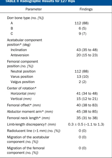
ranged from 0.31 to 0.47. One hundred and twelve hips (88%) were Dorr type A, six (5%) were type B, and nine (7%) were type C. As seen on the postoperative radiographs, 112 femoral stems (88%) were neutral, thirteen (10%) were in varus (<5°), and two (2%) were in valgus (<5°). The average inclination and anteversion of the acetabular component were 43° (range, 35° to 48°) and 20° (range, 15° to 23°), respectively. All hips had osseous in-tegration of the acetabular and the femoral components (Figs. 1-A and 1-B), and no hip exhibited any aseptic loosening of either component (Table II).
Osteolysis
No hip displayed femoral or acetabular osteolysis on the radiographs.
Survivorship
No femoral or acetabular component was revised because of aseptic loosening. One acetabular component (0.8%) was re-vised because of recurrent dislocation. Kaplan-Meier survi-vorship analysis32
revealed that the rate of survival of the femoral component at sixteen years was 100% (95% confi-dence interval, 94% to 100%), with loosening or revision considered the end point for failure, and the rate of survival of the acetabular components was 99% at sixteen years, with loosening or revision considered the end point for failure. Complications
A deep early postoperative infection developed in one hip (0.8%), and open debridement and exchange of the femoral head and acetabular liner were performed. Intravenous antibiotics
TABLE I Clinical Results
Parameter Preop. Postop.
Harris hip score* (points) 41 (9 to 53) 95 (71 to 100) Pain score* (points) 12 (0 to 31) 43 (38 to 44) Function* (points) 30 (0 to 40) 52 (30 to 56) WOMAC score* (points) 69 (39 to 85) 16 (3 to 30)
Thigh pain (n= 96) – 6 (6%)
UCLA activity score* (points)
3 (1 to 4) 8 (5 to 10)
Clicking or squeaking sound
– None
*The values are given as the mean and the standard deviation, with the range in parentheses. WOMAC= Western Ontario and McMaster University Osteoarthritis Index, and UCLA= University of California, Los Angeles.
TABLE II Radiographic Results for 127 Hips
Parameter Findings
Dorr bone type (no. [%])
A 112 (88) B 6 (5) C 9 (7) Acetabular component position* (deg) Inclination 43 (35 to 48) Anteversion 20 (15 to 23) Femoral component position (no. [%]) Neutral position 112 (88) Varus position 13 (10) Valgus position 2 (2) Center of rotation* Horizontal (mm) 41 (34 to 48) Vertical (mm) 15 (12 to 21) Femoral offset* (mm) 40 (38 to 83) Abductor moment arm* (mm) 45 (38 to 85) Femoral neck length* (mm) 35 (31 to 38) Limb-length discrepancy† (mm) 0.3± 0.5 (–1.1 to 1.3) Radiolucent line (>1 mm) (no. [%]) 0 (0)
Migration of the acetabular component (no. [%])
0 (0)
Migration of the femoral component (no. [%])
0 (0)
*The values are given as the mean, with the range in parentheses. †The values are given as the mean and the standard deviation, with the range in parentheses.
Fig. 1-A
Fig. 1-B
Radiographs of a man with osteonecrosis of the left femoral head. Fig. 1-A A preoperative anteroposterior view of both hips, made when the patient was twenty-eight years old, shows a collapsed left femoral head and Dorr type-A femoral bone. Fig. 1-B An anteroposterior view of both hips made fifteen years after the hip arthroplasty shows the metaphyseal fitting stem without any distal contact between the stem and the endosteal surface of the femoral canal. The acetabular and femoral components are solidly fixed in a satisfactory position with no evidence of osteolysis. Grade-3 calcar resorption is evident.
were administered for six weeks with no further infection noted.
Dislocation occurred in one hip (0.8%) on the twelfth day postoperatively and was treated successfully with closed reduction and the use of an abduction brace for three months. One hip had a recurrent dislocation, and the acetabular com-ponent was revised. There was no further dislocation in these two hips.
Fifteen (12%) of the 127 hips had a grade-1 or 2 heterotopic ossification. No hip had a grade-3 or 4 heterotopic ossification.
Discussion
T
he number of midterm or long-term studies on the current generation of cementless total hip replacements with an alumina-on-alumina ceramic bearing in patients younger than thirty years of age is limited. The midterm results of cementless metaphyseal fitting anatomic total hip replacement with an alumina-on-alumina bearing in our patients younger than thirty years of age demonstrated an extremely low prevalence of thigh pain and no loosening or osteolysis on radiographs.Clohisy et al.34
noted the mean Harris hip score for their patients twenty-five years of age or younger at the time of final follow-up was 83 points. They speculated that functional lim-itations resulting from systemic disease, severe hip deformities preoperatively, and previous hip procedures hampered the functional status in some of their young patients after total hip arthroplasty. In contrast, our patients had high hip scores re-sulting from isolated instances of unilateral or bilateral hip disease and the absence of systemic disease, severe hip defor-mities preoperatively, or previous hip procedures.
The most common potential causes of thigh pain are instability of the femoral stem and a tight distal fill by a rigid femoral stem17-21,34,35
. The low prevalence of transitory thigh pain in our current as well as previous series16
can be attributed to the axial and torsional stability of the stems, resulting from rigid proximal fixation of the stem and the absence of, or minimal, contact between the tapered polished distal part of the stem and the inner cortex of the femur.
The mechanical fixation of a current generation of ce-mentless total hip prostheses in young patients is quite encour-aging17,21,35-38
. It has been reported in the previous studies39,40 that the IPS stem had performed well, with a low prevalence of thigh pain or aseptic loosening. In another previous study of the IPS stem with ceramic-on-ceramic bearings in patients younger than forty-five years with an intermediate activity level (5 or 6 points according to Tegner and Lysholm activity score41
), no hip had a mechanical failure. We showed in the present study that this hip system had a low rate of mechanical failure (0% at 14.6 years) despite a higher activity level. We believe that eight factors were responsible for the results in this study of young patients: (1) the proximal canal-filling design of the femoral stem (including pronounced lateral flare, anteroposterior buildup, and short and narrow polished distal end of the stem), (2) a surgical technique that optimized fill, (3) the strong trabecular bone in young pa-tients, (4) patients who were small and light, (5) the absence of comorbidities, (6) faster mobilization than older patients, (7) the
utilization of an alumina-on-alumina ceramic articulation, and (8) relatively consistent and optimal alignment of the acetabular component, avoiding impingement.
Recent studies of the third-generation alumina bearings have found little or no osteolysis41-45. In our current as well as our previous series16
, we found no osteolysis. We postulate that ex-tremely low wear and the scant damage of the optimally posi-tioned articular surfaces were insufficient to cause osteolysis.
The reported prevalence of squeaking with ceramic bear-ings has ranged from 1% to 21%46-48
. In our previous studies16 , 2% (two) of ninety-three hips had a clicking or squeaking sound. Contributing factors to the absence of squeaking hips in the current series may be related to the lower height and weight of the patients and the optimal acetabular cup position.
Fracture of the alumina femoral head or acetabular liner has been reported in the literature43,44
. The absence of a ceramic head or liner fracture in the current series is attributed to op-timal cup orientation and opop-timal interlocking of the alumina head and taper of the stem.
There are several strengths in this study. First, drawing patients from a single center allows specific coordination of surgical technique or implant use throughout the study. Sec-ond, a large volume of patients younger than thirty years of age were treated and followed. Third, the follow-up was sufficient to determine the prevalence of radiographic osteolysis and loosening. Fourth, the performance of this stem and ceramic bearing was investigated in a group of patients with a high activity level. Finally, activity level data were collected for the patients and can be analyzed as a risk factor for failure.
There is a limitation in this study. The performance of all operations by a single surgeon may introduce a bias into in-terpretation of the data. However, our findings were similar to other published results42-45,49
. The similarity of the results be-tween the current series and other studies appears to mitigate single-surgeon bias.
Our results with the use of alumina-on-alumina ceramic bearings in patients thirty years of age or younger suggest that cementless acetabular and femoral components provide out-standing midterm fixation and substantial pain relief well into the second decade after surgery and provide a high rate of survivorship without evidence of osteolysis.
Appendix
A description of the clinical and radiographic evaluation is available with the online version of this article as a data supplement at jbjs.org.n
Young-Hoo Kim, MD Jang-Won Park, MD Jun-Shik Kim, MD
The Joint Replacement Center,
Ewha Womans University MokDong Hospital,
911-1, MokDong, YangChun-Ku, Seoul, South Korea 158-710. E-mail for Y.-H. Kim: [email protected]
References
1. Hooper GJ, Rothwell AG, Stringer M, Frampton C. Revision following cemented and uncemented primary total hip replacement: a seven-year analysis from the New Zealand Joint Registry. J Bone Joint Surg Br. 2009;91:451-8.
2. Gr¨ubl A, Chiari C, Gruber M, Kaider A, Gottsauner-Wolf F. Cementless total hip arthroplasty with a tapered, rectangular titanium stem and a threaded cup: a mini-mum ten-year follow-up. J Bone Joint Surg Am. 2002;84:425-31.
3. McAuley JP, Moore KD, Culpepper WJ 2nd, Engh CA. Total hip arthroplasty with porous-coated prostheses fixed without cement in patients who are sixty-five years of age or older. J Bone Joint Surg Am. 1998;80:1648-55.
4. Epinette JA, Manley MT, D’Antonio JA, Edidin AA, Capello WN. A 10-year minimum follow-up of hydroxyapatite-coated threaded cups: clinical, radiographic and sur-vivorship analyses with comparison to the literature. J Arthroplasty. 2003;18: 140-8.
5. McLaughlin JR, Lee KR. Total hip arthroplasty with an uncemented femoral component. Excellent results at ten-year follow-up. J Bone Joint Surg Br. 1997; 79:900-7.
6. Hallan G, Lie SA, Furnes O, Engesaeter LB, Vollset SE, Havelin LI. Medium- and long-term performance of 11,516 uncemented primary femoral stems from the Norwegian arthroplasty register. J Bone Joint Surg Br. 2007;89:1574-80. 7. Garcia-Cimbrelo E, Cruz-Pardos A, Cordero J, Sanchez-Sotelo J. Low-friction arthroplasty in patients younger than 40 years old: 20- to 25-year results. J Arthro-plasty. 2000;15:825-32.
8. Kobayashi S, Eftekhar NS, Terayama K, Joshi RP. Comparative study of total hip arthroplasty between younger and older patients. Clin Orthop Relat Res. 1997; 339:140-51.
9. Kim YH, Kook HK, Kim JS. Total hip replacement with a cementless acetabular component and a cemented femoral component in patients younger than fifty years of age. J Bone Joint Surg Am. 2002;84:770-4.
10. Emery DF, Clarke HJ, Grover ML. Stanmore total hip replacement in younger patients: review of a group of patients under 50 years of age at operation. J Bone Joint Surg Br. 1997;79:240-6.
11. Dowd JE, Sychterz CJ, Young AM, Engh CA. Characterization of long-term femoral-head-penetration rates. Association with and prediction of osteolysis. J Bone Joint Surg Am. 2000;82:1102-7.
12. Elfick AP, Hall RM, Pinder IM, Unsworth A. Wear in retrieved acetabular com-ponents: effect of femoral head radius and patient parameters. J Arthroplasty. 1998;13:291-5.
13. Tipper JL, Firkins PJ, Besong AA, Barbour PSM, Nevelos J, Stone MH, Ingham E, Fisher J. Characterisation of wear debris from UHMWPE on zirconia ceramic, metal-on-metal and alumina ceramic-on-ceramic hip prostheses generated in a physio-logical anatomical hip joint simulator. Wear. 2001;250:120-8.
14. Lewis PM, Al-Belooshi A, Olsen M, Schemitch EH, Waddell JP. Prospective ran-domized trial comparing alumina ceramic-on-ceramic with ceramic-on-conventional polyethylene bearings in total hip arthroplasty. J Arthroplasty. 2010;25:392-7. 15. Lusty PJ, Tai CC, Sew-Hoy RP, Walter WL, Walter WK, Zicat BA. Third-generation alumina-on-alumina ceramic bearings in cementless total hip arthroplasty. J Bone Joint Surg Am. 2007;89:2676-83.
16. Kim YH, Choi Y, Kim JS. Cementless total hip arthroplasty with ceramic-on-ceramic bearing in patients younger than 45 years with femoral-head osteonecrosis. Int Orthop. 2010;34:1123-7.
17. Kim YH, Kim JS, Park JW, Joo JH. Comparison of total hip replacement with and without cement in patients younger than 50 years of age: the results at 18 years. J Bone Joint Surg Br. 2011;93:449-55.
18. Kim YH, Kook HK, Kim JS. Total hip replacement with a cementless acetabular component and a cemented femoral component in patients younger than fifty years of age. J Bone Joint Surg Am. 2002;84:770-4.
19. Kim YH, Kim JS, Yoon SH. Long-term survivorship of the Charnley Elite Plus femoral component in young patients. J Bone Joint Surg Am. 2007;89:449-54. 20. Kim YH, Oh SH, Kim JS, Koo KH. Contemporary total hip arthroplasty with and without cement in patients with osteonecrosis of the femoral head. J Bone Joint Surg Am. 2003;85:675-81.
21. Kim YH, Oh SH, Kim JS. Primary total hip arthroplasty with a second-generation cementless total hip prosthesis in patients younger than fifty years of age. J Bone Joint Surg Am. 2003;85:109-14.
22. Harris WH. Traumatic arthritis of the hip after dislocation and acetabular frac-tures: treatment by mold arthroplasty. An end-result study using a new method of result evaluation. J Bone Joint Surg Am. 1969;51:737-55.
23. Bellamy N, Buchanan WW, Goldsmith CH, Campbell J, Stitt LW. Validation study of WOMAC: a health status instrument for measuring clinically important patient
relevant outcomes to antirheumatic drug therapy in patients with osteoarthritis of the hip or knee. J Rheumatol. 1988;15:1833-40.
24. Zahiri CA, Schmalzried TP, Szuszczewicz ES, Amstutz HC. Assessing activity in joint replacement patients. J Arthroplasty. 1998;13:890-5.
25. Kim YH, Kim JS, Oh SH, Kim JM. Comparison of porous-coated titanium femoral stems with and without hydroxyapatite coating. J Bone Joint Surg Am. 2003;85: 1682-8.
26. Sutherland CJ, Wilde AH, Borden LS, Marks KE. A ten-year follow-up of one hundred consecutive M¨uller curved-stem total hip-replacement arthroplasties. J Bone Joint Surg Am. 1982;64:970-82.
27. DeLee JG, Charnley J. Radiological demarcation of cemented sockets in total hip replacement. Clin Orthop Relat Res. 1976;121:20-32.
28. Gruen TA, McNeice GM, Amstutz HC. ‘‘Modes of failure’’ of cemented stem-type femoral components: a radiographic analysis of loosening. Clin Orthop Relat Res. 1979;141:17-27.
29. Engh CA, Bobyn JD, Glassman AH. Porous-coated hip replacement. The factors governing bone ingrowth, stress shielding, and clinical results. J Bone Joint Surg Br. 1987;69:45-55.
30. Kim YH, Kim JS, Cho SH. A comparison of polyethylene wear in hips with cobalt-chrome or zirconia heads. A prospective, randomised study. J Bone Joint Surg Br. 2001;83:742-50.
31. Brooker AF, Bowerman JW, Robinson RA, Riley LH Jr. Ectopic ossification fol-lowing total hip replacement. Incidence and a method of classification. J Bone Joint Surgy Am. 1973;55:1629-32.
32. Kaplan EL, Meier P. Nonparametric estimation from incomplete observation. J Am Statist Assn. 1958;53:457-81.
33. Dorr LD. Total hip replacement using APR system. Tech Orthop. 1986;1:22-34. 34. Clohisy JC, Oryhon JM, Seyler TM, Wells CW, Liu SS, Callaghan JJ, Mont MA. Function and fixation of total hip arthroplasty in patients 25 years of age or younger. Clin Orthop Relat Res. 2010;468:3207-13.
35. Bourne RB, Rorabeck CH, Ghazal ME, Lee MH. Pain in the thigh following total hip replacement with a porous-coated anatomic prosthesis for osteoarthrosis. A five-year follow-up study. J Bone Joint Surg Am. 1994;76:1464-70.
36. Restrepo C, Lettich T, Roberts N, Parvizi J, Hozack WJ. Uncemented total hip arthroplasty in patients less than twenty-years. Acta Orthop Belg. 2008;74:615-22. 37. Springer BD, Connelly SE, Odum SM, Fehring TK, Griffin WL, Mason JB, Masonis JL. Cementless femoral components in young patients: review and meta-analysis of total hip arthroplasty and hip resurfacing. J Arthroplasty. 2009;24(6 Suppl):2-8. 38. Petsatodes GE, Christoforides JE, Papadopoulos PP, Christodoulou AG, Karataglis D, Pournaras JD. Primary total-hip arthroplasty with the autophor 900-s fully porous coated stem in young patients seven to seventeen years of follow-up. J Arthroplasty. 2005;20:436-42.
39. Kim YH. Cementless total hip arthroplasty with a close proximal fit and short tapered distal stem (third-generation) prosthesis. J Arthroplasty. 2002;17:841-50. 40. Kim YH. The results of a proximally-coated cementless femoral component in total hip replacement: a five- to 12-year follow-up. J Bone Joint Surg Br. 2008;90: 299-305.
41. Tegner Y, Lysholm J. Rating systems in the evaluation of knee ligament injuries. Clin Orthop Relat Res. 1985;198:43-9.
42. Capello WN, D’Antonio JA, Feinberg JR, Manley MT, Naughton M. Ceramic-on-ceramic total hip arthroplasty: update. J Arthroplasty. 2008;23(7 Suppl):39-43. 43. Bierbaum BE, Nairus J, Kuesis D, Morrison JC, Ward D. Ceramic-on-ceramic bearings in total hip arthroplasty. Clin Orthop Relat Res. 2002;405:158-63. 44. Yoo JJ, Kim YM, Yoon KS, Koo KH, Song WS, Kim HJ. Alumina-on-alumina total hip arthroplasty. A five-year minimum follow-up study. J Bone Joint Surg Am. 2005;87:530-5.
45. Lusty PJ, Tai CC, Sew-Hoy RP, Walter WL, Walter WK, Zicat BA. Third-generation alumina-on-alumina ceramic bearings in cementless total hip arthroplasty. J Bone Joint Surg Am. 2007;89:2676-83.
46. Jarrett CA, Ranawat AS, Bruzzone M, Blum YC, Rodriguez JA, Ranawat CS. The squeaking hip: a phenomenon of ceramic-on-ceramic total hip arthroplasty. J Bone Joint Surg Am. 2009;91:1344-9.
47. Keurentjes JC, Kuipers RM, Wever DJ, Schreurs BW. High incidence of squeaking in THAs with alumina ceramic-on-ceramic bearings. Clin Orthop Relat Res. 2008;466:1438-43.
48. Walter WL, Waters TS, Gillies M, Donohoo S, Kurtz SM, Ranawat AS, Hozack WJ, Tuke MA. Squeaking hips. J Bone Joint Surg Am. 2008;90 Suppl. 4:102-11. 49. Murphy SB, Ecker TM, Tannast M. Two- to 9-year clinical results of alumina ceramic-on-ceramic THA. Clin Orthop Relat Res. 2006;453:97-102.