E
쁘땐 rn걷얀낸멘띤흔츠쁘민흔 ScÎence Vol. 10,No.낀De성mb회,2이3l
Chest Tuberculosis: Radiological Classification According to Current Concepts
Sun Young Jeong
,
Jeong Jae Kim,
Gukmyung Choi,
Seung Hyung Kim,
and 1m Kyung Hwang Departmenl이RadiOIOgy‘Jeiu NationalUniversitySChool이Medicine.Jeju,Korea(Received November 29‘2013; l1<>visedDecember 6. 2013; Accepted December 13. 2013)
Abstract
Tuberculosis(TB) is one of the most impo미ant infectious diseases. causing high mortality and morbidity wortdwide. The tradit띠nal imaging concepl 이primary and reactivation T8 recenlly has been challenged on the basis 01 DNAfinger printing; the
radiological findings are closelv rela1ed에th the patien1’s immune status rather than the elapsed time after the infection. In this
regard,we need 10 use new radiological classification instead of using the inappropriale pre이。us termino[ogies (primary and
reactivation TB). In this paper,we will classify lhe imaging
’
indings of chest TB as follows; parenchymal TB (air-space consolidation,local nodular lesion and Iinear density’ca이ty,tuberculoma,fibrosis-scar-destructi 。이,tracheobronchial TB,mi1iary TB,Iymph node T8,pleural T8,and pericardial TB. (J Med Ufe Sci 2013;10(2):163-174)Key Words : chest tuberculosis,current concepts,radiological classification
。
r due to reinfection밍"e considered to have postprimary TB까"8dition외ly,it was believed tbat tbe clinical,patboiogic,
and radiologic m밍1ifestations of posφrun밍y 1'8 infection were quite d.istinctfrom those of prim밍y TB infection:
‘
71.Fα:al,。
r patchy heterogeneous consolidation or reticulonodu1ar。
pacities in the upper 1000s and cavitation were considered as the manifestation of postprimary TB. whereas hilar or mediastinal 1ymph nodes enlargement and homogeneous외rspace consolidation ₩'ere considered as the manifestation
。
fprim밍YTB8JHowever,this concept has been recently challenged on tbe basis of DNA fmgerprinling. DNA fmgerprint pattern with restriction fragment length polymorphism(RFLP) an밍ysis of M. ωb잉"Culosisisolates can give c1inicians an insight into ilie transrnission of 1'8. lsolates from patients infected with epiderniological1yunre1ated strains of TB have difTerent RFLP patterns,whereas those from patients with epidemiologically linked strains generally have identical따매
patterns. Therefore,c1ustered cases of TB,defmed as those in which the isolates have identical or closely related genotypes. have usually been transmitted recently. In contrast ,cases in which the isolates have distinctive genotypes generally are a reactivation of infection'
앙'"
l1<>centstudies based on DNA fmgerprinting showed tbat the radiographic features are often sirnilar in patients wh
。
have a primary clisease and those who have POstprimary TBI2,i3J 꺼lerefore,time from acquisition of infection to thedevelopment of clinical disease does not reliably predict the radiographic appearance of TB까,e only independ잉lt predicror Introduction
Tuberculosis (TB) is an airbome infectious disease caused by Mycobacterium tubercuJosis and is a major cause of morbidity and mort외ity worldwide'... In 2012,an eslimated 8.6 million people developed TB and 1.3 miIlion died from the disease'시 Most cases occur in Southeast Asia and Africa
Patients with active pulmonary TB may be a앙mptornatic, have mild or progressive dry cough,or present with rriultiple 양mptoms including fever. fatigue,wei앙1t 10ss,night sweats and a cough that produces bloody sputum
“
Jf TB is detected early and fully σ'eated,people with the disease quick1y OOcornenoninfectious and evenwally cured π1e prompt diagnosis of TB is essential for community public health infection control. Unfortunately. acid-fast bacilli are found in the spuwm in a limited nurnber of patien앙 with active pulmonary TB. Therefore,the imaging diagnosis would provide an appropriate therapy for infected patients before tbe defmitive diagnosis by tbe bacteriology""
Correspondence to : Sun YoungJeong,MD
Department01Radiology,Jeju NationalUniversitySch。이이
Medicine‘102 Jejudaehakno,690-756,Jeju,Korea E-mail:sy7728.ieong@엉mail,com
ìhis research was supported by Ihe 2013 scientificpromotion
α
。
gram ω때ed by Je띠NationalUniversily."this study was prese미ed at 3rd WorldCongress이Thoraciclmagingin 8eo비.Koreain June‘2013:
Patients who develop disease after initial exposure are considered to have primary 1'8‘밍1d others who develop
disease as a result of reactivation of a previous focus of TB
Sun YoungJeong,Jeong Jae Kim,Guk:myungChoi,Seung 1ψung Kîm,뻐d Im K인mg Hwang
。
f the radiographic appearance is the integrity of the host s nnmune re야XJnse3M),Severely imrnunocompromised patients showa tendency to have the primary form of TB→ψm야mdenopathy,
whereas immunocompetent patients tend to have the postprimary form←parenchymal granulomatous inflammation
with slowly progressive nodularity and cavitation8)
η1e radiographic fmdings carmot be simply devided into pnmarγ and postprim잉γ forms of TB,and the traditional
classification of TB into primary and postprimarγ should be avoided
h뼈s regard,we need to use the accurate descriptive terrninologγ , instead of using the inappropriate previous terminologies (primarγ and reactivation τ'B). We suggest a radiol。밍cal classification of chest TB as follows;
•Parehehymal TB •Air-space consolidation
• Focal nodular lesion 없d linear density
• Cavilò' •Tuberculoma
• Fibrosis,scar ,and desφ니ction
• Tγacheobronchial TB • Mili잉γ TB
• Lympb node TB
• Pleural and chest w:머1 TB • Peric앙dial τB
Radio얘ical Classification and IlIustration01 Chest Tuberculosis 1. Parenchymal T8
1)셰r-space consolidation
It is related to parenchymal granulomatous inflammation3)
Particularly in patients with impaired T-cell function , coalescence and enlargement of multiple foci of such inflammation lead to extensive consolidation. In imrnunocompetent patients , foc외
。
r patchy consolidation with surrounding nodules may be seen in the upper lobes and the superior segments of the lower lobes71.(Fig. 1 & Fig,2)"
•..
-‘
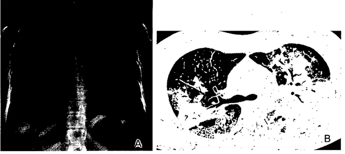
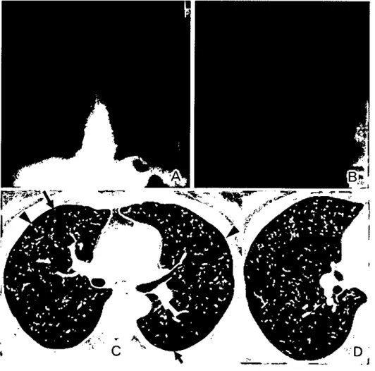
Figure 1. Pulmonary tuberculosis in a 24-year-old wom밍1.
CI>잉t radiograph(A) and bi명1 π없ution cr sc뼈B) show e섰:ensive air-spaæ
∞
nsolidation in both야)per lung zones니페
2) Focal nodular tesìon and Iìnear densìly
The centrilobular nodules and branching linear/ nod비ar
。
pacities (tree-in-bud pattern) are due to the presence of caseation necrosis 업1d granω。
matous infl잉nmation withinand surrounding the tenninal and respσatαγ bronchioles and alveolar ducts8.14.151(fig.3).Co혀escence ar clustering of sm삶1 nodules lead to the formation of a large nodule (s
。
←ealled galro앙 Slgn에(fig.4)
164
3) Cavìty
With erosion into airways and subsequent evacuation of necrotic materials , a cavity can be formed within a parenchymal lesion (Fig. 5) η1e expulsed necro디c materi따
frequently spreads via the bronchi to other paπ:S of the lung (endobronchial spread)71. Therefore , cavitation is an important sign of an active disease
Chest 'fuberculosis: Radiological Classification Acoording 10 Current Concepts
.
B
Fi밍πe 2. Pulmonary tuberculosis in a 35-year-old woman
α1est radi땅"IlhWar 펴뼈1-rosolution cr scan(B)잉lOWfocal
∞
nsolidation와잉surrounding small nod띠ar lesions in the ri앙11때,.,
.1야g‘i「r*
C
•
‘
Fi밍lfe 3. P띠mOIl8lγ tuberc 버。sis presenting with sm퍼I n여ules and branφing 비lear structures in a 24-year-old WOm8Il. ATar 뿜엄d view of ch잉1 radiogroph 허lOWSperibror쉽뻐I sm외I nod비ar lesions밍1dfiα:al conrotidation in the ri양11u끼,.,.lobe
B-c‘μmgwu 피ow images of transv먼ε"hi 명,-π""hrtion cm .()-mn section 1hickness) scans ob때1ed at leve1s oflI삶lCa떠~)ar엄ærina (C) demonslI없e bra찌피1ll tinear structuræ ar1d&T빼1nod버es(tree해14>.업~.ar1d 녔>IIar cx:rs뼈어.ioninthe κ>SIeriarægnmt ciri양111쩌lOr났Je.
Sun Young Jeong,Jeong Jae Kim. Gukmyung Choi. Seung바ung Kim‘and 1m Kyung Hwang
C
B
1
“;.
,."
ι‘
,~
Fi밍Jre 4. PulmOnary tuooπ 띠。sis 강'owlng CT g피axy sign in a 53-year-old man A Chest mdiograph 양lOWSfmel엉ticulonodular
。
αlcitiesinbila 따I예비J]Jer lung wnesB & C. Lung쩨0001ν irnages of lmnsvezæ 비앙l-resolution cr scans (1.0-τnm section tl삐mess) obtsined at lev하s of aortic arch 03) and _81 하,(C). reSPeCtiVeIY.demonstrate lung lesions
∞
nsisling of small∞
ntrilobular n여ules and br밍피피 11:linear 왜uctures (,밍πws. forming∞
-cal1,최 뽕Jaxy잉gn) in bo1h upper lobes. similar lesions 81견。
k영rved in SUPeriOrse,맑lent of left lower 1000‘C
B
•
‘--
“
Fi밍Jre 5. Active p비monary tubeπ 버。SIS wl상1 cavi~ in a 44-year-old man.
A chest mdiograph 양lOWS
∞
W벼ry 1esion in the ri양,t upper lungwne 밍1d nOdUIar opacities in bilateral upper lung wnes. B. μmgwindowimage of tmnsv잉-sehi앙,-resolution cr scans (1.D--mm seclion th때mess) obtsined at 1양01 of great
"'"훌
Is demonstrates ca씨1Bry lesions inboth u]야ler lobes. C. l.ung window image of 1mnSVeIæ 비양'-re 헤ution cr scans (1.Q--mm seclion뻐ckness) obtain어at lev머of subcarina demonstrates cavitmγ Il(성ules in the left φαn' 10벼와1d dense 10m띠ar
∞
n잉idation and tree-in'←bud 잉gns in the 꺼영,t u]찌ler 1000.Chest Tuberculosis: Radiolo밍cal Cl잃S퍼m
디。
nA∞。
rding to Currant Concepts4) Tuberculoma
"Tuberculoma'’refers to a wel1-delimited ,round or ov외
focus of parenchymal TBl~. 'I\Iberculomas may show cenσ'al necrosis ,cavitation ,뻐d saælliæ n여띠es (Fig. 6) α1 CT.
following intravenous administration of contrast ,
tuberculomas often show ring-like enhancement. 에ng-like e띠lancement corresponds hisωlogically to the granulomatous inflammatory tissue capsule. whereas the nonenhancing area corresponds to the central necrotic material1l. Only calcified lesions should be considered inac디vel1l
’、
‘
、、
첫靈
후녁
ξ
@L/B
‘앓좋
f
찮
홈
t\\
\
、
4
i ‘ι、
,
、\‘“
x
•
?t
“
.r
-ν“→..•
::
....
γ i‘
1’ / f'D
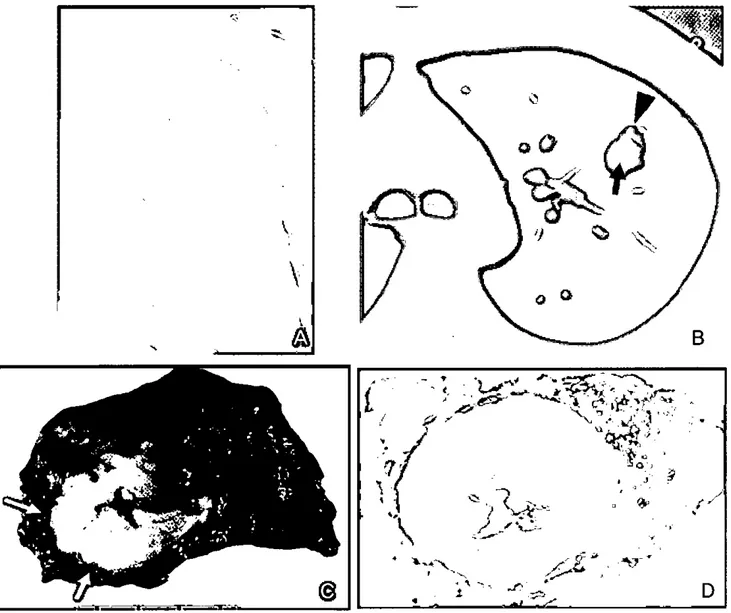
Figure 6. Tubercu10ma in a 41-year-old man. (Courtesy: Kyoung s
∞
Lee. MD. of sam밍mg Medical Center. Seo버.Korea) A Chest radiogr뼈1하lOWSan ov머 n여버e in Jeft up야rr lung ZOIle. B ωng window image of transv잉'sccr
=
(5.o-mm section thickness) ob때빼at level of main bro띠li demonstrates a n<때ule∞뼈ining centra.l ca씨tation (arrow).찌∞nota SU!TOUI피ing saællite nod비e(",
π 얘1e!1d). C. Photogr빼1 of surgiæl resection 허:>ecimendemonstrates nod비e consi밟ng of∞
ntral æ앙~tion n∞
usis (tan y피low밍
W>'"
얘surrounding 에떠멍1∞
us oonn∞
tive fJbrous∞
lpsu1e (anm연,). D. Low-n갱gnif∞
tion이)Qtomicrograph rev∞
ls빼æl tuben:uloma oomposed of well-녕efmed∞
ntral fi∞
US of neaosi.s 밍1d SU!TOUI녕ingi벼lammatorγ ar1d fibrous∞없
~eSun Young‘leong,Jcong Jac Kim,GukmyungChoi,Seung HyungKim.and 1m싸ung Hwang
5) Fìbrosìs,scar,and destruction
Healing of parenchymal TB is associated with more marked fibrosis and calcification71(Fig.7). Cicatrization atelectasis is conunon 따t.er ca'씨어ry disease‘밍ld m밍파ests as atelectasis of the upper lobe. retraction of the hilum‘
compensatory lower lobe hyperinflation‘and mediastinal shift toward the fibrotic lungl!
’
Apical pleural thickeningassociated with fibrosis may reveal proliferation of extrapleural fatW tissue and peripheral atelectasis on C~9) Complete destruction of a whole lung or a mBjor part of a lung is not uncommon in the end stages of tuberculosis Such damage results from a combinati.on of parenchymal and 밍rway involvemene81(Fig.8)
Fi밍lre 7. Healed ’rB lesion in 8 38-ye밍~old WOman.
Chest radiograph하10WSmultiple뼈renchym외cal이icationsand Iinearfibroticbar념s in the 10ftupper lung rone.
짧많
l
.
Figure 8. Chronic destructive 'IB lesion m외nIY involving ri엉lt upper lobe in a 7()-year-old man.
A chest ra이땅'8j)h히m
、
NSmarl<edvolume d∞
"C8SCin ri영lt u찌Jer lung zone、
.vith따뻐펴 elevation of the ri영lt hi뼈nandtr. 뼈E외deviation뼈tethe
π
영1t api,엄I p1밍I히lhicl‘
:eningand ri앙lt αJS!ophrenicangle blunling.Fαoal p1el1l,
피thicl<eningin the 10ftapex and똥앤ml nodular야>acitiesin the 10ft띠JpeTlung zone are어50 not떠 B. Lung windaw irr엽geoftrnnsven;ehi 앙l-r잉sohrtioncr æan O.G-mmsection thiclσ"",,)
ob뻐εd at 100 of great"'"증여s demonstmtæ traction bmnchiectasiswith v이unre d∞않.sc in the ri영lt upper 1000.C. Coronalrefom
‘
atμld cr æan (2.G-mmsection thickrε",)demonstmtæ extrap1euralfat pro1if잉ation in the ri앙lt쩍JeX.-Chest Tuberculosis: Radiological Classification According to Current Concepts
2. Tracheobronchial T8
Traeheobronchial TB has been reported in 10-20% of patients with pulmonarγ TB. 1ρng segmental circumferenti.떠
W외]1hiekening end luminal n외TOWing of Ü1e cenσal 밍rways
can be seen in botl1 active and fibrotic s녕ges. However. in
patients with active disease ,CT scans show irregular and 1hick-walled aIrways. a patrem that is reversible <Fig. 9)‘
whereas patients with flrotic disease generally had smooth narrowing of 없rways and minimal wall thickening ,a pattern
that is not reversible""(Fig.1Ol
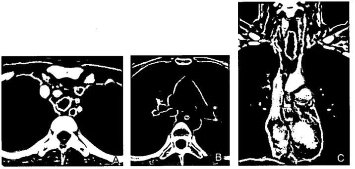
Figure 9. Active stage of σachea-bronebial TB in an 18-y'않,-old girI.
A & B. Mediastinal w이1야w images of transv잉"se enhen<εd cr scans (2.5-mm section따ckness) obùlined at lev마s of great vess허S어)and main bronchi田)‘respective\Y. demonstrate in않버따W밍1 thickening ln d녕외U삶1ea와‘dri양lt main bror떼1US
C.Cα"Onal r성orrnatted rned생S미영1window image demonstrates irre,밍larw 허1 thickening ln d뼈1 tmchea.
Fi밍Jre 10. Fibrotie stage of bron상j외TB involving 1eft m머n bronchus in a 23-year-old woman.
A&B ‘M때$뼈1 window images of transverse ul뻐1hanced cr scans (2.5-mm soc띠1 thickness) ob보Ined at I얀아s of main bror잉j 어〕 and 끼명lt upper lobar brOI잉1US ffi). 1얻;pectiveIy. demonstrate conoen미e w외1 thickening of 1없 main bronchus. Also note obti띠"ted bror찌뼈1 lumen ln이sta1 portion of lefì main bronchus. C. 3D vo!urn<Hendering 띠1Bgedis이oses marl<ed lurninaI l1BIl"OWingand obtitemtion of lefì main 1>>π임1US.
Sun YoungJeong.Jeong Jae Kim‘GukmyungChoi,ScungHyungKim,and 1m앙ung Hwang
3. Miliary TB
M피aγ TB refers to widespread dissemination of TB by
hematogenous spread. The characteristic radiographic 뼈d
high-resoluti.on CT fmdings consist of innurnerable,1- t
。
3-mm diameter nodules randomly distributed throughout both lungs'"(Fig.ll)';'D
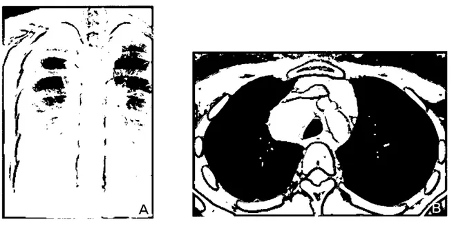
Figure 11. Miliary TB in a 40-year-old woman‘
A chest radiogrnph해ows diffuse granul앙or ground←밍!lS301힐~fy in both Iungs. B. Tm뿜tlld view of chest rndiograph허lOWSmore cl
∞
rsmall n<성ules of 2-3 mm in diameter‘rrul띠ry nodules. C & D. Lung window irnages of transveræ 매양1-'"
∞
lution cr sæns O,o-mmsection thicknessl ob떠뼈d at lev퍼of bro,잉lUS intem뼈ius (C) and tar흉엄d view 01뼈inod at 100 of끼명1t middle lobar bro띠1US(D) demo잉trnlemiliarγ n여ules of random dis이b띠ion: rx쳐ules disbibuted in
∞
ntri10bularocation(밍πWS),에ong미eura (;외m애1eadsl.어펴 뼈ng fissure (curvedarrows)4. Lymph node TB
During the stage of active disease ,T8 organisms frequently spread to the regional Iymph nodes. where the ensuing granulomatous inf1amrnatory reaction results in Iymph node enlargement. πloracic lymphadenopathy is most commonly unilateral and 1
∞
ated in the hi1um or paratracheal region. On CT‘the enlarged nodes frequently have lowattenuation 뻐d show peripheral (rirn) enhancement. The
fonner corresponds to the central necroti.c portion of the node,and the latter.
ω
the surrounding inf1ammatory 디ssue1,211(Fig.12). In TB infection. there is considerable difference in the prevalence of radiologic fmdings in infants and children compared with those in adults π1e most common abnonnality in infants and children consists of Iymph node enlargement,which is seen in 90% ω 95% of cases1.22l-Chest Tuberculosis:RadiologicalClassificationAccordingto Current Concepts
/
.
「
훌룹『훌‘、”、'.
A
Figure 12. Tubeπ버ous Iymphadeniûs in a 28-y'않r-old wom.an A α_r 뼈iograph강'lOWSbilaterals매erior med떠돼nal widening
B,M어.iastina1window irr멍ge of trnnsversc e미뀐næd ar scan (5-1πn section thickness) derα:mstrateser뻐rgOO\yrnph n<성es in the ri영1\ lφper
맹_
area and biia떠'll!jJl연애∞Jlar areas with æntral llOO"Oticlow attenuation밍념pen야eral enhancingrim.5. Pleural and chesl wall TB
Pleural엉Tusion.앙pically w피ateral. occurs in 15-20% of 1'8 patient낭a 쩌야1。빼 plew'll! efl냉ion is usua1Jyass
∞
iated withparenchymal abnonnalities. it may be the only radiologic m밍rifestation of 1'8. Pleural eπUSJon
∞
n be caused by rupture。
f a tuberculous cavity into tbe pleural space. This may-
해.ι
‘ø
좋
、
훨
1용
l
‘‘
‘」
,
A
~--result in the formation of tubercuous empyema and,
。
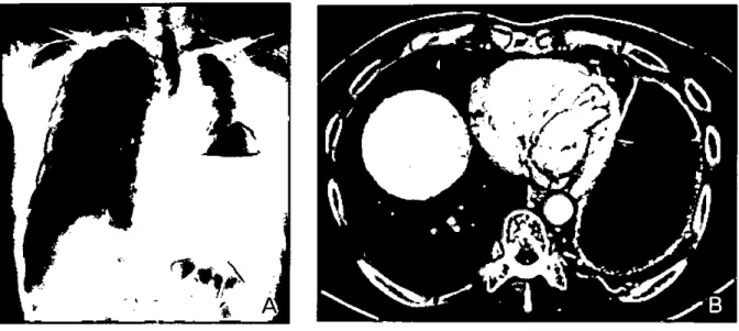
ccasional1y,a bronchopleural fistuJa with pleural air-fluid levelS,1까Fig.l3 & Fig.14). Empyema necessitatis resul\ from leakage of TB empyema U1rou앙1 the parietal pleura with discharge of its contents into the subcutaneous tissues of the ches\ wallV'(Fig.l5)Figure 13. l.oculated empy,잉na in a 51-year-old man.
A Chest radiograph양10WSm여erate plouralefTusion에α1 oonvexborrlerin the ri양1\ hemi1horax,repw풍nting ernpyema
B. M어ias며1회 뼈x10w image of transverne enhanæd ar scan잉6꺼1Jn section 1hickness)양10WSI
∞
]!a뼈d미OUraIeffusion with뻐따'" plO1띠'll!1hi야æningintheri 양1\ hemi1horax,rejJlε;enting잉npyema.Sun Young Jeong,Jeong Jae Kim‘Gukmyung Choi,Seur냉Hyung Kim. and 1m J(yung Hwang
rJ~
획}
꾀
Figure 14. Bron야lopleur떠flSlula in a 62-year-old woman.
A Chest mdiogrnph shom air꺼u버lev밍in the left hemithomx. which밍g홈tsthe αs 강Jilily of btπ엉lOpleurai flSillla fö영lt upper 띠11( 7.one강lOWSmultiple SmaJ] PeribrOnChiaJnod매ar opacities. AISO note vohnne d
∞
-ease with f1btα:aJcified ParenC.l\YITl8Ichan양 in the left때pe.r lung zonc ar얘
。
rcumferen떠1 pleur피 뻐ckening or 잉fusion in the I여t upper hemith뼈x.B.M 떠iastir뻐애1dow image of transv힌똥αh 밍lCed cr scan (2.o-mm secIion thickness) 의10
‘
NSl∞비ated pleuml effusion with diffuse plew밍thicl‘잉
ling and 외r-fluid level in the lefthemithomx. representing empyerna
、
Ni1hbronchopleuraJ flSiulai
•
•....
、‘
A
Figure 15. Empyema necessitatis in a 30-year-old woman
A Lungw 뼈ow imagos of Iransv
밍""
enhanæd cr scan (5.o-mm secIion thiclmess) obt외n<엉at ventriαlar, level demonstrales 뼈E 떼lYlll외tubereulous 1성oκ ∞때sting of n여비g 외1d tree-in-bud signs. in ri잉lt lower lobe
B. M어iaStinaJ window image of transver용 enhanæd cr scan (5.o-mm secIion thiclmes) 야tair뼈at level of supra-!쩌>atic때잉lOTvena cava dernonstrntes Iow-attenu와ion lesion써th rim enhancement (뼈uws) having both intrathoracic and chest WaiI α)mponents of di9:영se
(empyerrι
,
neæssit 뼈5)-Chest Tubcrculosis: Radiological Classifi
∞
tion According to Current Concepts6. Pericardial T8
'I\tbercu1ous pericarditis develops secondary to contiguous spread from mediastinal nodes,lungs ,spine ,or sternum ,or
/
during nωi밍"y dissemination25l. Pericardial 1'8 presents as
pericardial effusion. thickening. or calcification on CT scans에(Fig.16).
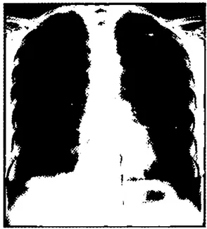
Figure 16. Cons1rictive pericardiüs as a sequelae of tuberculOUB perica펴iüs.
A &B.Posterosnterior
ω
어피Iater머03) chest rsdiogl뼈15양lOWthick며lC:a떠iaI∞
lcifiαmon (arrows), 머。ng tbe anterior ar엄d빼1TQglJl삶icS펴àæ of tbeheart
Sun Young‘Jeong,Jeong Jae Kim. GukmyungChoi,se따19lψ니ng Kim,뼈d 1mKyungHwang
1) Cegielski JP,Chin DP,Espinal MA,et 려 η1e glob외
tubercu10sis situation: progress 밍1d problems in the 20th
centurγ,prospects for the 21st centurγ lnfect Dis ClinN Am. 2002: 16: 1-58
2) Corbett EL,Watt CJ,Walker N,et a1. The growing burden of tuberculosis: global trends and interactions with the HIV epidemic. Arch Intern med. 2003; 163 1009-1021
3) Jeong YJ,Lee KS. p1이hnona:ry tuberculosis: up-t。←date
imaging and management. Am J Roentgenol. 2008; 191 834-844
4) World Health Organization. Glob81 tuberculosis report 2013
、
NWW.who.int!tb/p니blications/global]ep。
πjgtbr13_executivesummarγ pdf. WHO
、
;vebsite. Accessed on November 25, 20135) NL Muller,Franquet T,Lee KS. Imaging of p띠monarγ infections. Philadelphia , PA: 1ippincott Williams & W뼈ins,2007
6) 1ee KS,1m JG. CT in adults with tubercu10sis of the chest: characteristic fmclings and role in management AmJ Roentgeno1.1995: 164: 1361-1367
7) Lee KS,Franquet T,Han J,Johkoh T. M매ler’s disease
。
f the lung: Radiologic 잉ld Pathologic Correlations. 2'띠ed. Philadelphia,PA: Lippincott Wiliarns & Wilkins,2012 8) N1 Muller,CI Silva. hnaging of the chest - volume I
Philadelphia,PA: Saunders/Elsevier ,2008
9) Barnes PF,Cave MD. Molecular epidemiology of tuberculosis. N Engl J Med 2003: 349: 1149-1156
10) Small PM,Hopewell PC,Singh SP,et a1. The epidemiology of tuberculosis in San Francisco: a popu1ation-based study using conventional 밍ld molec띠ar
methods. N En링J Med 1994: 330: 1703←1709
11)Alland D,K려.kut GE,Moss AR,et a1. Transmission of tuberculosis in New York City: an analysis by DNA fmgerprinting 밍띠 conventional epidemiologic methods. N
Engl J Med 1994: 330: 1710-1716
12)Jones BE,Ryu R,Yang Z,et a1. Chest radiographic findings in patients with tuberculosis with recent or remote infection. Am J Respir Cn"t Care Med 1997; 156 1270-1273
References
174
131Geng E,Kreiswirtb B,Burzynski J,èt al. ClinièaI' and radiographic correlates of primarγ and reactivation tuberculosis:. a molecular epidemiology study. JAMA2005;
293: 2740-2745
14)1m JG,ltoh H,Lee KS,et aI. CT-pa1hology correlation of pulmon81γ tuberculosis. Cn"tReγ Diagn Imaging 1995: 36: 227-285
15)Lee JY,Lee KS,Juug KJ,et aI. p1미.lmonarγ tuberculosis
CT and pathologic correlaiion. J Comput Assist Tomogr
2000: 24: 691-698
16)Murayama S,Murakami J,Hashimoto S,et al Noncalcified pulmona:ry tubercu1omas: CT enhancement pattems with histologica1 correlation. J Thorac imaging
1995: 10: 91-95
17)Andronikou S,Vanhoenacker FM,De Backer AI,et al Advances in Imaging Chest Tuberculosis: Blurring of Differences .Between Children and Adults. Clin Chest Med. 2009 Dec: 3이4): 717-744
18)Kim HY,Song KS,Goo JM,et aI ηloracic sequelae and complications of tubercu1osis. Radiogra.phics2001; 21(4) 839-858
19)1m JG,Webb WR,Han MC,et al. Apical opacity associated with pulmonarγ tuberculosis: high-resolution CT fmdings. Radiolo양 1991: 178: 727-731
20) Moon VVK,1m JG,Yeon 10.1,et a1. Tuberculosis of 당1e central airways: CT findings of active and fibrotic disease. AmJ Roenl;genol.1997: 169: 649-653
21) 1m JG,Song KS,Kang HS,et at. Mediastiual tubercuIous lymphadenitis: CT manifestations. Ji녕diologγ 1987: 164 115-119
22) Leung AN,Muller NL,Pineda PR,et a1. Primary tuberculosis in childhood: radiographic manifestations
Radiology 1992: 182: 87-91
23) Ebsæin DM,Kline LR,Albelda SM,et aI. Tuberculous ple니r외effusions. Chest. 1987: 91: 106-109
3'1)Choi JY,Hong KT,Oh YW,et aI. CT manifes\ations of late sequelae in patients with tubercu10us plewiiis. Am J
Roentgenol. 2001: 176: 441-445
25) Golden MP,Vikrarn HR. Extrap버mona:ry tuberculosis 없l
overview. AmFarn Physician2005: 72: 1761-8
26) Mayosi BM,Burgess LJ,Doubell AF. Tuberculous pericarditis. Circu1ation2005: 112: 3608-16