A Study on the Expression and Regulation
of Plasminogen Activators
in Brain Glial Cells
Department of Medical Sciences
The Graduated School, Ajou University
ACKNOWLEDGEMENTS
제 가 석 사 2년 을 마 치 고 , 드 디 어 졸 업 을 하 게 되 었 습 니 다 . 여 기 까 지 저 를 이 끌 어 주 신 지 도 교 수 이 신 주 일 로 교 수 님 , 심 사 위 원 이 셨 던 조 은 혜 교 수 님 , 최 경 숙 교 수 님, 저 의 실 험 을 도 와 주 셨 던 박 은 정 박 사 님 께 감 사 의 말 을 전 합 니 다. 그 외 에 도 항 상 제 옆 에 서 조 언 을 아 끼 지 않 았 던 약 리 학 교 실 모 든 박 사 과 정 의 명 순 언 니 , 희 영 언 니 , 온 순 언 니 , 경 애 언 니 조 교 인 수 정 언 니 , 경 진 언 니, 동 기 였 던 우 혁 오 빠 , 지 형 언 니 , 석 사 과 정 의 희 정 이 , 새 봄 이 , 지 영 이 , 떨 어 져 있 지 만 함 께 했 던 것 처 럼 도 움 을 주 었 던 현 실 언 니 외 에 도 열 거 하 지 못 한 소 중 한 분 들 께 도 고 마 움 을 전 합 니 다 . 더 불 어 항 상 같 이 해 주 었 던 동 기 지 훈 이 에 게 도 고 마 움 을 전 합 니 다 . 마 지 막 으 로 이 세 상 에 서 가 장 소 중 하 신 부 모 님 과 가 족 모 두 에 게 도 , 이 글 을 빌 어 다 시 한 번 감 사 하 다 는 말 을 전 하 고 싶 습 니 다 .A Study on the Expression and Regulation
of Plasminogen Activators in Brain Glial Cells
by
SooYoung Park
A Dissertation Submitted to The Graduated School of Ajou University in Partial Fulfillment of the Requirements for the Degree of
MASTER OF MEDICAL SCIENCES
Supervised by
Ilo Jou, M.D., Ph.D.
Department of Medical Sciences
The Graduated School, Ajou University
-ABSTRACT-A Study on the Expression and Regulation of
Plasminogen Activators in Brain Glial Cells
Urokinase- and tissue-type plasminogen activators (uPA and tPA, repectively) are major components of the fibrinolytic system. Recently, non-fibrinolytic functions of them such as chemotaxis, migration and adhesion as well as their roles as signaling molecules for gene transcription, cytoskeleton organization and differentiation are reported. They are also detected in the central nervous system cells, but the exact cellular sources or the functions of them in the central nervous system are largely unknown. In this study, we monitor mRNA expression and the activities of uPA in cultures of microglia as well as uPA and tPA in astrocyte cultures from rat brain. Our data show that activities and transcription of PAs are differentially regulated by the microglial activators that we tested (lipopolysaccharides, gangliosides, thrombin). Moreover those of tPA were detected only in primary astrocytes from rat brain. These results from a basis for stimuli-specific activation of glial cells.
TABLE OF CONTENTS
TITLE PAGE ---1 ABSTRACT---2 TABLE OF CONTENTS---3 LIST OF FIGURES--- 5 ABBREVIATIONS--- 6 .INTRODUCTION--- 7. MATERALS AND METHODS A. Reagent---9
B. Preparation of microglia and astrocyte--- 9
C. RNA Isolation and Reverse Transcription Polymerase Chain Reaction (RT-PCR)--- 10
D. Zymography of urokinase- and tissue-type plasminogen activators--- 10
E. A coupled photometric assay for plasminogen activator--- 11
. RESULT A. Differential regulations of expressions of urokinase type plasminogen activators in microglia and astrocyte by microglial activators.--- 14
B. Activities of urokinase plasminogen activators in microglia and astrocyte by microglial activators ---17
C. Differential regulations of expressions of tissue type plasminogen activators in microglia and astrocyte by microglial activators--- 20
D. Activities of tissue type plasminogen activators(tPA) in primary astrocyte by microglial activators --- 22
E. Transcript of PAI-1 in microglia and astrocyte by microglial activators--- 25
. DISCUSSION--- 28
. CONCLUSION --- 31
BIBLIOGRAPHY---32
LIST OF FIGURES
Fig 1. Suggested arrangement for simultaneous measurement of PA activity and PAI activity---13
Fig 2. mRNA expressions of uPA, uPAR by in microglia and astrocyte by
microglial activators--- 15
Fig 3. Zymogram showing the activities of urokinase plasminogen activator by microglial activators--- 18
Fig 4. mRNA expressions of tPA in microglia and astrocytes by microglial activators --- 21
Fig 5. Zymogram showing the activities of tissue plasminogen activator by microglial activators--- 23
Fig 6. PAI-1 mRNA expressions in microglia and astrocytes by microglial activators--- 26
ABBREVIATIONS
PA: plasminogen activator
uPA: urokinase plasminogen activator tPA: tissue type plasminogen activator
uPAR: urokinase plasminogen activator receptor PAI-1: plasminogen activator inhibitor-1
Mic-CM: microglia-conditioned media Ast-CM: astrocyte-conditioned media
Z-Lys-SBzl: thiobenzyl-benzyloxycarbonyl-L-lysinate DTNB: 5,5'-Dithiobis(2-nitrobenzoic acid)
. INTRODUCTION
Plasminogen activators (PAs) are major components of fibrinolytic system. This system comprises plasminogen/plasmin, urokinase-/tissue-type plasminogen activators (tPA, uPA, respectively), plasminogen activator receptor (uPAR), and plasminogen activator inhibitors (PAIs).1 Plasminogen activators (PAs) are like plasmin, serine proteases, thus synthesized and released as zymogens, then converted to active proteolytic enzymes. Urokinase plasminogen activator (uPA) is activated when bound to uPA receptor (uPAR) on the cell surface and transformed into two chain active uPA.2 In contrast, tPA is transformed into active tPA without binding to receptor.3 The activities of PAs are also regulated by binding to plasminogen activator inhibitors (PAIs). Among several subtypes of plasminogen activator inhibitors, plasminogen activator inhibitor type 1 (PAI-1) is a major inhibitor of tPA and uPA, whereas plasminogen activator inhibitor-2 (PAI-2) exhibits inhibitory activity mainly toward u-PA and less effective against t-PA.4,5 Accordingly, the activities of components of fibrinolytic plasminogen/plasmin system are finely regulated. Recently, there have been reports that the activities of PAs are detected in brain and the synthesis and release of plasminogen/plasmin system components are attributed to a number of central nervous system cells, including endothelial cells, neurons, astrocytes, and microglia, in vivo as well as in vitro.6,7 In ischemia and excitotoxicity models, tPA induction and subsequent plasmin generation have been implicated in neuronal loss, 8-10and the significant increase in uPA, uPAR and PAI-1 in macrophages of the acute lesion of multiple sclerosis delineate the focal
proteolysis which promotes adhesion and migration of activated microglia and inflammatory cells through CNS parenchyma.11 But, there're increasing contradictory evidences that the plasmin system is induced and degrades amyloid- aggregates12 or direct infusion of tPA does not cause neuronal loss.13 In particular, a recent report that mice lacking tPA, uPA, or plasminogen genes showed delayed functional recovery after sciatic nerve crush imply protective roles of PAs.1 4 These contradictory roles of brain plasminogen / plasminogen activator system in brain pathology and physiology could be the result of integration of separate brain cells' reaction and interactions to stimuli.
In this study, we tried to find the cellular sources and regulations of plasminogen activators using cultured glial cells. uPA was detected in both of microglia and astocytes, while tPA was observed only in astrocytes. Moreover, the activities and the expression of uPA and tPA were differentially regulated by each of glial activators. These form the basis of stimuli-specific activation of glial cells, and thus explain the different roles of PAs in brain pathology.
. MATERALS AND METHODS
A. Reagents
Thrombin and lipopolysaccharide (LPS) were purchased from Sigma Chemicals (St. Louis, MO), and gangliosides mixture was purchased from Matreya (Pleasant Gap, PA). Interferon gamma was purchased from Calbiochem (San Diego, CA). Oligonucleotide primers were purchased from Bioneer ( Seoul, Korea).
Gelatin and casein were phrchased from Sigma Chemicals (St. Louis, MO), and fibrinogen and plasminogen was purchased from Calbiochem (San Diego, CA).
B. Preparation of microglia and astrocyte
Primary microglia were cultured from the cerebral cortices of 1-3 day Sprague-Dawley rats as previously described. Briefly, the cortices were triturated into single cells in minimal essential media containing 10 % fetal bovine serum (Hyclone, Logna, UT) and plated into 75cm2 T-flasks (0.5 hemisphere/flask) for 2 weeks. Then, microglia were detached from the flasks by mild shaking and applied to a nylon mesh to remove astrocytes and cell clumps. Cells were plated in 6-well plates (5 x 10⁵ cells/well) or 60 mm dishes ( 8x10⁵ cells/dish) or 100 mm dishes ( 2x10⁶ cells/dish). One hour later, the cells were washed to remove unattached cells before being used in experiments. Primary astrocytes were prepared using trypsin after microglia were removed. Detached astrocytes were seeded in 100 mm or 60 mm dishes. BV2 immortalized murine microglia cells were from Dr. EJ Choi. The BV2 cell line was grown in Dulbecco's Modified Eagle Medium and supplemented with
5% fetal bovine serum.
C. RNA Isolation and Reverse Transcription Polymerase Chain Reaction (RT-PCR)
Total RNA was isolated using RNAzolT MB (TEL-TEST Inc., friendwood, TX), and cDNA was prepared using reverse transcriptase that originated from Avian Myeloblastosis Virus (TaKaRa, Japan) according to the manufacturer's instructions. PCR was performed with 30 cycles of sequential reactions: 94 for 30 sec, 55 for 30 sec, and 72 for 30 sec. The sequences for PCR primers were as follows. (F) 5'-ACT GTG GCT GTC AGA ACG G and (R) 5'-CTC TGG TTG TCG GGG TTC for u-PA; (F ) 5'-AAT GGT GGC CCA GTT CTG-3' and (R) 5'-AGG GTC AGG AGC AGG GAG-3' for u-PAR., (F)
5'-ATGAGATCAGTACTGCGGACGCCATCTTTG-3' and (R)
5'-GCACGGAGATGGTGCTACCATCAGACTTGT-3' for PAI-1, and (F)5'-AAAGCTGACATGGGAATATTG-3',
(R)5'-ATGTTGTCTTGGATCCAGTTC-3' for tPA; (F) 5'-TCC CTC AAG ATT GTC AGC AA-3' and (R) 5'-AGATCCACAACGGATACATT-3' for GAPDH:(F) 5'-CATGTTTGAGACCTTCAACACCCC-3' and (R)
5'-GCCATCTCCTGCTCGAAGTCTAG-3' for Actin. PCR products were separated by electrophoresis in a 1.5 % agarose gel and detected under UV light.
D. Zymography of urokinase- and tissue type plasminogen activator
for more than overnight, then a total of 1ml of the media was used directly or freezed at 70 . To confirm the presence or changes of uPA activity, media (supernatants) were subjected to casein zymography as described.15 Briefly, serum-starved cells were stimulated with gangliosides 50 ug/ml, LPS 100 ng/ml or thrombin 20 U/ml for the indicated times. The harvested medium Cell lysates or media were concentrated 200~500 fold if necessary, using Rapid-CONTM Protein concentration Kit. Then, the media was obtained and electrophoresed on 10 % SDS-PAGE gels containing plasminogen (0.04 mg/ml), casein (0.1 mg/ml), fibrinogen (2.4 mg/ml) and thrombin (0.1 unit/ml) at 4 . After electrophoresis, the gels were gently shaken at room temperature for 1 hour in 2.5 % Triton X-100 to remove SDS, then incubated in developing buffer (50 mM Tris, 0.2 M NaCl, 5 mM CaCl2, 0.02 % Brij 5 w/v or 1 % Triton X-100) at 37 in a humidified chamber for
16 hrs to reactivate them. Then the gel were stained for 1 hr in 0.5 % solution of Coomasie blue and destained with 70 % methanol and 10 % acetic acid.
E. A coupled photometric assay for plasminogen activators
This assay consists of two successive steps.
Step 1 generates plasmin by plasminogen activators in samples, and step 2 dertermine the plasmin activity gernerated in step 1 using spectrophotometer.
Step 1, plasminogen activation is carried out at 37 in 96 well flat-bottomed microtiter plates. All reagents are diluted before the assay in 0.1mol/liter Tris.Gly, pH 8.5, containing 0.5 mg/ml BSA. The incubation mixture consist of 5 ul of the sample to be measured or PA standard and 0.002 U plasminogen, 1 ng/ml fibrinogen
fragment. Fibrinogen fragments are omitted when only uPA activities are determined. This step 1 incubation lasts usually 1 h,37 .
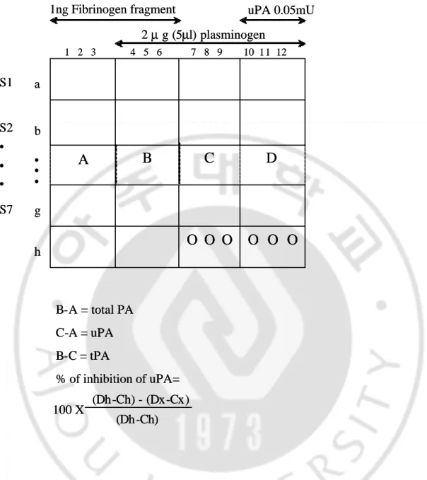
Next, step 2 determines activities of plasminogen activator. The reagent mixture for this determination contains 100 parts Pi-NaCl buffer (0.2 mol/liter NaCl, pH 7.5) and one part each of Triton X-100 at 0.1 % in water, DTNB 22 mM/L, Z-Lys-SBzl 20 mM/L. Aliquot ( 270 ul ) of this mixture is added to each well and the reaction allowed to proceed for 15 min at 20 . Optimal density is mesured at 405nm.16-18 PA and PAI activity was simultaneously measured by following method (Fig. 1). Five microliters of sample is added to the 12 wells of a line. The first three wells (A) receive no plasminogen and meawure the spontaneous hydrolysis of the substrate in contract with sample conditioned medium. The first 6 wells receive fibrinogen fragments and are used for the dertermination of total PA (tPA and uPA) activity (B minus A). In the last 6 wells, no fibrinogen fragment are added and the value in wells 7 to 9 (C) minus the value in wells 1 to 3 (A) thus represents endogenous uPA activity (C minus A). tPA activity is the difference between total PA activity and uPA activity (B minus C). uPA is added in the last 3 wells, and the difference in absorbance readings between wells 10 to 12 (D) and receives no sample, the last 6 wells of this line are used to give the 100 % value of the activity of the uPA (Dh - Ch). They also constitute an internal standard for comparison between plates in a same assay.
B-A = total PA C-A = uPA B-C = tPA % of inhibition of uPA= 100 X (Dh-Ch) - (Dx-Cx) (Dh-Ch)
D
C
B
A
O O O
O O O
1 2 3 4 5 6 7 8 9 10 11 12 • • • S2 • • • S7 S1 1ng Fibrinogen fragment a b g h 2 µ g (5µl) plasminogen uPA 0.05mU B-A = total PA C-A = uPA B-C = tPA % of inhibition of uPA= 100 X (Dh-Ch) - (Dx-Cx) (Dh-Ch)D
C
B
A
O O O
O O O
1 2 3 4 5 6 7 8 9 10 11 12 • • • S2 • • • S7 S1 1ng Fibrinogen fragment a b g h 2 µ g (5µl) plasminogen uPA 0.05mUFig. 1 Suggested arrangement for simultaneous measurement of PA activity and
. RESULT
A. Differential regulations of expressions of urokinase type plasminogen activators in microglia and astrocyte by microglial activators.
To define the cellular sources of uPA, we firstly examined mRNA expressions of them using RT-PCR. To stimulate microglia and astrocyte, we treated cells with gangliosides 50 ng/ml, thrombin 20 U/ml or LPS 100 ng/ml, according to our previous experiments.19,20 Inclusion of gangliosides or thrombin markedly induced the transcript of uPA in 3 hours, while inclusion of LPS suppressed it in (prim) microglia, BV2 cells (Fig. 2A, B) and prim.astrocyte(Fig. 2C). As uPA is known to be activated when it is bound to uPAR, we examined if the expression of uPAR behaves parallel to that of uPA (Fig.2). Inclusion of LPS or thrombin increased the expressions of uPAR, while inclusion of gangliosides did not show any changes of them in (prim) microglia, BV2 cells (Fig.2A, B) as well as prim. astrocyte (Fig. 2C). These results show that the expressions of uPA and uPAR were regulated separately.
uPA uPAR ACTIN Thrombin Control LPS Gmix
A. Primary
Microglia
C. Primary
Astrocyte
uPA uPARB. BV2 cells
uPA uPAR GAPDH GAPDH uPA uPAR ACTIN Thrombin Control LPS GmixA. Primary
Microglia
C. Primary
Astrocyte
uPA uPARB. BV2 cells
uPA uPAR GAPDH GAPDHFig. 2 mRNA expressions of uPA, uPAR in microglia and astrocyte by microglial
activators.
To stimulate microglia and astrocyte, we treated cells with gangliosides 50 ng/ml, thrombin 20 U/ml or LPS 100 ng/ml.
Inclusion of gangliosides and thrombin markedly induced the transcript of uPA in 3 hours, while inclusion of LPS suppressed it in primary microglia and BV2 cells and primary microglia. The other side, inclusion of LPS or thrombin increased the expressions of uPAR, while inclusion of gangliosides did not show any changes of them in primary microglia, BV2 cells as well as prim. astrocyte.
B. Activities of urokinase plasminogen activators(uPA) in microglia and astrocyte by microglial activators.
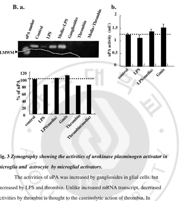
Since proteases including urokinase type plasminogen activators are tightly regulated, we next examined the activities of uPA in the microglia-conditioned media (Mic-CM) and astrocyte conditioned media (Ast-CM), using casein zymography which detect only active- and PAI-bound uPA, not inactive pro-uPA. The casein zymogram of freshly prepared Mic-CM and Ast-CM showed a lytic bands above the LMW human uPA (33 kDa), since the molecular weight of uPA from rat brain is reported to be about 48 kDa.21 With zymograms, PAI-bound uPA can be detected, but nearly no activity was detected above the uPA lytic band in our experimental conditions(data not shown). The activity of uPA decreased with inclusion of LPS in BV2 cell and prim. astrocyte, while it increased with inclusion of gangliosides (Fig. 3A, a , B, a), which is thought to result from decreased or increased mRNA transcript, respectively (Fig.2). Decreased activities by thrombin are described to the caseinolytic action of thrombin, since the addition of thrombin to control media also showed blocked activity (Fig 3A, a , B, a, lane 7). LPS preparations are reported to have contaminants as LPS-associated proteinases,22 but were adding of LPS to Mic-CM from control cells did not show proteolytic activities.(Fig 3A, a , B, a, lane 4). Moreover, as a zymogram done with cell lysates showed similar patterns with that of supernatants (data not shown), we concluded that the activities of secreted uPA reflect that of synthesized uPA. In experiments to confirmed these data , we were examined by photometric assay for plasminogen activator. These data showed similar pattern to change in zymogram.(Fig 3.A, b, B, b) The other side, thrombin
don' t measured due to caseinolytic action of thrombin. Media+Thrombin uPA marker
A.a
Control LPS Media+LPS Gangliosides Thrombin
LMWM HMWM % of uPA 0 20 40 60 80 100 120 140 control LPS LPS(media) Gmix Thrombin Thrombin(media) 0 0.2 0.4 0.6 0.8 1 1.2 1.4 1.6 Control LPS LPS(media) Gmix uPA activity (mU )
b.
Fig. 3 Zymography showing the activities of urokinase plasminogen activator in
microglia and astrocyte by microglial activators.
The activities of uPA was increased by gangliosides in glial cells. but decreased by LPS and thrombin. Unlike increased mRNA transcript, decreased activities by thrombin is thought to the caseinolytic action of thrombin. In
experiments to confirmed these data , we were examined by photometric assay for plasminogen activator. These data showed similar pattern to change in zymogram.
0 0.5 1 1.5 2 control LPS LPS(media) Gmix uPA activity (mU )
b.
Control LPS Media+LPS Gangliosides Thrombin Media+Thrombin
uPA marker control LPS LPS(media) Gmix Thrombin Thrombin(media) 0 20 40 60 80 100 120 % of uPA
B. a.
LMWMC. Differential regulations of expressions of tissue type plasminogen activators in microglia and astrocyte by microglial activators.
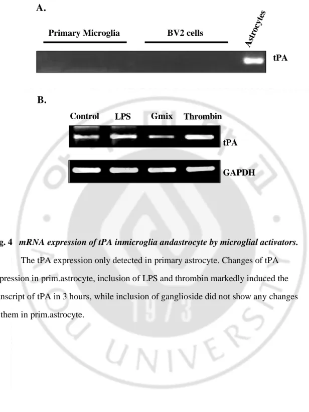
Next, we examined whether microglia and astrocytes express the transcripts of tPA. But, we could not detect tPA transcript in either control or stimulated
primary microglia from rat or BV2 cells (Fig. 4A). As distinct from (primary) microglial cells, transcripts of tPA were observed in primary astrocytes, only whose expressions increased with inclusion of LPS or thrombin, not with that of
gangliosides(Fig. 4).
Fig. 4 mRNA expression of tPA inmicroglia andastrocyte by microglial activators.
The tPA expression only detected in primary astrocyte. Changes of tPA expression in prim.astrocyte, inclusion of LPS and thrombin markedly induced the transcript of tPA in 3 hours, while inclusion of ganglioside did not show any changes of them in prim.astrocyte.
tPA
Astrocytes
Primary Microglia BV2 cells
GAPDH tPA Thrombin Control LPS Gmix
A.
B.
D. Activities of tissue type plasminogen activators(tPA) in primary astrocyte by microglial activators.
tPA activities were detected in Ast-CM from cultured primary astrocyte from rat around the molecular weight of 92 kDa. Inclusion of LPS or thrombin for 24~48 hr suppressed the activities of tPA, while inclusion of gangliosides showed little changes. (Fig 5A) In experiments to confirmed these data , we were examined by photometric assay for plasminogen activator. These data showed similar pattern to change in zymogram.(Fig 5B) The other side, thrombin don' t measured due to caseinolytic action of thrombin. But, decreased activity by LPS or thrombin are contradictory to the results with RT-PCR (compare Fig. 4). Previous report show that, tPA/PAI-1 complex was mediated cellular events.23 So, we tested the involvement of PAIs.
tPA Control LPS tPA marker Media+Thrombin Thrombin Ganglioside 75 80 85 90 95 100 105 control LPS Gmix Thrombin Thrombin(media) tPA of %
A.
B.
0 0.05 0.1 0.15 0.2 0.25 Control LPS LPS(media) Gmix tPA activity (mU )Fig. 5 Zymogram showing the activities of tissue type plasminogen activator (tPA) by microglial activators.
Unlike increased mRNA transcript, the activities of tPA suppressed by LPS and thrombin. Decreased activities by thrombin is thought to the caseinolytic action of thrombin. In experiments to confirmed these data , we were examined by photometric assay for plasminogen activator. These data showed similar pattern to change in zymogram.
E. Transcript of PAI-1 in primary microglia, BV2 cells and primary astrocytes.
Since the activity of urokinase-, tissue type plasminogen activators could also be regulated by binding to plasminogen activator inhibitors(PAIs), and PAI-1 is known to be a major inhibitors of uPA and tPA, we observed transcript levels of PAI-1 with RT-PCR analysis. Transcripts of PAI-1 were increased with inclusion of LPS or thrombin in (primary) microglia and prim.astrocyte (Fig.6A). And as expected, expressions of PAI-2 showed no significant changes(data not shown). In a study to confirm these data, were measured by a coupled photometric assay (Fig. 6B). So decreased tPA activity by zymogram in LPS-, thrombin-treated astrocytes, could be the results of increased expression and binding of tPA to PAI-1. In conclusion, tPA is transformed into active tPA without binding to receptor. Thus, PAI-1-bound tPA was increased with inclusion of LPS or thrombin.
a. Primary Microglia b. BV2 cells c. Primary Astrocyte GAPDH PAI-1 ACTIN PAI-1 GAPDH PAI-1
A
B.
a. Prim. Microglia PAI fold 0 0.2 0.4 0.6 0.81
1.2 1.4 1.6 Control LPS LPS(media)Gmixb.
Prim. Astrocyte 0 0.51
1.5 2 2.5 3 3.5 4 Control LPS LPS(media)Gmix PAI fold thrombin Ctrl LPS GmixFig 6 PAI-1 mRNA expressions in microglia and astrocytes by microglial
activators. .
Transcripts of PAI-1 were increased with inclusion of LPS or thrombin in (primary) microglia and prim.astrocyte. In experiments to confirmed these data , we were examined by photometric assay for plasminogen activator. These data showed similar pattern to change in RT-PCR.
. DISCUSSION
Microglia are among the representative immune effector cells in the brain. When brain is exposed to external stimuli, microglia are immediately activated and proliferate then migrate to damaged areas. Several materials are known to activate microglia in vitro and in vivo, and lipopolysaccharides, pro-inflammatory cytokines such as interferon- are among them.
We report lipopolysacharide, gangliosides24 and thrombin2 5 as another potent microglial activators, thus release cytokines and inflammatory mediators from microglia. In our previous study to compare the activation profiles among several microglial activators, no significant differences were noticed. Namely, all three activate and induce the release of nitric oxides, proinflammatory cytokines including IL-1, TNF- , as well as adhesion molecules and chemokines.
As LPS-, thrombin-, or gangliosides-activated microglia showed marked morphological changes distinct from resting microglia, we thought of the involvement of proteases, especially plasminogen activators. Consistent with the morphological changes, gangliosides treatment increased the mRNA expressions(Fig. 2) as well as the activities of uPA(Fig. 3). LPS, in spite of the characteristic morphological changes, decreased mRNA expressions and activities of uPA (Fig. 2A and B, Fig. 3A). Decreased uPA activity by zymogram is considered to reflect the decreased synthesis of uPA or increased binding of it to PAI-1. This is consistent with former report that microglia isolated from rat brain secrete a uPA and inclusion of LPS markedly decrease the uPA activity. 21 Different from the transcript
of uPA, that of uPAR was markedly induced by LPS, but not by gangliosides. uPAR is a monocyte-activation marker (CD87), whose surface expression is found only on activated monocyte, while quiescent monocytes exhibit little or no uPAR in vivo.26 These demonstrated endogenously expressed uPAR,27that increased in activated human cultured microglia. In our experiments, uPAR expression was also seen in control cells and markedly increased by LPS (Fig. 2A and B). These results reveal that uPA and uPAR are regulated separately, and uPAR could, independently of uPA, contribute to inflammatory functions of microglia. The fact that LPS treatments suppressed mRNA expressions and activities of uPA does not necessarily mean that LPS has an anti-inflammatory or anti-migration effects. There are reports that uPAR can act independently of uPA28 and uPA-deficient mice are resistant to microbial infection, while uPAR-deficient mice are susceptible to neuronal damages. While the transcripts of uPA and uPAR increased markedly with thrombin, the activities of uPA nearly abolished (Fig.3). It is likely that thrombin cut the active site of secreted uPA, thus, uPA lose the caseinolytic activity, since the inclusion of thrombin to Mic-CM from control cells for 30 min also abolished uPA activity (compare lane 3 and 4 in Fig. 3A). The changes in the expression of uPA and uPAR by the activators in primary astrocytes showed very similar pattern to those in microglia. Namely, inclusion of gangliosides and thrombin increased the expression of uPA, while inclusion of LPS and thrombin increased the expression of uPAR.
Transcripts as well as the activities of tPA were detected in primary astrocytes (Fig.4), but the activities and the transcripts showed inconsistent patterns (Fig.5, 6). Namely, transcript of tPA increased with inclusion of LPS or thrombin
(Fig.5), while the activities of them decreased with inclusion either of them (Fig.6). Those result from increased expression and binding of them to PAI-1 in primary astrocytes.
. CONCLUSION
1. mRNA expression and the activities of uPA were detected in primary microglia and astrocytes, but those of tPA were detected only in primary astrocytes from rat brain.
2. Increased uPA activities by gangliosides treatment are due to the increase in synthesisis in primary microglia and astrocyte.
3. mRNA expression of uPA increased with thrombin treatment.
4. mRNA expression of tPA in primary astrocytes increased with treatment of LPS and thrombin , but the activities shown in zymogram were decreased, possibly due to the increased binding with PAI-1.
Thus, all three microglial activators we tested differentially regulate the expressions and the activities of plasminogen activators in microglia and astrocytes.
BIBLIOGRAPHY
1. Henkin J, Marcott P, Yang HC (1991) The plasminogen-plasmin system. Prog Cardiovasdis 34:135-164
2. Yao Wang(2001), The role and regulation of urokinase-type plasminogen activator receptor gene expression in cancer invasion and metastasis, John Wiley & Sons, Inc. Med Res Rev, 21, No.2, 146-170
3. Siao C-J, Tsirka SE. 2002. Tissue plasminogen activator mediates microglial activation via its finger domain through annexin II. J Neurosci 22:3352-3358
4. Nuala A. Booth BSc, PhD, Reader (1999), Fibrinolysis and thrombosis. Baillieres Best Pract Res Clin Haematol. 1999 Sep;12(3):423-33.
5. Kruithof E. KO, Baker MS, Bunn CL. 1995. Biological and clinical aspects of plasminogen activator inhibitor type 2. Blood 86:4007-4021.
6. Walker DG, Lue L-F, Beach TG. 2002. Increased expression of the urokinase plasminogen-activator receptor in amyloid peptide-treated human brain microglia and in AD brain. Brain Res 926:69-79
7. Beschorner et al.,2000, Lesion-associated accumulation of uPAR/CD87 -expressing infiltrating granulocytes, activated microglial cells/macrophages and upregulation by endothelial cells following TBI and FCI in humans. Neuropathol Appl Neurobiol. 2000 Dec;26(6):522-7.
8. Tsirka SE, Rogove AD, Bugge TH, Degen JL, Strickland S. 1997. An extracellular proteolytic cascade promotes neuronal degeneration in the mouse hippocampus. J Neurosci 17:543-552
9. Tsirka SE, Gualandris A, Amaral DG, Strickland S. 1995. Excitotoxin-induced neuronal degeneration and seizure are mediated by tissue plasminogen activators. Nature 377:340-344
10. Flavin MP, Zhao G, Ho LT. 2000. Microglial tissue plasminogen activator triggers neuronal apoptosis in vitro. Glia 29: 347-354
11. Gveric D, Hanemaaijer R, Newcombe J, van Lent NA, Sier CFM, Cuzner ML. 2001. Plasminogen activators in multiple sclerosis lesions. Brain 124:1978-1988
12. Judianne Davis, Matthew R. Wagner, Weibing Zhang, Feng Xu and William E.Van Nostrand Amyloid β-Protein Stimulates the Expression of Urokinase-type Plasminogen Activator (uPA) and Its Receptor (uPAR) in Human Cerebrovascular Smooth Muscle Cells, J Biol Chem. 2003 May 23;278(21):19054-61.
13. Tirka SE. Clinical implications of the involvement of tPA in neuronal cell death.J Mol Med. 1997 May;75(5):341-7
14. Tucker HM, Kihiko M, Caldwell JN, Wright S, Kawarabayashi K, Price D, Walker D, Scheff S, McGillis JP, Rydel RE, Estus S. 2000. The plasmin system is induced by and degrades amyloid- aggregates. J Neurosci 20:3937-3946
15. Park CG, Goh YJ, Kook SH, Park HK, Kim BJ (1997), Increased expression of urokinase type plasmonogen activator(u-PA), plasminogen activation inhibitor-1(PAI-1),and collagenases in Caco-2 cells infected by salmonella typhimurium, J Korean Med Sci, 1997 Feb;12(1):23-31
16. Pierre Leprince, Bernard Rosister, and Gustave Moonen A colorimetric assay for the simultaneous measurement of plasminogen activators and
plasminogen activator inhibitor in serum free conditioned media from cultured cells. ANALYTICAL BIOCHEMISTRY 177, 341-346(1989).
17. Pierre Leprince,Coleman and George D.J.Green, A coupled photometric assay for plasminogen activator:Method in enzymology,Vol.80:408-414, (1981)
18. Beth Y. Karlan , Ann S.clark , A highly sensitive chromogenic microtitier plate assay for plasminogen activator which quantitatively discriminates between the urokinase and tissue type activators, Academic press, vol 142, NO.1 (1987)
19. Kim OS, Park EJ, Joe E, Jou I. 2002. JAK-STAT signaling mediates gangliosides-induced inflammatory response in brain microglial cells. J Biol
Chem 277:40594-40601
20. Pyo HK, Joe E, Jung SY, Lee SH, Jou I. 1999. Gangliosides activate cultured rat brain microglia. J Biol Chem 274:34584-34589
21. Nakajima K, Tsuzaki N, Shimojo M, Hamanoue M, Kohsaka S. 1992. Microglia isolated from rat brain secrete a urokinase-type plasminogen activator. Brain Res 577:285-292
22. Min D, Moore AG, Bain MA, Breit SN, Lyons JG. 2002. Activation of macrophge promatrix metalloporteinase-9 by lipopolysacharide-associated proteinases. J Immunol. 168:249-2455
23. Camani C, Gavin O, Kruithof EK.Cellular degradation of free and inhibitor-bound tissue-type plasminogen activator--requirement for co-receptor? Thromb Haemost. 2000 Feb;83(2):290-6.
24. Beschorner et al., 2000,Lesion-associated accumulation of uPAR/CD87- expressing infiltrating granulocytes, activated microglial cells/macrophages and upregulation by endothelial cells following TBI and FCI in humans. Neuropathol Appl Neurobiol. 2000 Dec;26(6):522-7.
25. Ryu JY, Pyo HK, Jou I, Joe E. 2000. Thrombin induces NO release from cultured rat microglia via protein kinase C, mitogen-activated protein kinase, and NF-B. J Biol. Chem 275:29955-29959
26. Chorner R, Schluesener HJ, Nguyen TD, Magdolen V, Luther T, Pedal I, Mattern R,Meyermann Schwab JM. 2000. Lesion-associated accumulation of uPAR/
CD87-expression infiltrating granulocytes, activated microglial cells/macrophages and upregulation by endothelial cells following TBI and FCI
in humans. Neuropathol Appl Neurobiol 26:522-527
27. Washington RA, Becher B, Balabanov R, Antel J, Dore-duffy P. 1996. Expression of the activation marker urokinase plasminogen-activator receptor
in cultured human central nervous system microglia. J Neurosci Res 45:392-399 28. Resnati M, Guttinger M, Valcamonica S, Sidenius N, Blasi F, Fazioli F. 1996.
Proteolytic cleavage of the urokinase receptor substitutes for the agonist-induced chemotactic effect. Embo J 15:1572-1582
Plasminogen activator (PA)
( : )
Plasminogen activators (PAs) urokinase-plasminogen activator(uPA) tissue type plasminogen activator(tPA)
(fibrine) (fibrinolytic system) .
zymogen plasminogen uPA, tPA
plasmin ,
uPA tPA plasminogen activator inhibitors(PAIs) . , (chemotaxis), (migration), (adhesion), , . . microglia astrocytes , LPS, Gmix, Thrombin
, uPA tPA . uPA
LPS , Gmix Thrombin .
uPA LPS , Gmix .
uPA uPA .
tPA LPS Thrombin , Gmix
. tPA LPS
. tPA PA
PAI . PAI
, LPS Thrombin tPA PAI
LPS . tPA
(microglia) , (astrocyte)
.
uPA tPA .