저작자표시-비영리-변경금지 2.0 대한민국 이용자는 아래의 조건을 따르는 경우에 한하여 자유롭게 l 이 저작물을 복제, 배포, 전송, 전시, 공연 및 방송할 수 있습니다. 다음과 같은 조건을 따라야 합니다: l 귀하는, 이 저작물의 재이용이나 배포의 경우, 이 저작물에 적용된 이용허락조건 을 명확하게 나타내어야 합니다. l 저작권자로부터 별도의 허가를 받으면 이러한 조건들은 적용되지 않습니다. 저작권법에 따른 이용자의 권리는 위의 내용에 의하여 영향을 받지 않습니다. 이것은 이용허락규약(Legal Code)을 이해하기 쉽게 요약한 것입니다. Disclaimer 저작자표시. 귀하는 원저작자를 표시하여야 합니다. 비영리. 귀하는 이 저작물을 영리 목적으로 이용할 수 없습니다. 변경금지. 귀하는 이 저작물을 개작, 변형 또는 가공할 수 없습니다.
A Dissertation for the Degree of Master of Science
Study on Lysophosphatidic Acid in
Activating Formation of Blastocoel in
Porcine Embryos In Vitro
Min-Young Shin
Department of Biotechnology
GRADUATE SCHOOL
JEJU NATIONAL UNIVERSITY
리소포스파티드산이 체외 돼지
배반포기의 포배강 형성에
미치는 연구
지도교수 박세필
신 민 영
이 논문을 이학 석사학위 논문으로 제출함
제주대학교 대학원
2018 년 2 월
Study on Lysophosphatidic Acid in
Activating Formation of Blastocoel in
Porcine Embryos In Vitro
Min-Young Shin
(Supervised by Professor Se-Pill Park)
A thesis submitted as a Qualified Dissertation
for the Degree of Master of Science
February, 2018
Department of Biotechnology
GRADUATE SCHOOL
JEJU NATIONAL UNIVERSITY
Approved as a Qualified Dissertation
for the Degree of Master of Science
Dissertation Committee:
Chairperson of the supervising committee
Professor Key-Zung Riu, Ph.D., College of Applied Life Sciences, Jeju National University
Professor Eun-Young Kim, Ph.D., College of Applied Life Sciences, Jeju National University
Professor Se-Pill Park, Ph.D., College of Applied Life Sciences, Jeju National University
Department of Biotechnology
GRADUATE SCHOOL
JEJU NATIONAL UNIVERSITY
i
CONTENTS
CONTENTS...ⅰ
LIST OF FIGURES...ⅱ
LIST OF TABLES... ...ⅲ
ABSTRACT...1
1. INTRODUCTION...2
2. MATERIALS AND METHODS...4
2.1. Oocyte collection and in vitro maturation (IVM)...4
2.2. Parthenogenetic activation and in vitro culture...4
2.3. TUNEL assay...5
2.4. Extraction of mRNA...6
2.5. Real-time RT-PCR with SYBR Green...6
2.6. Western blot...8
2.7. Time-lapse monitoring system...8
2.8. Statistical analysis...9
3. RESULTS...10
3.1. Effect of LPA treatment on in vitro developmental capacity of porcine embryos..10
3.2. Effect of LPA treatment on the timing of each developmental stage in porcine embryos..14
3.3. Effect of LPA treatment on the kinetics of development of porcine embryos...17
3.4. Effect of LPA treatment on the quality of embryos at the blastocyst stag...20
4. DISCUSSION... .22
REFERENCES...28
ABSTRACT IN KOREAN...35
ii
LIST OF FIGURES
Figure 1. Effects of lysophosphatidic acid (LPA) on in vitro porcine blastocyst formation after
parthenogenetic activation from day 0 to 7 (top row) or from day 4 to 7 (bottom row). Scale bars, 200 μm.
Figure 2. Time-lapse imaging of porcine embryos developing in vitro. (A) Embryos from each
treatment group at the same developmental stage, cultured from day 4 to 7 with or without 1 µM LPA. (B) Quantity of control (Con) and LPA-treated embryos at different developmental stages (morula, early blastocyst [Ea BL], and expanded blastocyst [Ex BL]) at 98.0, 115.5, and 138.3 h after activation.
Figure 3. Morphological parameters of in vitro-developed porcine embryos. (A) Embryos of the
same developmental time cultured from day 4 to 7, with or without 1 µM LPA. (B) Diameter of control (Con) and LPA-treated embryos at 94.5, 101, 118, and 136 h after activation. Values are means ± standard error of the mean. (*p < 0.05).
Figure 4. Expression of components involved in compaction and cell-cell communication. (A)
Abundance of TJP1 and Connexin 43 protein, or (B) GJC1, CDH1, and TJP1 mRNA in control (Con) and blastocysts treated with 1 µM LPA from days 4 to 7. Values are means ± standard error of the mean. (*p < 0.05, **p < 0.01).
Figure 5. Apoptosis in in vitro-cultured Day-7 blastocysts. (A) Images of fragmented DNA, as
detected with the terminal deoxynucleotidyl transferase dUTP nick-end labeling assay, versus total nuclei. Scale bars, 100 µm. (B) Total cell number and (C) apoptotic index. (D) Relative transcript abundance of genes involved in cell reprogramming (POU5F1 and DPPA5) and apoptosis (BCL2L1, BAK, and CASP3) in control (Con) and blastocysts treated with 1 µM LPA from Days 4 to 7. Values are means ± standard error of the mean. (*p < 0.05; **p < 0.01).
Figure 6. Mechanism of action of lysophosphatidic acid (LPA). LPA activates Ras and Rho family
GTPases to control cell migration, proliferation, and morphogenesis as well as signaling via classic second messenger pathways.
iii
LIST OF TABLES
Table 1. Primers used for real-time RT-PCR
Table 2. Effects of lysophosphatidic acid (LPA) on the in vitro development of porcine
parthenogenetic oocytes
Table 3. Effects of lysophosphatidic acid (LPA) on in vitro development of porcine parthenogenetic
embryos
Table 4. The timing of each developmental stage in porcine parthenogenetic embryos using
1
ABSTRACT
Modifications to culture media, which include the addition of various factors, are important for the in vitro production (IVP) of oocytes and embryos. In this study, we investigated the effects of lysophosphatidic acid (LPA) on porcine embryo development. Porcine parthenogenetic embryos were cultured with 0, 0.1, 1, and 10 µM LPA (0 LPA, 0.1 LPA, 1 LPA, and 10 LPA) for 7 days or cultured in basic medium until day 4 and then treated with LPA from day 4 to 7. We found no difference in the in vitro development of embryos cultured with LPA for 7 days. However, the rates of blastocyst and over-expanded blastocyst formation were higher in the 0.1 and 1 μM LPA-treated groups from day 4 to 7 than in the other groups. Moreover, the timing for the formation of early blastocysts was shorter and the embryo size was larger in LPA-treated embryos than in the control. In addition, the expression of connexin 43 and gap junction and cell adhesion-related gene (GJC1 and CDH1) increased in treated embryos compared with control. Although there was no difference in the total cell number in blastocysts between groups, the apoptotic index was lower in the LPA-treated group than in the control. Furthermore, BCL2L1 increased but BAK decreased in the LPA-treated group. These results indicate that the addition of LPA to the medium from day 4 to 7 improves blastocyst formation and would be helpful in the development of preimplantation embryos.
Key words: Lysophosphatidic acid, In vitro culture, Porcine, Embryo, Time-lapse monitoring system
2
1. INTRODUCTION
Maintaining embryo viability and development in vitro is crucial for a number of applications in the field of animal biotechnology (Reed et al., 1992). The developmental capacity and quality of blastocysts cultured in vitro are low compared with in vivo. Many investigators have attempted to overcome the shortcomings of the in vitro production (IVP) system by aiming for successful full-term pregnancies after the transfer of blastocysts to recipients (Kikuchi et al., 2002). However, the increased interest in using the pig as a model system requires that new improvements are made in the currently available in vitro culture system (Long et al., 1999), which can improve the developmental competence of oocytes and embryos (Boruszewska et al., 2016).
To enhance the efficiency of in vitro embryo development, investigators have the option of adding factors into the culture medium or adjusting the culture conditions and/or culture medium. Previous studies report that the addition of serum, such as bovine serum albumin (BSA) and fetal bovine serum (FBS), and serum replacements (SR) can improve the efficiency of embryo development (McKiernan et al., 1992; Thompson et al., 1998; Moore et al., 2007). In fact, serum and serum derivatives greatly affect the development, morphology, and metabolism of embryos (Carolan et al., 1995; Thompson, 1997). However, the undefined mixture in serum may result in highly variable developmental rates in vitro and affect the high incidence of apoptosis observed in in vitro produced embryos (Gjorret et al., 2001; Wrenzycki et al., 1999). Currently, glucose, carbohydrates, and fatty acids are included in porcine embryo medium (Petters et al., 1990; Wirtu et al., 2004; Hochi et al., 1999; Khandoker et al., 1999). Serum phospholipids can also improve the quality of oocytes and/or embryos (Amini et al., 2016).
3
Lysophosphatidic acid (LPA) is the common name for monoacyl-sn-glycero-3-phosphate, a phospholipid derivative that can also act as a signaling molecule. It has numerous biological actions and is produced extracellularly (principally from lysophosphatidylcholine) in the serum and plasma. The diverse actions of LPA on most mammalian cells, together with the finding that LPA is a major bioactive constituent of serum, led to the identification of its cognate receptors (Eichholtz et al., 1993; Contos et al., 2000). Since its initial characterization as a growth factor, the list of cellular responses caused by LPA has expanded considerably, and it now includes many non-proliferative effects, ranging from the stimulation of cell migration and survival to the growth of neurites (Moolenaar et al., 2004). LPA activates Ras and Rho family GTPases to control cell migration, proliferation, and morphogenesis as well as signaling via classic second messenger pathways (Xu et al., 2007). Previous studies involved the addition of LPA to the maturation medium of oocytes from the golden hamster (Hinokio et al., 2002), mouse (Jo et al., 2013), and pig (Zhang et al., 2015). In addition, LPA was added into the medium of mouse (Kobayashi et al., 1994) and bovine (Torres et al., 2014) embryos to enhance their developmental competence.
In this study, we investigated the rate and kinetics of blastocyst development and examined the expression of genes involved in cell adhesion and apoptosis during the in vitro culture of porcine parthenogenetic embryos. Based on the results of this study, we conclude that LPA treatment markedly improved the efficiency of embryo development.
4
2. MATERIALS & METHODS
2.1. Oocyte collection and in vitro maturation (IVM)
Porcine ovaries were collected from gilts at a local slaughterhouse and transported to the laboratory in saline supplemented with 75 mg/mL penicillin G and 50 mg/mL streptomycin sulfate within 2 h at 32–35°C. Cumulus oocyte complexes (COCs) were aspirated from follicles of 2–8 mm diameter with an 18-gauge needle and a disposable 10 mL syringe. COCs with a minimum of two layers of cumulus cells were selected and washed three times in HEPES-buffered tissue culture medium (TCM)-199 containing 0.1% (w/v) BSA. Groups of 50 COCs were matured in 500 μL of TCM-199 (Gibco, Grand Island, NY, USA) containing Earle’s salts, 0.57 mM cysteine, 10 ng/mL epidermal growth factor (EGF), 0.5 μg/mL follicle stimulating hormone (FSH), 0.5 μg/mL luteinizing hormone (LH), and 10% (v/v) porcine follicular fluid under mineral oil for 44 h at 38.8°C in 5% (v/v) CO2 and 95%
(v/v) air.
2.2. Parthenogenetic activation and in vitro culture
Following maturation, cumulus cells were removed by mechanical pipetting in the presence of 1 mg/mL hyaluronidase for 2–3 min. The denuded oocytes were activated with 5 μM Ca2+
-ionomycin for 5 min. After 3 h of culture in porcine zygote medium-5 (PZM-5) containing 7.5 μg/mL cytochalasin B (Sigma), embryos were washed three times in PZM-5 containing 0.4% (w/v) fatty acid-free (FAF)-BSA and cultured in the same medium at 38.8°C in a humidified atmosphere of 5% (v/v) CO2 and 95% (v/v) air. The dose- and
time-5
dependent effects of LPA on the development of parthenogenetic porcine embryos were investigated. First, the embryos were cultured in porcine IVC (P-IVC) medium (PZM-5 medium containing 4 mg/mL FAF-BSA) supplemented with 0, 0.1, 1, and 10 μM LPA (0, 0.1, 1, and 10 LPA, respectively) from day 0 to 7, with day 7 corresponding to blastocyst development. In other experiments, embryos were cultured in basic P-IVC medium for 4 days, followed by incubation in P-IVC medium with 0, 0.1, 1, and 10 μM LPA (Con, 0.1 LPA, 1 LPA, and 10 LPA) until day 7. The experiment was independently repeated four or five times.
2.3. Terminal deoxynucleotidyl transferase dUTP nick end labeling (TUNEL) assay
To detect fragmented DNA, blastocysts were fixed with 4% (w/v) paraformaldehyde (PFA) in phosphate-buffered saline (PBS) overnight at 4°C. After fixation, blastocysts were permeabilized by incubating in 0.1% Triton X-100 for 30 min at 38.8°C and incubated with fluorescein-conjugated dUTP and terminal deoxynucleotidyl transferase (In Situ Cell Death Detection Kit, Roche, Manheim, Germany) in the dark for 1 h at 38.8°C. The total number of mitotic and apoptotic cells was scored. Nuclei were stained with Hoechst 33342 (1 μg/mL) for 30 min, and embryos were washed in 0.1% BSA-PBS. Blastocysts were mounted onto glass slides and examined under an inverted Olympus IX-71 microscope (Tokyo, Japan). The experiment was independently repeated six times, and at least 30 blastocysts were examined per group.
6 2.4. Extraction of mRNA
For quantitative real-time reverse transcription-polymerase chain reaction (RT-PCR) analysis, mRNA was isolated from blastocysts using the Dynabeads mRNA Purification Kit (Dynal, Oslo, Norway) according to the manufacturer’s instructions. Briefly, in each treatment group, blastocysts were resuspended in 20 μL of lysis/binding buffer [100 mM Tris-HCl, pH 7.5, 500 mM LiCl, 10 mM EDTA (pH 8.0), 1% (w/v) LiDS, and 5 mM DTT] and vortexed at room temperature for 5 min to completely lyse the embryos. A 30 μL aliquot of an oligo(dT)25 magnetic-bead suspension was added, and the samples were incubated at
room temperature for 5 min. The hybridized mRNA and oligo(dT) beads were washed twice with 300 μL of wash buffer A [10 mM Tris-HCl, pH 7.5, 0.15 M LiCl, 1 mM EDTA, and 1% (w/v) LiDS] and then washed once with 150 μL of wash buffer B [10 mM Tris-HCl, pH 7.5, 0.15 M LiCl, and 1 mM EDTA]. mRNA was eluted from the beads in 10 μL of double-distilled DEPC-treated water.
2.5. Quantitative real-time RT-PCR with SYBR Green
mRNA was isolated as described above, and cDNA was synthesized using an oligo(dT)20
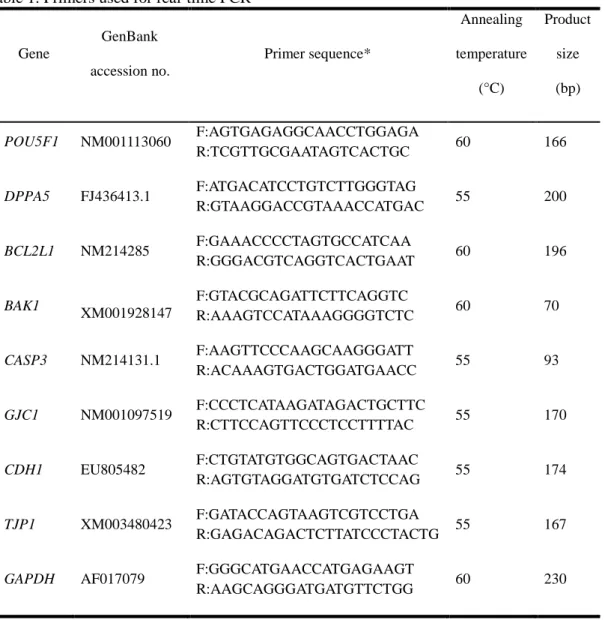
primer and Super Script II reverse transcriptase (Invitrogen, Carlsbad, CA, USA). Real-time RT-PCR was performed using an Applied Biosystems Step One Plus Real-Time PCR System (Warrington, UK) in a final reaction volume of 20 µL containing the SYBR Green PCR Master Mix (Applied Biosystems), The primers used for real-time RT-PCR are listed in Table 1. The PCR conditions were as follows: 10 min at 94°C, followed by 39 cycles of 30 s at 60°C and 55 s at 72°C, and a final extension of 5 min at 72°C. Samples were then cooled to 12°C. The relative gene expression was analyzed by the 2-△△Ct method (Livak and
7
Schmittgen, 2001) after normalization against the GAPDH mRNA level. The experiment was independently repeated three times.
Table 1. Primers used for real-time PCR
Gene GenBank accession no. Primer sequence* Annealing temperature (°C) Product size (bp) POU5F1 NM001113060 F:AGTGAGAGGCAACCTGGAGA R:TCGTTGCGAATAGTCACTGC 60 166 DPPA5 FJ436413.1 F:ATGACATCCTGTCTTGGGTAG R:GTAAGGACCGTAAACCATGAC 55 200 BCL2L1 NM214285 F:GAAACCCCTAGTGCCATCAA R:GGGACGTCAGGTCACTGAAT 60 196 BAK1 XM001928147 F:GTACGCAGATTCTTCAGGTC R:AAAGTCCATAAAGGGGTCTC 60 70 CASP3 NM214131.1 F:AAGTTCCCAAGCAAGGGATT R:ACAAAGTGACTGGATGAACC 55 93 GJC1 NM001097519 F:CCCTCATAAGATAGACTGCTTC R:CTTCCAGTTCCCTCCTTTTAC 55 170 CDH1 EU805482 F:CTGTATGTGGCAGTGACTAAC R:AGTGTAGGATGTGATCTCCAG 55 174 TJP1 XM003480423 F:GATACCAGTAAGTCGTCCTGA R:GAGACAGACTCTTATCCCTACTG 55 167
GAPDH AF017079 F:GGGCATGAACCATGAGAAGT
R:AAGCAGGGATGATGTTCTGG 60 230
8 2.6. Western blot
Blastocysts (50 blastocysts per sample) were solubilized in 20 μL of 1 sodium dodecyl sulfate (SDS) sample buffer [62.5 mM Tris-HCl, pH 6.8, 2% (w/v) SDS, 10% (v/v) glycerol, 50 mM DTT, and 0.01% (w/v) bromophenol blue or phenol red] and heated for 5 min at 95°C. For western blotting, proteins were resolved on 5–12% Tris-SDS polyacrylamide gel electrophoresis (PAGE) gels for 1.5 h at 80–120 V. Samples were then transferred to Hybond ECL nitrocellulose membranes (Amersham, Buckinghamshire, UK) at 400 mA for 1.5 h in transfer buffer (25 mM Tris, pH 8.5, 200 mM glycine, and 20% methanol). After blocking for 1 h with 5% non-fat milk (w/v) in PBS, the membranes were incubated overnight with anti-p44/42 Mitogen-activated protein kinase (MAPK) antibody (1:300, Cell Signaling Technology, Danvers, MA, USA); anti-ZO1 tight junction protein antibody (1:50, Abcam, Cambridge, UK); or Connexin 43 antibody (1:300, Cell Signaling Technology, Danvers, MA, USA) diluted in blocking solution [1×PBS, 5% nonfat milk (w/v)]. Membranes were then washed three times in PBST [1×PBS, 0.2% (v/v) Tween® 20] and incubated for 1 h with anti-rabbit immunoglobulin G–horseradish peroxidase (IgG-HRP; Cell Signaling Technology) diluted 1:2000 in blocking solution. Antibody binding was detected, after three washes, with chemiluminescent reagents (Invitrogen). The experiment was independently repeated three times.
2.7. Time-lapse monitoring system
The micro-wells of a specially designed Primo Vision culture dish (16 micro-wells in a 4
4 matrix) for time-lapse observation were filled with 10–30 μl of PZM-5 medium supplemented with 4 mg/ml fatty-acid-free BSA and covered with 2.5–3 ml of mineral oil.
9
The dish was incubated at 38.8˚C in 5% CO2 for at least 3 h before use. After
parthenogenetic activation, oocytes were placed into each micro-well and cultured individually. A digital Primo Vision time-lapse microscope (Cryo Innovation, Newport Beach, CA, USA) was used inside an incubator (38.8˚C and 5% CO2 in 95% air). The
camera was set to take a single picture and a total scan of all embryos every 5 min for 7 days. Retrospective analysis of each embryo’s development was performed by Primo Vision Software using all recorded images. This allowed for determination of the times of all the key developmental events. The time of cell division was defined as the first observed frame when the newly formed blastomeres were completely separated by confluent cell membranes (Knez et al., 2013). Twenty-six embryos were examined per group.
2.8. Statistical analysis
The general linear model (GLM) procedure within Statistical Analysis System Software (Cary, NC, USA) was used to analyze data from all experiments. Significant differences were determined by Tukey’s multiple range tests. A paired Student’s t-test was used to compare the relative gene expression between the groups. P-values of < 0.05 were considered statistically significant.
10
3. Results
3.1. Effect of LPA treatment on the in vitro developmental potential of porcine embryos
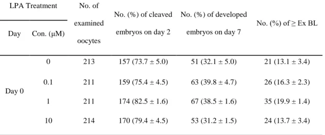
To determine the optimal concentration of LPA to be treated, parthenotes were cultured in IVC medium supplemented with LPA at various concentrations (0, 0.1, 1, and 10 μM) for 7 days. As indicated in Table 2, there was no difference in the cleavage rate between control and LPA-treated parthenotes (control, 73.7% ± 5.0%; 0.1 LPA, 75.4% ± 4.5%; 1 LPA, 82.5% ± 1.6%; and 10 LPA, 79.4% ± 4.5%) on day 2. On day 7, to confirm the acceleration of parthenogenetic embryo development by treated LPA, we evaluated the development of blastocysts cultured by treating LPA, and classified each blastocyst as expanded, hatching, or hatched. There were no differences in the rates of blastocyst formation (control, 32.1% ± 5.0%; 0.1 LPA, 39.8% ± 4.7%; 1 LPA, 38.5% ± 1.6%; and 10 LPA, 31.2% ± 1.5%) and over-expanded blastocyst formation (control, 13.1% ± 3.4%; 0.1 LPA, 16.3% ± 2.3%; 1 LPA, 19.9% ± 1.4%; and 10 LPA, 13.7% ± 3.4%) at all concentrations of LPA. The developmental morphology of blastocysts when LPA treated on day 0 after activation was shown in Figure 1, and morphologies did not significantly differ between the groups.
As shown in Table 3, 722 embryos were cleaved 4 days after parthenogenetic activation. Embryos were randomly divided into four groups and cultured in IVC medium supplemented with 0, 0.1, 1, and 10 μM LPA until day 7. On day 7, the blastocyst formation rate was significantly higher in the 1 μM LPA-treated group than in the control and 0.1 and 10 LPA-treated groups (control, 32.6% ± 2.9%; 0.1 LPA, 40.3% ± 2.7%; 1 LPA, 48.6% ± 1.8%; and 10 LPA, 35.0% ± 2.3%, p < 0.05). Furthermore, the percentage of expanded, hatching, or hatched blastocysts was significantly higher in the 0.1 and 1 μM LPA-treated
11
groups than in the control and 10 LPA groups (control, 16.2% ± 2.2%; 0.1 LPA, 23.2% ± 2.3%; 1 LPA, 27.1% ± 3.1%; and 10 LPA, 17.9% ± 3.4%, p < 0.05). As shown in Figure 1, the developmental morphology was best in blastocysts cultured with 1 μM LPA from day 4 to 7.
12
Table 2. Effects of lysophosphatidic acid (LPA) on the in vitro development of porcine
parthenogenetic oocytes (r = 4)
LPA Treatment No. of examined oocytes No. (%) of cleaved embryos on day 2 No. (%) of developed embryos on day 7 No. (%) of ≥ Ex BL Day Con. (μM) Day 0 0 213 157 (73.7 ± 5.0) 51 (32.1 ± 5.0) 21 (13.1 ± 3.4) 0.1 211 159 (75.4 ± 4.5) 63 (39.8 ± 4.7) 26 (16.3 ± 2.3) 1 211 174 (82.5 ± 1.6) 67 (38.5 ± 1.6) 35 (19.9 ± 1.4) 10 214 170 (79.4 ± 4.5) 53 (31.2 ± 1.5) 24 (13.7 ± 3.4)
Abbreviation: Ex BL, Expanded blastocyst.
Table 3. Effects of lysophosphatidic acid (LPA) on in vitro development of porcine
parthenogenetic embryos (r = 5)
LPA Treatment No. of examined embryos on day 4 No. (%) of developed embryos on day 7 No. (%) of ≥ Ex BL Day Con. (μM) Day 4 0 181 59 (32.6 ± 2.9)a 29 (16.2 ± 2.2)a 0.1 181 73 (40.3 ± 2.7)b 42 (23.2 ± 2.3)b 1 181 88 (48.6 ± 1.8)c 49 (27.1 ± 3.1)b 10 179 63 (35.0 ± 2.3)a 32 (17.9 ± 3.4)a
Significant differences from the control are indicated (a-cp < 0.05). Abbreviation: Ex BL, Expanded blastocyst.
13
Figure 1. Effects of LPA on in vitro porcine blastocyst formation after parthenogenetic
14
3.2. Effect of LPA treatment on the timing of each developmental stage in porcine parthenogenetic embryos
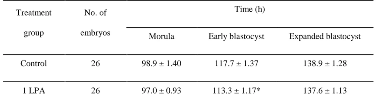
We examined the embryo developmental timing of each stage (morula, early blastocyst, and expanded blastocyst) from day 4 to 7 after activating. To achieve this, we observed each embryo every 5 minutes using time-lapse monitoring system to capture images. We found no significant differences in the timing of the morula stage (control, 98.9 ± 1.40 h and 1 LPA, 97.0 ± 0.93 h) and the expanded blastocyst stage (control, 138.9 ± 1.28 h and 1 LPA, 137.6 ± 1.13 h) between the groups (Table 4, p < 0.05). However, LPA-treated early blastocysts developed faster than untreated blastocysts (control, 117.7 ± 1.37 h and 1 LPA, 113.3 ± 1.17 h).
In addition, we evaluated the quality of porcine embryos at each developmental stage (morula, early blastocyst, and expanded blastocyst) and quantified the number of embryos reaching each stage at 98.0, 115.5, and 138.3 h after activation (Figure 2). We found that LPA-treated embryos reached morula (control, 98.9 h and 1 LPA, 97.0 h), early blastocyst (control, 117.7 h and 1 LPA, 113.3 h), and expanded blastocyst (control, 138.9 h and 1 LPA, 137.6 h) stages faster than untreated embryos. As shown in Table 4, the kinetics of early blastocyst development were significantly faster in the LPA-treated group than in the control (Figure 2A).
We also examined the number of embryos reaching each stage at a specific time. After 98 h of activation, we counted 16 embryos at the 2- to 8-cell stage and 10 embryos at the morula stage in the control group. In the LPA-treated group, there were 9 embryos at the 2- to 8-cell stage and 17 embryos at the morula stage. After 115.5 h of activation, which represents the time of early blastocyst development, we counted 9 embryos at the early blastocyst stage and 17 embryos that did not yet reach this stage in the control. In the LPA-treated group, there
15
were 17 embryos at the early blastocyst stage and 9 embryos that did not yet reach this stage. At 138.3 h after activation, we counted 13 embryos at the expanded blastocyst stage and 13 embryos that did not yet reach this stage in the control. In the LPA-treated group, there were 16 embryos at the expanded blastocyst stage and 10 embryos that did not reach this stage (Figure 2B).
Table 4. The timing of each developmental stage in porcine parthenogenetic embryos using
time-lapse monitoring system
Treatment group
No. of embryos
Time (h)
Morula Early blastocyst Expanded blastocyst
Control 26 98.9 ± 1.40 117.7 ± 1.37 138.9 ± 1.28
1 LPA 26 97.0 ± 0.93 113.3 ± 1.17* 137.6 ± 1.13
Values are means ± standard error. *p < 0.01 (relative to control for the indicated embryo stage).
16
Figure. 2. Time-lapse imaging of porcine embryos developing in vitro. (A) Embryos from
each treatment group at the same developmental stage, cultured from day 4 to 7 with or without 1 µM LPA. (B) Quantity of control (Con) and LPA-treated embryos at different developmental stages (morula, early blastocyst [Ea BL], and expanded blastocyst [Ex BL]) at 98.0, 115.5, and 138.3 h after activation.
17
3.3. Effect of LPA treatment on the kinetics of development of porcine parthenogenetic embryos
The effects of LPA on the kinetics of development were evaluated more carefully by comparing the stages of embryos at 101, 118, and 136 h after activation. At 118 h post-activation, LPA-treated embryos had expanded faster than untreated embryos, possessing larger diameters (165 µm versus 170 µm in control versus 1 µM LPA, respectively; p < 0.05) (Figure 3).
The levels of Tight junction protein ZO-1 (TJP1) and Connexin 43 in LPA-treated and untreated embryos were measured to assess the quality cell-cell contacts within the blastocyst. No difference was observed in TJP1 abundance between groups, whereas Connexin 43 abundance was significantly higher (4.7-fold increase) in the LPA-treated group than in the control group (p < 0.01) (Figure 4A). Transcript abundance of GJC1 (Gap junction protein gamma 1) and CDH1 (Cadherin 1) were significantly higher in LPA-treated embryos than in untreated embryos, whereas no significant difference was observed for TJP1 (Figure 4B).
18
Figure. 3. Morphological parameters of in vitro-developed porcine embryos. (A) Embryos
of the same developmental time cultured from day 4 to 7, with or without 1 µM LPA. (B) Diameter of control (Con) and LPA-treated embryos at 94.5, 101, 118, and 136 h after activation. Values are means ± standard error of the mean. (*p < 0.05).
19
Figure. 4. Expression of components involved in compaction and cell-cell communication.
(A) Abundance of TJP1 and Connexin 43 protein, or (B) GJC1, CDH1, and TJP1 mRNA in control (Con) and blastocysts treated with 1 µM LPA from days 4 to 7. Values are means ± standard error of the mean. (*p < 0.05, **p < 0.01).
20
3.4. Effect of LPA treatment on the quality of embryos at the blastocyst stage
To examine the effect of LPA on the quality of blastocysts, untreated and LPA-treated blastocysts at day 7 were stained with terminal-uridine nick-end labeling (TUNEL) and Hoechst 33342. There was no significant difference in the total cell number of expanded blastocyst between groups (Figure 5A, 5B). However, the apoptotic index was significantly lower in the LPA-treated group at day 4 than in the control (control, 5.7% ± 1.1% and 1 LPA, 3.3% ± 0.6%, p < 0.05) (Figure 5A, 5C).
The relative mRNA levels of genes involved in embryo development and apoptosis were examined (Figure 5D). Although there were no differences in the mRNA levels of POU5F1, DPPA5, and CASP3 between groups, BCL2L1 expression increased and BAK expression decreased in the LPA-treated group compared with the control.
21
Figure. 5. Apoptosis in in vitro-cultured Day-7 blastocysts. (A) Images of fragmented DNA,
as detected with the terminal deoxynucleotidyl transferase dUTP nick-end labeling assay, versus total nuclei. Scale bars, 100 µm. (B) Total cell number and (C) apoptotic index. (D) Relative transcript abundance of genes involved in cell reprogramming (POU5F1 and DPPA5) and apoptosis (BCL2L1, BAK, and CASP3) in control (Con) and blastocysts treated with 1 µM LPA from Days 4 to 7. Values are means ± standard error of the mean. (*p < 0.05, **p < 0.01).
22
4. DISSCUSTION
The quality of embryos produced in vitro is consistently inferior to those produced in vivo. Adjustments to the culture medium can, however, improve the efficiency of embryo development. Here, we investigated if LPA affects the developmental competence and kinetics of porcine embryos, as well as the frequency of DNA fragmentation in blastocysts. Addition of 1 μM LPA to in vitro culture media, beginning on day 4 of culture, improved the rate of blastocyst formation; the kinetics of early blastocyst development; and the expression of tight junction- and gap junction-related protein and genes (Connexin 43, GJC1, and CDH1); whereas the apoptotic index decreased, which agrees with the elevated expression of the anti-apoptotic gene BCL2L1 and the decrease in expression of the pro-apoptotic gene BAK.
Culture conditions often govern the cleavage, compaction, survival, and development of embryos (Im et al., 2004). Many studies have contributed to the optimization of culture systems – e.g., by addition of specific factors – to produce healthy blastocysts of higher quality. For example, the addition of γ-linolenic acid into the medium of bovine in vitro-produced embryos promoted the developmental competence of zygotes and increased the cleavage rate until day 2 (Gaja et al., 2010). Treatment with stem cell-derived bioactive materials also accelerated the development of porcine in vitro- fertilized embryos (Lee et al., 2015). We found that rates of blastocyst development and over-expanded blastocyst development were higher when embryos were treated with LPA starting on day 4 post-activation (Figure 1, Table 2). By contrast, addition of serum can result in opposing changes – e.g., suppressing pre-compacted embryo development but enhancing blastocyst development (Pinyopummintr and Bavister, 1994). Addition of serum at later stages of embryonic development, however, can markedly decrease the timing between early morula,
23
compact morula, and early blastocyst stages, as well as stimulate embryonic cell division and accelerate embryonic development (Robl et al., 1981; Holm et al., 2002).
A time-lapse monitoring system, which captured images of developing embryos at regular intervals (Kirkegaard et al., 2012), was used to observe embryos cultured under stable culture conditions as well as those that have been induced to cleave after activation (Kovacs, 2014). Using such a system, we found that LPA administration on day 4 of culture promoted the development of embryos to the early blastocyst stage, increased the number of embryos reaching the next developmental stage, and increased embryo size (Table 3, Figures 2B and 3B). This protocol is consistent with the addition of growth factors into the medium of day-3 embryos, which promoted their development to the blastocyst stage via autocrine/paracrine signaling, leading to the formation of good blastocysts (Kawamura et al., 2012).
Time-lapse analysis also revealed faster developmental kinetics from the morula to the early blastocyst stage in embryos treated with LPA. This acceleration may have been influenced by the abundance of gap junctions and adhering junctions in the embryo, given that components of both were elevated in this group of embryos. Blastomeres in early embryos are linked together by gap junctions, intercellular structures that allow the transfer of ions and small molecules directly from one cell to the next, thereby providing improved synchrony in developmental processes among the blastomeres (Buehr et al., 1987; Lee et al., 1987; Becker et al., 1995). The gap junction is a hexamer of protein subunits called connexins (Houghton et al., 2002). Connexin 43 is the main connexin isoform in humans (Bloor et al., 2004) and bovine blastocysts (Rizos et al., 2003). Human embryos predominantly express Connexin 43, whose abundance increases during preimplantation and development (Hardy et al., 1996). Here, we confirmed that changes in the Connexin 43 accelerated the kinetics of embryo cleavage, suggesting that intercellular communication via gap junctions may control porcine embryonic development. Gap junction gamma-1 (GJC1),
24
another member of the connexin family, allows for the transfer of metabolites from cumulus cells to the oocyte by controlling the opening and closing of gap junction and ion channels, thereby improving the developmental competence of oocytes (Sugimura et al., 2014); our results suggest that GJC1 also participates in porcine embryo development.
Compaction, which involves the conversion of an embryo with eight clearly defined blastomeres into a morula, whose cell outlines are not readily distinguishable, is mediated between blastomeres by the adhesion protein E-cadherin (Vestweber et al., 1987; Ohsugi et al., 1997). The elevated CDH1 transcript abundance in the LPA-treated group, compared to the control group (Figure 4) is consistent with the improved morphology of the compacted embryos (Figure 2A).
Formation of the trophoblast and inner cell mass and the expansion of the blastocoel are important events in the development of the early preimplantation mammalian embryo (Ducibella et al., 1975); cell junctions may have specific and indispensable roles in these processes (Schlafke et al., 1967). For example, tight junctions between trophectoderm cells create a barrier that is essential for the maintenance of cell polarity and the regulation of paracellular permeability. Tight junctions can control the inner cell mass microenvironment via trophectoderm-mediated transport of ions, amino acids, energy substrates, growth factors, and other metabolites (González-Mariscal et al., 2003; Thomas et al., 2004). Such stage-specific function is consistent with changes in expression of tight junction proteins such as ZO-1α+, which is expressed by late morulae, suggesting its involvement in late stages of development (Sheth et al., 1997, 2000), and ZO-1, which is utilized by the trophectoderm in bovine and murine embryos (Barcroft et al., 1998; Sheth et al., 1997). Although LPA promoted compaction and blastocoel formation, no changes were observed in TJP1 abundance (Figure 4A), suggesting that the beneficial effects of LPA do not directly affect tight junction composition.
25
No difference was observed in total cell number or in the expression of cell lineage markers POU5F1 and DPPA5 in day-7 blastocysts between groups (Figures 5A and 5D). DPPA5 and POU5F1 regulate stem cell pluripotency during early embryogenesis (Kim et al., 2005). POU5F1, a developmentally regulated transcription factor, is activated prior to the 8-cell stage (Palmieri et al., 1994).
Apoptosis is an excellent indicator of cell stress, and suboptimal culture conditions can trigger the apoptotic cascade and abnormal embryo development (Makarevich et al., 2008; Brill et al., 1999; Elmore, 2007). Excess apoptosis can affect blastocyst maturation, induce early embryonic death, and cause fetal anomalies (Brison et al., 1997). Apoptosis is controlled by several conserved genes. For example, the BCL2 family includes several subgroups of anti-apoptotic and pro-apoptotic members: BCL2L1 suppresses apoptosis (Shimizu et al., 1999) while BAK and CASP3 promote apoptosis (Suyama et al., 2002). The intrinsic apoptotic pathway involved the formation of a pore in the mitochondrial membrane via BAK/BAX, resulting in the release of cytochrome c into the cytoplasm via BCL2L1 (Shamas-Din et al., 2013). Another pro-apoptotic gene, CASP3, functions in the final stages of apoptosis as an executioner caspase, meaning that it is cleaved and activated by late apoptotic events (Riedl et al., 2004). In our study, the apoptotic index was lower in the LPA-treated group than in the control group (Figure 5A-C), which is consistent with the changes in BCL2L1 (higher) and BAK (lower) transcript abundance in the LPA-treated group (Figure 5D). These specific changes to BAK and BCL2L1 expression suggest that the intrinsic pathway is active during porcine embryo development, which confirm previous studies that implicate interactions between BCL-2 proteins as critical for embryo development (Yang et al., 2002; Park et al., 2014).
In conclusion, the addition of LPA into the medium can affect the developmental potential of porcine parthenogenetic embryos by accelerating the kinetics of blastocyst formation,
26
resulting in the production of more good-quality blastocysts. A major focus of many investigations is promoting the development of healthy blastocysts, especially those with high rates of hatching, and improving embryo transplantation potential (Balaban et al., 2000) because transfer of this class of embryos is associated with improved rates of implantation and pregnancy (Yoon et al., 2001; Khorram et al., 2000) as well as the live birth (Chimote et al., 2013). LPA was previously shown to stimulate fibroblast DNA synthesis and proliferation, thus acting as a growth factor (Kobayashi et al., 1994); similar signaling could promote cell divisions in embryos, based on our findings that LPA improved the developmental potential of parthenogenetic porcine embryos beyond the 8-cell stage. Taken collectively, our results indicate that LPA improves the quality of in vitro produced embryos, and we recommend its use in assisted reproductive technologies to produce healthy embryos.
27
Figure 6. Mechanism of action of lysophosphatidic acid (LPA). LPA activates Ras and Rho
family GTPases to control cell migration, proliferation, and morphogenesis as well as signaling via classic second messenger pathways.
28
REFERENCES
Amini E, Asadpour R, Roshangar L, Jafari-Joozani R. 2016. Effect of linoleic acid supplementation on in vitro maturation, embryo development and apoptotic related gene expression in ovine. Int J Reprod Biomed (Yazd). 14(4): 255-262.
Arav A, Aroyo A, Yavin S, Roth Z. 2008. Prediction of embryonic developmental competence by time-lapse observation and 'shortest-half' analysis. Reprod Biomed Online 17(5):669-75.
Balaban B, Urman B, Sertac A, Alatas C, Aksoy S, Mercan R. 2000. Blastocyst quality affects the success of blastocyst-stage embryo transfer. Fertil Steril 74(2):282-287.
Barcroft LC, Hay-Schmidt A, Caveney A, Gilfoyle E, Overstrom EW, Hyttel P, Watson AJ. 1998. Trophectoderm differentiation in the bovine embryo: characterization of a polarized epithelium. J Reprod Fertil 114(2):327-339.
Becker DL, Evans WH, Green CR, Warner AE. 1995. Functional analysis of amino acid sequences in connexin 43 involved in intercellular communication through gap junctions. J Cell Sci 108:1455-1467.
Bloor DJ, Wilson Y, Kibschull M, Traub O, Leese HJ, Winterhager E, Kimber SJ. 2004. Expression of connexins in human preimplantation embryos in vitro. Reprod Biol Endocrinol 2:25.
Boruszewska D, Sinderewicz E, Kowalczyk-Zieba I, Grycmacher K, Woclawek-Potocka I. 2016. Studies on lysophosphatidic acid action during in vitro Preimplantation embryo development. Domest Anim Endocrinol 54:15-29.
Brill A, Torchinsky A, Carp H, Toder V. 1999. The role of apoptosis in normal and abnormal embryonic development. J Assist Reprod Genet 16(10):512-9.
Brison DR, Schultz RM. 1997. Apoptosis during mouse blastocyst formation: evidence for a role for survival factors including transforming growth factor alpha. Biol Reprod 56(5):1088-1096.
29
Buehr M, Lee S, McLaren A, Warner A. 1987. Reduced gap junctional communication is associated with the lethal condition characteristic of DDK mouse eggs fertilized by foreign sperm. Development 101(3):449-459.
Carolan C, Lonergan P, Van Langendonckt A, Mermillod P. 1995. Factors affecting bovine embryo development in synthetic oviduct fluid following oocytes maturation and fertilization in vitro. Theriogenology 43(6):1115-1128.
Chimote NM, Chimote NN, Nath NM, Mehta BN. 2013. Transfer of spontaneously hatching or hatched blastocyst yields better pregnancy rates than expanded blastocyst transfer. J Hum Reprod Sci 6(3):183-188.
Contos JJ, Ishii I, Chun J. 2000. Lysophosphatidic acid receptors. Mol Pharmacol 58(6):1188-1196.
Ducibella T, Albertini DF, Anderson E, Biggers JD. 1975. The preimplantation mammalian embryo: characterization of intercellular junctions and their appearance during development. Dev Biol 45(2):231-250.
Eichholtz T, Jalink K, Fahrenfort I, Moolenaar WH. 1993. The bioactive phospholipid lysophosphatidic acid is released from activated platelets. Biochem J 291(3):677-680. Elmore S. 2007. Apoptosis: a review of programmed cell death. Toxicol Pathol 35(4):495-516.
Gaja A, Meng CL, Sato M, Nakajima T, Kubota C, Kojima T. 2010. γ-LA-supplementation to IVC for IVP bovine embryos. Asian-Australas J Anim Sci 23(1):25-32.
Gjorret JO, Avery B, Larson LI, Schellander K, Hyttel P. 2001. Apoptosis in bovine blastocysts produced in vivo and in vitro. Theriogenology 55: 321.
González-Mariscal L, Betanzos A, Nava P, Jaramillo BE. 2003. Tight junction proteins. Prog Biophys Mol Biol 81(1):1-44.
Hardy K, Warner A, Winston RM, Becker DL. 1996. Expression of intercellular junctions during preimplantation development of the human embryo. Mol Hum Reprod 2(8):621-632.
30
Hinokio K, Yamano S, Nakagawa K, Iraharaa M, Kamada M, Tokumura A, Aono T. 2002. Lysophosphatidic acid stimulates nuclear and cytoplasmic maturation of golden hamster immature oocytes in vitro via cumulus cells. Life Sci 70(7):759-767.
Hochi S, Kimura K, Hanada A. 1999. Effect of linoleic acid-albumin in the culture medium on freezing sensitivity of in vitro-produced bovine morulae. Theriogenology 52(3):497-504. Holm P, Booth PJ, Callesen H. 2002. Kinetics of early in vitro development of bovine in vivo- and in vitro-derived zygotes produced and/or cultured in chemically defined or serum-containing media. Reproduction 123:553-565.
Houghton FD, Barr KJ, Walter G, Gabriel HD, Grümmer R, Traub O, Leese HJ, Winterhager E, Kidder GM. 2002. Functional significance of gap junctional coupling in preimplantation development. Biol Reprod 66(5):1403-1412.
Im GS, Lai L, Liu Z, Hao Y, Wax D, Bonk A, Prather RS. 2004. In vitro development of preimplantation porcine nuclear transfer embryos cultured in different media and gas atmospheres. Theriogenology 61(6):1125-35.
Jo JW, Jee BC, Suh CS, Kim SH. 2013. Addition of lysophosphatidic acid to mouse oocyte maturation media can enhance fertilization and developmental competence. Hum Reprod 29(2):234-241.
Kawamura K, Chen Y, Shu Y, Cheng Y, Qiao J, Behr B, Pera RA, Hsueh AJ. 2012. Promotion of human early embryonic development and blastocyst outgrowth in vitro using autocrine/paracrine growth factors. PLoS One 7(11):e49328.
Khandoker M, Tsujii H. 1999. Effect of exogenous fatty acids on in vitro development of rat embryos. Asian-Australas J Anim Sci 12:169-173.
Khorram O, Shapiro SS, Jones JM. 2000. Transfer of nonassisted hatched and hatching human blastocysts after in vitro fertilization. Fertil Steril 74(1):163-165.
Kikuchi K, Onishi A, Kashiwazaki N, Iwamoto M, Noguchi J, Kaneko H, Akita T, Nagai T. 2002. Successful piglet production after transfer of blastocysts produced by a modified in vitro system. Biol Reprod 66(4):1033-1041.
31
Kirkegaard K, Agerholm IE, Ingerslev HJ. 2012. Time-lapse monitoring as a tool for clinical embryo assessment. Hum Reprod 27(5):1277-1285.
Kim SK, Suh MR, Yoon HS, Lee JB, Oh SK, Moon SY, Moon SH, Lee JY, Hwang JH, Cho WJ, Kim KS. 2005. Identification of developmental pluripotency associated 5 expression in human pluripotent stem cells. Stem Cells 23(4):458-62.
Knez K, Tomazevic T, Vrtacnik-Bokal E, Virant-Klun I. 2013. Developmental dynamics of IMSI-derived embryos: a time-lapse prospective study. Reprod Biomed Online 27:161-171. Kobayashi T, Yamano S, Murayama S, Ishikawa H, Tokumura A, Aono T. 1994. Effect of lysophosphatidic acid on the preimplantation development of mouse embryos. FEBS lett 351(1):38-40.
Kovacs P. 2014. Embryo selection: the role of time-lapse monitoring. Reprod Biol Endocrinol 12:124.
Lee S, Gilula NB, Warner AE. 1987. Gap junctional communication and compaction during preimplantation stages of mouse development. Cell 51:851-860.
Lee SE, Moon JJ, Kim EY, Park SP. 2015. Stem cell–derived bioactive materials accelerate development of porcine in vitro–fertilized embryos. Cell Reprogram 17(3):181-190.
Lemmen JG, Agerholm I, Ziebe S. 2008. Kinetic markers of human embryo quality using time-lapse recordings of IVF/ICSI-fertilized oocytes. Reprod Biomed Online 17(3):385-91. Long CR, Dobrinsky JR, Johnson LA. 1999. In vitro production of pig embryos: Comparison of culture media and boars. Theriogenology 51(7):1375-1390.
Makarevich AV, Chrenek P, Massanyi P, Lukac N, Pivko J. 2008. Apoptosis detection as a tool for the determination of animal embryo quality. Slovak J Anim Sci 41(4):153-159. McKiernan SH, Bavister BD. 1992. Different lots of bovine serum albumin inhibit or stimulate in vitro development of hamster embryos. In Vitro Cell Dev Biol 28A:154-156. Moolenaar WH, van Meeteren LA, Giepmans BN. 2004. The ins and outs of lysophosphatidic acid signaling. Bioessays 26(8):870-881.
32
Moore K, Rodríguez-Sallaberry CJ, Kramer JM, Johnson S, Wroclawska E, Goicoa S, Niasari-Naslaji A. 2007. In vitro production of bovine embryos in medium supplemented with a serum replacer: effects on blastocyst development, cryotolerance and survival to term. Theriogenology 68(9):1316-1325.
Ohsugi M, Larue L, Schwarz H, Kemler R. 1997. Cell-junctional and cytoskeletal organization in mouse blastocysts lacking E-cadherin. Dev Biol 185(2):261-71.
Palmieri SL, Peter W, Hess H, Schöler HR. 1994. Oct-4 transcription factor is differentially expressed in the mouse embryo during establishment of the first two extraembryonic cell lineages involved in implantation. Dev Biol 166(1):259-267.
Park SH, Cho HS, Yu IJ. 2014. Effect of bovine follicular fluid on reactive oxygen species and glutathione in oocytes, apoptosis and apoptosis-related gene expression of in vitro-produced blastocysts. Reprod Domest Anim 49(3):370-377.
Petters RM, Johnson BH, Reed ML, Archibong AE. Glucose, glutamine and inorganic phosphate in early development of the pig embryo in vitro, J Reprod Fertil 89(1):269-275. Pinyopummintr T, Bavister BD. 1994. Development of bovine embryos in a cell-free culture medium: effects of type of serum, timing of its inclusion and heat inactivation. Theriogenology 41(6):1241-1249.
Reed ML, Illera MJ, Petters RM. 1992. In vitro culture of pig embryos. Theriogenology 37:95-109.
Riedl SJ, Shi Y. 2004. Molecular mechanisms of caspase regulation during apoptosis. Nat Rev Mol Cell Biol 5(11):897-907.
Rizos D, Gutiérrez-Adán A, Pérez-Garnelo S, De La Fuente J, Boland MP, Lonergan P. 2003. Bovine embryo culture in the presence or absence of serum: implications for blastocyst development, cryotolerance, and messenger RNA expression. Biol Reprod 68(1):236-243.
Robl JM, Davis DL. 1981. Effects of serum on swine morulae and blastocysts in vitro. J Anim Sci 52(6):1450-1456.
33
Schlafke S, Enders AC. 1967. Cytological changes during cleavage and blastocyst formation in the rat. J Anat 102(1):13-32.
Shamas-Din A, Kale J, Leber B, Andrews DW. 2013. Mechanisms of action of Bcl-2 family proteins. Cold Spring Harb Perspect Biol 5(4):a008714.
Sheth B, Fesenko I, Collins JE, Moran B, Wild AE, Anderson JM, Fleming TP. 1997. Tight junction assembly during mouse blastocyst formation is regulated by late expression of ZO-1 α+ isoform. Development 124(10):2027-2037.
Sheth B, Moran B, Anderson JM, Fleming TP. 2000. Post-translational control of occludin membrane assembly in mouse trophectoderm: a mechanism to regulate timing of tight junction biogenesis and blastocyst formation. Development 127(4):831-840.
Shimizu S, Narita M, Tsujimoto Y. 1999. Bcl-2 family proteins regulate the release of apoptogenic cytochrome c by the mitochondrial channel VDAC. Nature 399(6735):483-487. Sugimura S, Ritter LJ, Sutton-McDowall ML, Mottershead DG, Thompson JG, Gilchrist RB. 2014. Amphiregulin co-operates with bone morphogenetic protein 15 to increase bovine oocyte developmental competence: effects on gap junction-mediated metabolite supply. Mol Hum Reprod 20(6):499-513.
Suyama E, Kawasaki H, Taira K. 2002. Identification of a caspase 3-independent role of pro-apoptotic factor Bak in TNF-alpha-induced apoptosis. FEBS Lett 528(1-3):63-69.
Thomas FC, Sheth B, Eckert JJ, Bazzoni G, Dejana E, Fleming TP. 2004. Contribution of JAM-1 to epithelial differentiation and tight-junction biogenesis in the mouse preimplantation embryo. J Cell Sci 117(23):5599-5608.
Thompson JG. 1997. Comparison between in vivo-derived and in vitro-produced pre-elongation embryos from domestic ruminants. Reprod Fertil Dev 9(3):341-354.
Thompson JG, Allen NW, McGowan LT, Bell AC, Lambert MG, Tervit HR. 1998. Effect of delayed supplementation of fetal calf serum to culture medium on bovine embryo development in vitro and following transfer. Theriogenology 49(6):1239-49.
34
Saulnier-Blache JS, Woclawek-Potocka I, Lopes-da-Costa L. 2014. Lysophosphatidic acid signaling in late cleavage and blastocyst stage bovine embryos. Mediators Inflamm 2014:678968.
Vestweber D, Gossler A, Boller K, Kemler R. 1987. Expression and distribution of cell adhesion molecule uvomorulin in mouse preimplantation embryos, Dev Biol 124(2):451-456. Wirtu G, Bailey TL, Chauhan MS, Parker NA, Dascanio JJ, Gwazdauskas FC, Ley WB. 2004. Xenogenous fertilization of equine oocytes following recovery from slaughterhouse ovaries and in vitro maturation. Theriogenology 61:381-391.
Wrenzycki C, Herrmann D, Carnwath JW, Niemann H. 1999. Alterations in the relative abundance of gene transcripts in preimplantation bovine embryos cultured in medium supplemented with either serum or PVA. Mol Reprod Dev 53:8-18.
Wrenzycki C, Herrmann D, Niemann H. 2003. Timing of blastocyst expansion affects spatial messenger RNA expression patterns of genes in bovine blastocysts produced in vitro. Biol Reprod 68(6):2073-80.
Xu KP, Yin J, Yu FS. 2007. Lysophosphatidic acid promoting corneal epithelial wound healing by transactivation of epidermal growth factor receptor. Invest Ophthalmol Vis Sci 48(2):636-643.
Yang MY, Rajamahendran R. 2002. Expression of Bcl-2 and Bax proteins in relation to quality of bovine oocytes and embryos produced in vitro. Anim Reprod Sci 70(3-4):159-169. Yoon HG, Yoon SH, Son WY, Im KS, Lim JH. 2001. High implantation and pregnancy rates with transfer of human hatching day 6 blastocysts. Fertil Steril 75(4):832-833.
Zhang JY, Jiang Y, Lin T, Kang JW, Lee JE, Jin DI. 2015. Lysophosphatidic acid improves porcine oocyte maturation and embryo development in vitro. Mol Reprod Dev 82(1):66-77.
35
ABSTRACT IN KOREAN
난모세포와 배아의 체외 생산에 다양한 인자를 첨가하는 배양액의 조성은 중요하다. 리소포스파티드산 (LPA)은 세포 증식, 분화, 이동, 생존 등 다양한 동물 세포에 영향을 줄 수 있는 성장 인자 및 호르몬 유사 활성을 나타내는 신호 분자로서 작용할 수 있는 인지질 유도체이다. 본 논문에서는 LPA 가 돼지 배아 발달에 미치는 영향을 조사하였다. 돼지 단위발생란을 0, 0.1, 1, 10 μM 의 LPA (0 LPA, 0.1 LPA, 1 LPA 및 10 LPA)를 첨가한 배양액에서 7 일동안 배양하거나 기본 배양액에서 4 일까지 배양 후 4 일째부터 LPA 를 첨가한 배양액에서 배양하였다. 7 일동안 LPA 를 처리한 배양액에서 발달한 배아의 발달률은 실험군 간의 차이가 없었다. 그러나 배양 4 일째부터 7 일까지 LPA 를 처리한 배양액에서 발달한 배아의 배반포 발달률과 확장배반포 단계 이상으로 발달한 배반포의 형성률은 다른 처리군보다 0.1 및 1 μM LPA 처리군에서 더 높았다. 또한 LPA 를 처리한 군에서 초기 배반포 단계까지 소요시간이 짧고, 배아의 크기가 컸다. LPA 처리군에서 connexin 43 단백질과 간극연접 및 세포부착 관련 유전자 (GJC1 및 CDH1)의 발현이 대조군에 비해 증가하였다. 대조군과 처리군 배반포 사이의 총 세포 수는 차이가 없었지만, LPA 처리군에서 대조군보다 세포사멸지수가 낮았고, BCL2L1의 발현은 증가하고 BAK 의 발현은 감소하였다. 이러한 결과는 LPA 를 배양 4 일부터 7 일까지의 배양액에 첨가하였을 때 배반포 형성이 증가하고 배아의 발달 능력이 높아진다는 것을 의미한다.36