DOI: https://doi.org/10.3339/jkspn.2020.24.2.91 ISSN 2384-0250 (online)
Clinical Characteristics and Long-Term Prognosis of
Alport Syndrome: A Retrospective Single-Center Study
Purpose: Alport syndrome (AS) is one of the most common inherited renal dis-eases caused due to mutations of genes encoding specific proteins of the type IV collagen family, and its major clinical manifestations include progressive renal failure, sensorineural deafness, and ocular abnormalities. We investigated the clinical characteristics and long-term prognosis of AS in Korean pediatric and adult populations.
Methods: We conducted a retrospective review of medical records of 33 children and adults who had been diagnosed or treated with AS from 1985 to 2019.
Results: The mean age of the 33 patients diagnosed with AS was 16.2±13.6 years, and the male-to-female ratio was 2:1. At the first visit, recurrent gross hematuria was the most common initial symptom. In 10 of 33 patients (30.3%), sensorineural hearing loss (SNHL) was diagnosed, but none had ophthalmic problems. Moreover, 11 of 33 patients (33.3%) had advanced to end-stage renal disease (ESRD), and a significant difference was observed in the age of the patients who progressed to ESRD based on the presence or absence of SNHL (P=0.035).
Conclusion: SNHL in AS can be an important prognostic factor for long-term de terioration of renal function. Further investigation is required to confirm the clinical course and the genetic characteristics of AS in Korea through prospective national cohort studies.
Key words: Alport syndrome, End-stage renal disease, Sensorineural hearing loss
Hea Min Jang, M.D.
1Hee Sun Baek, M.D.
1Sun-Hee Park, M.D.
2Yong-Lim Kim, M.D.
2Chan-Duck Kim, M.D.
2Hee-Yeon Jung, M.D.
2Jang-Hee Cho, M.D.
2Man Hoon Han, M.D.
3Yong Jin Kim, M.D.
3Min Hyun Cho, M.D.
1Department of Pediatrics1, Internal
Medicine2, Pathology3, Kyungpook
National University, School of Medicine, Daegu, Republic of Korea
Corresponding author: Min Hyun Cho, M.D.
Department of Pediatrics, Kyungpook National University Hospital
130 Dongdeok-ro, Jung-gu, Daegu 41944, Republic of Korea Tel: +82-53-200-5719 Fax: +82-53-425-6683 E-mail: [email protected] Received: 10 August 2020 Revised: 17 September 2020 Accepted: 4 October 2020
This is an open-access article distributed under the terms of the Creative Commons Attribu tion Non-Commercial License (http:// crea tivecom mons.org/licenses/by-nc/4.0/) which permits unrestricted non-commercial use, distribution, and reproduction in any medium, provided the original work is properly cited.
Copyright © 2020 The Korean Society of Pediatric Nephrology
Introduction
Alport syndrome (AS) is one of the most common inherited renal diseases caused due to mutations of genes encoding specific proteins of the type IV collagen family1). The type IV collagen family is expressed in the basement
membrane of the kidney as well as the ear and eye2). The major features of AS
are progressive renal failure, sensorineural deafness, and ocular abnorma lities. Clinically, hematuria is the most common symptom of AS and gene rally manifests as microscopic or recurrent gross hematuria13). Proteinuria is
absent in the early stages, but it gradually increases with age, and all males with Xlinked AS (XLAS) progress to endstage renal disease (ESRD)1,2).
AS has three genetic patterns. The majority, comprising approximately 80–85%, show the Xlinked dominant form caused due to mutations of the COL4A5 gene. Autosomal recessive or dominant forms caused due to muta tion of the COL4A3 or COL4A4 gene have also been reported46). XLAS can
be suspected when a young male has a severe course of ill ness. Untreated males with XLAS progress to ESRD before the age of 30 years and are frequently accompanied by hea ring loss. However, females with XLAS exhibit various disease severities depending on the relative activities of the mutant and the normal X chromosome. Autosomal reces sive AS (ARAS) should be suspected in young females who do not have a family history with typical clinical features and severe disease, such as ESRD and deafness2,7). The aims
of this study were to identify the genetic and clinical cha racteristics of patients with AS in Korea and to identify the clinical indicators that can predict longterm prognosis
Materials and methods
1. Study design and patients
We retrospectively reviewed the medical records of 33 patients who had been diagnosed or treated with AS at our hospital from 1985 to 2019. AS was diagnosed through in vestigations such as family history, audiogram, ophthalmic examination, renal biopsy, and genetic study in patients with hematuria or AS family history. Clinical details such as age, sex, familial history, serum creatinine, urinalysis, and estimated glomerular filtration rate (eGFR) were col lected. Renal biopsy was performed in 27 of the subjects, and particularly diffuse thickening and multilamellation of the glomerular basement membrane (GBM) on an elec tron microscope (EM) were used as diagnostic criteria. Genetic testing was performed in 18 of the subjects, and the conventional Sanger sequencing for the COL4A5 gene or nextgeneration sequencing (NGS) was performed. Hea ring abnormality was defined as sensorineural hearing loss (SNHL) of moderate abnormality (41–55 dB) in pure tone audiometry by audiogram. Ophthalmic abnormalities were characterized by anterior lenticonus and maculopathy, which are the characteristic of AS. Renal function was eva luated according to the eGFR value, and ESRD was defined when the eGFR value was less than 15 mL/min/1.73 m2.
2. Statistical analysis
The mean value of each category is reported as mean± SD. The Student’s ttest or the Wilcoxon ranksum test was used for analyzing continuous variables and nonparametric
distributions. The Chisquare test and Fisher’s exact test were used for analyzing categorical variables. The signifi cance level was defined as P<0.05. All statistical analyses were performed using PASW Statistics ver. 18.0 (SPSS Inc., Chicago, Illinois, USA).
Results
The mean age of the 33 patients who first visited our hos pital was 12.7±14.0 years, and the maletofemale ratio was 2:1. Of the 33 patients, 26 (78.8%) were diagnosed before the age of 18 years, with the mean age at diagnosis being 16.2± 13.6 years. At the first visit, recurrent gross hematuria was the most common initial symptom (57.6%), followed by proteinuria with persistent microscopic hematuria (PMH) (30.3%) and PMH only (6.1%) (Table 1).
For confirming the diagnosis, 27 (81.8%) patients under went renal biopsy, genetic tests were conducted in 18 pati ents (54.5%), and 15 patients (45.5%) underwent both. In the genetic tests, the conventional Sanger sequencing was performed in 14 patients (77.8%) to identify COL4A5 gene mutation, and the remaining 4 patients underwent NGS. Based on the test results, COL4A5 gene mutation was con firmed in 7 patients (38.9%), and 1 patient with COL4A4 gene mutation was confirmed through NGS. In the remai ning patients, the COL4A5 gene mutation could not be confirmed by Sanger sequencing or the results of the mu tation were unknown.
A total of 18 patients (54.5%) had a family history of kid ney disease, such as AS, chronic kidney disease, proteinuria, or microscopic hematuria. On the basis of the results of genetic tests and family history, the specific genetic patterns could be confirmed in only 9 patients, of whom 7 patients had an Xlinked pattern, and 2 patients had an autosomal recessive genetic pattern.
Of the 33 patients, 11 (33.3%) progressed to ESRD, with the mean age at ESRD diagnosis being 24.1±10.9 years. The mean duration of progression to ESRD was 14.0±5.8 years from the diagnosis of AS. The 8 patients (72.7%) with ESRD underwent kidney transplantation, and 3 of them are on hemodialysis. None of the patients were diagnosed with ophthalmic problems, but there were 10 patients (30.3%) with SNHL. The mean age at the diagnosis of SNHL was
21.5 years (Table 1 and 2).
We divided all the patients with AS into 26 pediatric pa tients diagnosed at age <18 years and 7 adult patients diag nosed at age ≥18 years. In pediatric patients, recurrent gross hematuria was the most common cause of the first visit, accounting for 73.1%, whereas proteinuria with PMH was the most common in adults (71.4%). In the diagnostic me thod, 23 of the 26 children (88.5%) were biopsied and 17 (65.4%) underwent genetic study, whereas in the adult group, 3 of 7 adults (42.9%) were biopsied and only 1 (14.3 %) underwent genetic study. However, only 17 of the 23 children who underwent biopsy actually met the diagnostic criteria of AS and were subjected to more genetic tests than adults (Table 3).
When compared with the presence or absence of protei nuria as an initial symptom, no significant difference was detected in ESRD progression between the two groups. In contrast, SNHL was found at a significantly higher frequ
ency in the ESRD group (P=0.003, Fisher’s exact test) (Table 4). A significant difference was observed in the age of the patients who progressed to ESRD according to the presence or absence of SNHL (P=0.035) (Fig. 1).
Discussion
AS is a hereditary kidney disease caused due to mutation of a specific protein of the type IV collagen family consti tuting the basement membrane. The disease is accompa nied by progressive renal failure, hearing loss, and ocular abnormalities, and its prevalence has been estimated at 1 in 5,000 people in the United States or Europe. AS accounts for 0.5% of adults and 12.9% of children among all patients with ESRD7,8). Most of the clinical symptoms in patients
with AS manifest as recurrent gross hematuria and PMH before the age of 5 years. Proteinuria is uncommon at a
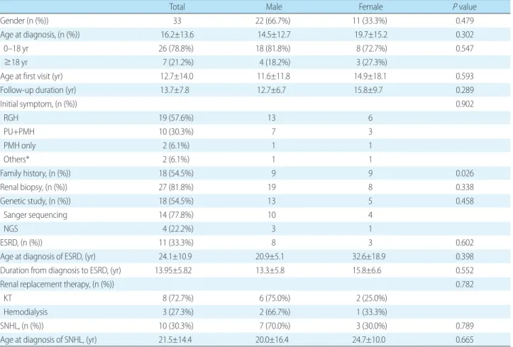
Table 1. General Characteristics and Laboratory Features of Patients with Alport Syndrome
Total Male Female P value
Gender (n (%)) 33 22 (66.7%) 11 (33.3%) 0.479
Age at diagnosis, (n (%)) 16.2±13.6 14.5±12.7 19.7±15.2 0.302
0–18 yr 26 (78.8%) 18 (81.8%) 8 (72.7%) 0.547
≥18 yr 7 (21.2%) 4 (18.2%) 3 (27.3%)
Age at first visit (yr) 12.7±14.0 11.6±11.8 14.9±18.1 0.593
Follow-up duration (yr) 13.7±7.8 12.7±6.7 15.8±9.7 0.289
Initial symptom, (n (%)) 0.902 RGH 19 (57.6%) 13 6 PU+PMH 10 (30.3%) 7 3 PMH only 2 (6.1%) 1 1 Others* 2 (6.1%) 1 1 Family history, (n (%)) 18 (54.5%) 9 9 0.026 Renal biopsy, (n (%)) 27 (81.8%) 19 8 0.338 Genetic study, (n (%)) 18 (54.5%) 13 5 0.458 Sanger sequencing 14 (77.8%) 10 4 NGS 4 (22.2%) 3 1 ESRD, (n (%)) 11 (33.3%) 8 3 0.602
Age at diagnosis of ESRD, (yr) 24.1±10.9 20.9±5.1 32.6±18.9 0.398
Duration from diagnosis to ESRD, (yr) 13.95±5.82 13.3±5.8 15.8±6.6 0.552
Renal replacement therapy, (n (%)) 0.782
KT 8 (72.7%) 6 (75.0%) 2 (25.0%)
Hemodialysis 3 (27.3%) 2 (66.7%) 1 (33.3%)
SNHL, (n (%)) 10 (30.3%) 7 (70.0%) 3 (30.0%) 0.789
Age at diagnosis of SNHL, (yr) 21.5±14.4 20.0±16.4 24.7±10.0 0.665
*Other includes seizure and familial history of Alport syndrome or other renal diseases.
Abbreviations: RGH, recurrent gross hematuria; PU, proteinuria; PMH, persistent microscopic hematuria; NGS, Next-generation sequencing; ESRD, end-stage renal disease; KT, kidney transplantation; SNHL, sensorineural hearing loss.
young age, but it gradually increases with age5,7,8). In our
study, the maletofemale ratio was 2:1, and 26 patients (78.8%) visited the hospital before the age of 18 years. At the first visit, recurrent gross hematuria was the most common initial symptom (57.6%). When the symptoms were divided into hematuria and proteinuria, hematuria was observed in
all patients, whereas proteinuria was found in only 33.3% of the patients. Jais et al. reported that 75.2% of patients with AS in Europe had proteinuria and 95% had hematuria9,10).
In Japan, Yamamura et al. reported proteinuria in 72.6% and hematuria in 97.9% of patients11). Furthermore, Chugh
et al. reported proteinuria and hematuria prevalence rates
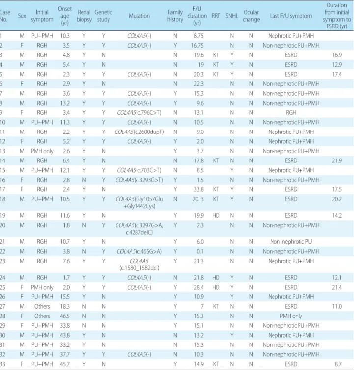
Table 2. Patient Characteristics Case
No. Sex symptomInitial Onset
age (yr)
Renal
biopsy Genetic study Mutation historyFamily F/U duration
(yr) RRT SNHL Ocular change Last F/U symptom
Duration from initial symptom to
ESRD (yr)
1 M PU+PMH 10.3 Y Y COL4A5(-) N 8.75 N N Nephrotic PU+PMH
2 F RGH 3.5 Y Y COL4A5(-) Y 16.75 N N Non-nephrotic PU+PMH
3 M RGH 4.8 Y N N 19.6 KT Y N ESRD 16.9
4 M RGH 5.4 Y N N 19 KT Y N ESRD 12.9
5 M RGH 2.3 Y Y COL4A5(-) N 20.3 KT Y N ESRD 17.4
6 F RGH 2.9 Y N N 22.3 N N Non-nephrotic PU+PMH
7 M RGH 3.6 Y Y COL4A5(-) Y 15.3 N N Non-nephrotic PU+PMH
8 M RGH 13.2 Y Y COL4A5(-) Y 9.6 N N Non-nephrotic PU+PMH
9 F RGH 3.4 Y Y COL4A5(c.796C>T) N 13.1 N N RGH
10 M PU+PMH 11.3 Y Y COL4A5(-) N 10.5 N N Non-nephrotic PU+PMH
11 M RGH 2.2 Y Y COL4A5(c.2600dupT) N 9.0 N N Nephrotic PU+PMH
12 F RGH 5.2 Y Y COL4A5(-) Y 2.0 N N Nephrotic PU+PMH
13 M PMH only 2.6 Y N Y 3.7 N N Non-nephrotic PU+PMH
14 M RGH 6.4 Y N N 17.8 KT N N ESRD 21.9
15 M PU+PMH 12.1 Y Y COL4A5(c.703C>T) N 8.5 Y N Nephrotic PU+PMH
16 F RGH 2.8 N Y COL4A5(c.3293G>T) Y 1.5 N N Non-nephrotic PU+PMH
17 F RGH 2.4 Y N Y 33.8 KT Y N ESRD 17.5
18 M PU+PMH 10.5 Y Y COL4A5(Gly1057Glu
+Gly1442Cys) N 20. 3 KT Y N ESRD 20.2
19 M RGH 11.6 Y N Y 19.9 HD N N ESRD 14.2
20 M RGH 1.8 N Y COL4A5(c.3297G>A,
c.4287delC) Y 2.3 N N Non-nephrotic PU+PMH
21 M RGH 10.7 Y N Y 6.0 N N Non-nephrotic PU
22 M RGH 3.8 N Y COL4A5(c.465G>A) Y 0.1 N N Non-nephrotic PU+PMH
23 M RGH 7.6 Y Y COL4A5
(c.1580_1582del) Y 21.3 N N Nephrotic PU+PMH
24 M RGH 1.7 Y Y COL4A5(-) N 21.8 HD Y N ESRD 12.1
25 F PMH only 2.0 Y Y COL4A5(-) Y 28.4 HD Y N ESRD 21.4
26 F PU+PMH 15.5 Y N Y 10.9 Y N Nephrotic PU+PMH
27 M Others 18.3 N N Y 7 KT N N ESRD 11.0
28 F Others 46.5 N N Y 15.3 N N PMH only
29 F PU+PMH 33.8 N N Y 15.1 N N Non-nephrotic PU+PMH
30 M PU+PMH 43.8 Y N N 13.2 Y N Nephrotic PU+PMH
31 M PU+PMH 33.2 Y N N 15.3 N N Non-nephrotic PU+PMH
32 M PU+PMH 37.7 Y Y COL4A5(-) N 10.3 N N Non-nephrotic PU+PMH
33 F PU+PMH 45.7 Y N Y 14.9 KT N N ESRD 8.7
Abbreviations: RGH, recurrent gross hematuria; PU, proteinuria; PMH, persistent microscopic hematuria; F/U, follow up; RRT, renal replacement therapy; SNHL, sensorineural hearing loss; ESRD, end-stage renal disease; KT, kidney transplantation; HD, hemodialysis.
of 31.7% and 96.8%, respectively12). Although there was a
slight difference, hematuria was the most common in the majority of cases.
AS is diagnosed in patients with hematuria based on EM results of renal biopsy showing a multilamellation of GBM, and family history, SNHL, and ocular abnormalities are helpful for diagnosis2,3,7). In our study, family history could
be confirmed in 54.5% of the patients, and 27 patients (81.8 %) underwent renal biopsy, whereas only 18 patients (24.2 %) were genetically tested. Genetic study was performed in 65.4% of pediatric patients, whereas among the adults, only 1 patient (14.3%) underwent genetic test. Moreover, among adults, the diagnosis was based on family history and clinical symptoms rather than renal biopsy or genetic testing. Inheritance could be confirmed in 9 patients based on family history and genetic study results, of whom 7 (77.8 %) had XLAS, 2 (22.2%) had ARAS confirmed, whereas ADAS was absent. In an earlier study of patients in China, Wei et al. reported that 89.7% of those with AS had XLAS and 10.3% had ARAS13).
In particular, the results of renal biopsy at a young age or in females can be ambiguous, which poses a limitation to diagnosis based on biopsy alone; moreover, the diagnosis
Fig. 1. Comparison of the time taken to proceed to ESRD accor-ding to the presence or absence of SNHL. Solid line indicates patients without sensorineural hearing loss (n=23). Dots indicate patients with sensorineural hearing loss (n=10) (P=0.035). Abbre-viations: ESRD, end-stage renal disease; SNHL, sensorineural hearing loss.
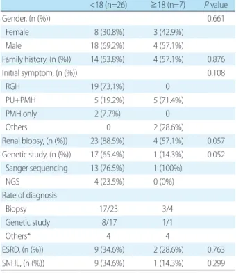
Table 3. Comparison of General Characteristics and Laboratory Features according to the Age of Diagnosis of Alport Syndrome
<18 (n=26) ≥18 (n=7) P value Gender, (n (%)) 0.661 Female 8 (30.8%) 3 (42.9%) Male 18 (69.2%) 4 (57.1%) Family history, (n (%)) 14 (53.8%) 4 (57.1%) 0.876 Initial symptom, (n (%)) 0.108 RGH 19 (73.1%) 0 PU+PMH 5 (19.2%) 5 (71.4%) PMH only 2 (7.7%) 0 Others 0 2 (28.6%) Renal biopsy, (n (%)) 23 (88.5%) 4 (57.1%) 0.057 Genetic study, (n (%)) 17 (65.4%) 1 (14.3%) 0.052 Sanger sequencing 13 (76.5%) 1 (100%) NGS 4 (23.5%) 0 (0%) Rate of diagnosis Biopsy 17/23 3/4 Genetic study 8/17 1/1 Others* 4 4 ESRD, (n (%)) 9 (34.6%) 2 (28.6%) 0.763 SNHL, (n (%)) 9 (34.6%) 1 (14.3%) 0.299
*Other includes seizure and familial history of Alport syndrome or other renal disease.
Abbreviations: NGS, Next-generation sequencing; RGH, recurrent gross hematuria; PU, proteinuria; PMH, persistent microscopic hematuria; ESRD, end-stage renal disease; SNHL, sensorineural hearing loss.
Table 4. Association between ESRD and Clinical and Laboratory Findings in Patients with Alport Syndrome
ESRD (-) (n=22) ESRD (+) (n=11) P value Gender, (n (%)) 0.602 Female 8 (36.4%) 3 (27.3%) Male 14 (63.6%) 8 (72.7%) Age at diagnosis, (n (%)) 0.763 0–18 yr 17 (77.3%) 9 (81.8%) ≥18 yr 5 (22.7%) 2 (18.2%) Initial symptom 0.789 Hematuria 15 (68.2%) 8 (72.7%) Proteinuria 7 (31.8%) 3 (27.3%) Initial eGFR 115.5±46.7 99.3±49.3 0.021 Initial UP/Cr 1.7±2.1 1.8±2.2 0.527 Family history, (n (%)) 13 (59.1%) 5 (45.5%) 0.458 Age at diagnosis (yr) 17.4±14.7 13.9±11.4 0.467
SNHL, (n (%)) 3 (13.6%) 7 (63.6%) 0.003
Age at diagnosis of SNHL (yr) 26.3±23.5 19.4±10.4 0.663 Abbreviations: ESRD, end-stage renal disease; SNHL, sensorineural hearing loss; eGFR, estimated glomerular filtration rate; UP/Cr, urine proteinu/ creatinine.
becomes further complicated due to the difficulty in con firming the exact family history2,7,14). Therefore, it is neces
sary to suspect AS if there is persistent hematuria of un known etiology at a young age. In our study, only 17 of the 23 pediatric patients who underwent renal biopsy were diagnosed by biopsy, implying that genetic testing was performed in more children than adults. Furthermore, as genetic patterns cannot be discriminated based on only the abnormalities observed in EM findings, genetic study is essential to confirm the exact genetic pattern2). Because it
is known that the majority of AS is caused due to mutations of the COL4A5 gene, performing conventional Sanger sequencing of COL4A5 is costeffective4); however, NGS
also has advantages as the possibility of mutation of the COL4A4 gene cannot be ruled out15). Moreover, the results
of genetic testing are more important because various phe notypes can be displayed depending on the mutation posi tion or the mutation type. Through NGS, it is easy to con firm large deletions and insertions in female patients, which are difficult to confirm by Sanger sequencing, and will help confirm inheritance in patients in whom it is difficult to confirm the pedigree. In countries with a large number of renal donations among relatives, such as Korea, it is believed that genetic testing for asymptomatic female carriers will help determine whether the donor has AS4). In the results
of this study, it can be confirmed that the diagnosis of AS in adults is based on clinical symptoms, so that biopsy or genetic testing was rarely performed. Therefore, since ge netic testing is important for identifying genetic patterns and predicting prognosis, it is essential to diagnose AS patients.
The prognosis for AS is poor, with 90% of males with XLAS and the majority of those with ARAS progressing to ESRD before age 30 years7,10,15). No specific treatments are
available till date, and recent studies have demonstrated that inhibition of the reninangiotensinaldosterone system using angiotensinconverting enzyme inhibitor or angio tensin receptor blocker slows the progression of the disease by reducing proteinuria. Dialysis or renal transplantation is performed when the patient reaches ESRD2,7). Most of the
patients with AS who progress to ESRD have a better prog nosis than those with other kidney diseases. However, 2–5 % of transplant patients are likely to have graft failure due to posttransplant antiGBM nephritis caused due to allo
antibodies to the donor kidney GBM7,16,17). The factors as
sociated with the prognosis of AS include gender, family history, age at onset, hearing loss, ocular abnormalities, and abnormalities in EM findings1,14). A study that analyzed the
risk factors associated with longterm prognosis of AS in Korea reported hypertension, edema, proteinuria, and GFR at the time of diagnosis as the important factors in predic ting prognosis1). Jais et al. reported that 78.3% of patients
with XLAS progressed to ESRD, and 90% of patients pro gressed to ESRD before age 40 years. In addition, SNHL was reported in 28% of patients with XLAS in Europe, and ophthalmic complications accounted for 15%9). On the
other hand, Wei et al. reported that 47 of 126 (37.3%) pa tients progressed to ESRD, 38.9% of those in the XLAS group and 23.1% of those in the ARAS group had renal failure. SNHL was reported in 59.8% of the patients, with males being more significantly affected than females13,18).
In the present study, 11 patients (33.3%) had ESRD, and the mean age at the time of ESRD diagnosis was 24.1±10.9 years. Moreover, there were 30.3% of patients with SNHL. In a previous study, Chugh et al. reported that there was no relationship between ESRD frequency and hearing loss and ophthalmic complications12). However, in our study,
SNHL was found to be more frequent in the ESRD group (P=0.003) and there was a significant difference in the age of the patients who progressed to ESRD based on the pre sence or absence of SNHL (P=0.035). Jais et al. reported that hearing loss and proteinuria were significantly related to ESRD progression9), whereas no significant relationship
was confirmed between proteinuria and ESRD in our study. The present study has some limitations. Due to its retro spective design and being conducted in a single center, it was difficult to identify the exact inheritance pattern and the type of gene mutation because the diagnostic processes of the enrolled patients were not consistent and most phy sicians to treat adults with AS tend to not perform the ge netic test for AS. However, by analyzing the clinical symp toms of pediatric and adult patients simultaneously, it was possible to analyze the diagnostic process and the longterm prognosis of AS in Korea. Moreover, our study confirmed that SNHL is an important prognostic factor for longterm deterioration of renal function. In addition, in order to confirm the correct inheritance related to the prognosis of AS, it is necessary to actively conduct genetic testing at
diagnosis. Therefore, further research is needed to confirm the clinical course and the genetic characteristics of AS in Korea through prospective national cohort studies.
Conflicts of interest
No potential conflict of interest relevant to this article was reported.
Ethics statement
This study was approved by the Institutional Review Board of the Kyungpook National University Hospital (IRB No. 202007054).
ORCID IDs
Hea Min Jang https://orcid.org/0000000282312950 Hee Sun Baek https://orcid.org/000000030940360X SunHee Park https://orcid.org/0000000209533343 YongLim Kim https://orcid.org/0000000213443455 ChanDuck Kim https://orcid.org/0000000246480324 HeeYeon Jung https://orcid.org/0000000302327202 JangHee Cho https://orcid.org/0000000270315214 Man Hoon Han https://orcid.org/000000018856553X YongJin Kim https://orcid.org/0000000298670752 Min Hyun Cho https://orcid.org/0000000279657587
References
1. Byun JY, Baek SY, Lee YM, Kim JH, Lee JS, Kim PK, et al. Risk Factor's Affecting long-term Outcome of Alport syndrome. J Korean Soc Pediatr Nephrol 2001;5:164-75.
2. Kashtan CE, Michael AF. Alport syndrome. Kidney Int 1996;50: 1445-63.
3. Flinter FA. Bobrow M, Chantler C. Alport's syndrome or hereditary nephritis? Pediatr Nephrol 1987;1:438-40.
4. Cheong HI. Genetic diagnosis of Alport syndrome. Korean J Pediatr 2019;62:164-5.
5. Bekheirnia MR, Reed B, Gregory MC, McFann K, Shamshirsaz AA, Masoumi A, et al. Genotype-phenotype correlation in X-linked Alport syndrome. J Am Soc Nephrol 2010;21:876-83.
6. Kruegel J, Rubel D, Gross O. Alport syndrome--insights from basic and clinical research. Nat Rev Nephrol 2013;9:170-8.
7. Nozu K, Nakanishi K, Abe Y, Udagawa T, Okada S, Okamoto T, et al. A review of clinical characteristics and genetic backgrounds in Alport syndrome. Clin Exp Nephrol 2019;23:158-68.
8. Barker DF, Hostikka SL, Zhou J, Chow LT, Oliphant AR, Gerken SC, et al. Identification of mutations in the COL4A5 collagen gene in Alport syndrome. Science 1990;248:1224-7.
9. Jais JP, Knebelmann B, Giatras I, De Marchi M, Rizzoni G, Renieri A, et al. X-linked Alport syndrome: natural history in 195 families and genotype-phenotype correlations in males. J Am Soc Nephrol 2000;11:649-57.
10. Jais JP, Knebelmann B, Giatras I, De Marchi M, Rizzoni G, Renieri A, et al. X-linked Alport syndrome: natural history and genotype-phenotype correlations in girls and women belonging to 195 families: a "European Community Alport Syndrome Concerted Action" study. J Am Soc Nephrol 2003;14:2603-10.
11. Yamamura T, Nozu K, Fu XJ, Nozu Y, Ye MJ, Shono A, et al. Natural History and Genotype-Phenotype Correlation in Female X-Linked Alport Syndrome. Kidney Int Rep 2017;2:850-5.
12. Chugh K, Sakhuja V, Agarwal A, Jha V, Joshi K, Datta B, et al. Here-ditary nephritis (Alport's syndrome)-clinical profile and inheri-tance in 28 kindreds. Nephrol Dial Transplant 1993;8:690-5. 13. Wei G, Zhihong L, Huiping C, Caihong Z, Zhaohong C, Leishi L.
Spectrum of clinical features and type IV collagen alpha-chain distribution in Chinese patients with Alport syndrome. Nephrol Dial Transplant 2006;21:3146-54.
14. Grunfeld JP. The clinical spectrum of hereditary nephritis. Kidney Int 1985;27:83-92.
15. Artuso R, Fallerini C, Dosa L, Scionti F, Clementi M, Garosi G, et al. Advances in Alport syndrome diagnosis using next-generation sequencing. Eur J Hum Genet 2012;20:50-7.
16. Kelly YP, Patil A, Wallis L, Murray S, Kant S, Kaballo MA, et al. Out-comes of kidney transplantation in Alport syndrome compared with other forms of renal disease. Ren Fail 2017;39:290-3. 17. Temme J, Kramer A, Jager KJ, Lange K, Peters F, Muller GA, et al.
Outcomes of male patients with Alport syndrome undergoing renal replacement therapy. Clin J Am Soc Nephrol 2012;7:1969-76.
18. Wang F, Ding J, Guo S, Yang J. Phenotypic and genotypic features of Alport syndrome in Chinese children. Pediatr Nephrol 2002; 17:1013-20.