INTRODUCTION
Polymer nanofiber mats prepared by the electrospinning of polymer solutions have unique properties, such as a high surface area-to-volume ratio and high porosity. Recently, the incorporation of metal nanoparticles into polymer nano-fibers has drawn a great deal of attention, because these metal nanoparticles can endow the polymer nanofibers with distinctive properties. Silver nanoparticles, a noble metallic nanomaterial, can be used as antibacterial materials (Jiang et al. 2004), antistatic materials, cryogenic superconducting materials (Hirano et al. 2003), biosensor materials (Ren and Tang 2002), etc. PVA is of particular interest for wound dressings since it is highly hydrophilic, has an inherent fibre- and film-forming ability, and can be easily cross-linked. Cross-linking of electrospun PVA mat in the solid state has been reported (Ding et al. 2002).
The incorporation of metal nanoparticles into polymer nanofibers can be achieved either by electrospinning poly-mer solutions containing the metal nanoparticles or by reducing metal salts or complexes in electrospun polymer nanofibers.
During the last few years, many methods have been em-ployed to prepare silver nanoparticles such as chemical (Turkevich et al. 1951), photochemical (Ohtaki et al. 1990), electrochemical (Reetz et al. 1994), radiolytic (Gutierrez et al. 1993), and sonochemical (Mizukoshi et al. 1997) reduc-tion. Of these techniques, the radiation-induced synthesis is one of the most promising strategies because there are some important advantages to the use of the irradiation techni-ques, as compared to conventional chemical and photoche-mical methods: (1) the process is simple and clean, (2) the gamma-ray irradiation has harmless feature, (3) controlled reduction of metal ions can be carried out without using excess reducing agent or producing undesired oxidation products of the reductant, (4) the method provides metal nanoparticles in fully reduced, highly pure and highly
─ ─ 129 ──
Preparation of Poly (Vinyl Alcohol) Nanofibers Containing Silver
Nanoparticles by Gamma-ray Irradiation
Yun-Hye Kim, Junwha Shin1, Min-Ho Youn1, Sung-Jun An1, Youn-Mook Lim1, Hui-Jeong Gwon1and Young-Chang Nho1,*
AMOTECH Co., Ltd., Kimpo 415-881, Korea
1Advanced Radiation Technology Institute, Korea Atomic Energy Research Institute, Jeongeup 580-185, Korea
Abstract -- PVA nanofibers containing silver nanoparticles were prepared by two methods. The first method was electrospinning of irradiated solution. The prepared PVA/AgNO3solution was irradiated by gamma-rays. And then the irradiated solution was electrospun. The second method was irradiation of electrospun nanofibers. Nanofibers prepared by electrospinning of unirra-diated PVA/AgNO3solution. The morphology of the nanofibers was observed with a SEM, TEM. When the irradiated PVA/AgNO3solution were electrospun, the average size of the Ag nano-particles was increased, but their number was decreased.
Key words : PVA nanofibers, Silver nanoparticles, Electrosrinning, Gamma-ray irradiation
* Corresponding author: Young-Chang Nho, Tel. +82-63-570-3060, Fax. +82-63-570-3069, E-mail. [email protected]
stable state and (5) no disturbing impurities like metal oxi-de are introduced.
Radiolytic reduction generally involves radiolysis of aqueous solutions that provides an efficient method to re-duce metal ions and form homo- and heteronuclear clusters of transition metals. In the radiolytic method, aqueous solu-tions are exposed to gamma-rays creating solvated elec-trons, eaq-. These solvated electrons, in turn, reduce the
metal ions and the metal atoms eventually coalesce to form aggregates as depicted by following reactions (Marignier et
al. 1985): H2ORadiationeaq-, H3O++, H∙, H2, OH∙, H2O2 eaq-++Mm++ →M(m-1)++ eaq-++Mm++ →M0 nM0→M 2→...Mn...→Magg
In another previous study (Hong et al. 2006), poly (vinyl alcohol)(PVA) nanofibers containing Ag nanoparticles were prepared by the short heat treatment of electrospun PVA/silver nitrate (AgNO3) nanofiber mats at 155�C for use in wound dressing applications. After the short heat treatment, the Ag++
ions therein were reduced to produce a large number of Ag nanoparticles. The polymer nanofibers containing Ag nanoparticles showed very strong antimi-crobial activities.
In this study, for the preparation of PVA nanofibers con-taining Ag nanoparticles for use in antimicrobial applica-tions, the following two methods were investigated. In the first method, the Ag++ions in PVA/AgNO3aqueous solu-tions was reduced by gamma-ray irradiation without any chemical reducing agents and, then, the resulting PVA solutions containing Ag nanoparticles were directly elec-trospun. In the second method, electrospun PVA/AgNO3 nanofibers were irradiated instead of heat treated for the controlled generation of Ag nanoparticles in the PVA nano-fibers. It was reported that PVA could be used as a reduc-ing agent as well as a stabilizer of Ag nanoparticles (Long-enberger et al. 1995), and it was therefore expected that it would enable the generation and agglomeration of the Ag nanoparticles in the PVA nanofibers to be controlled.
MATERIALS AND METHODS
MaterialsPVA (poly(vinyl alcohol)) was purchased from Aldrich
Chemical Company (St. Louis, MO, USA, Mw 85,000-124,000, 99++% hydrolyzed). Silver nitrate was received from INVISHO PRECIOUS METALS CO.
Preparation of polymer solution containing silver ion
PVA (8 wt%) was dissolved in deionized (DI) water using autoclave (1.5 kgf cm-2, 20 min.). Silver nitrate (0.34 g, 1.7 wt%) added to the PVA solution (20 ml, silver nitrate 1× 10⁻³ M). And prepared solution was diluted by centuple (silver nitrate 1×10⁻¹ M).
Electrospinning process
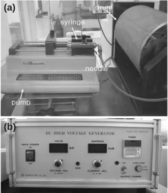
Fig. 1 shows the electrospinning apparatus. A syringe pump (see Fig. 1(a), FABRIQUE AUXETATS UNIS, Kd Scientific, USA.) was utilized to supply polymer solution at a constant flow of 0.003 ml min-1during electrospinning. DC-high-voltage generator (see Fig. 1(b)), Model cps-40KO3VIT, CHUNGPA EMT CO.) provided a voltage of 15 kV to draw the nanofibers from the prepared solution. The electrospun fibers were collected on a grounded drum. Needle size was 28G and working distance (the distance between the needle tip-to-collector) was 10 cm.
Fig. 1. Electrospinning apparatus.
(a)
Formation of silver nanoparticles
PVA nanofibers containing silver nanoparticles were pre-pared by two methods. First method was electrospinning of irradiated solution. The prepared PVA/AgNO3solution was
irradiated by gamma-rays at irradiation dose rate of 2 kGy hr-1for 2.5 h. And then the irradiated solution was
electro-spun. Second method was irradiation of electrospun nanofi-bers. Nanofibers prepared by electrospinning of unirradiated PVA/AgNO3solution. The prepared nanofibers were
irradi-ated with gamma-rays at irradiation dose rate of 2 kGy hr-1
for 2.5 h.
Characterization
The morphology of the nanofibers was observed with a scanning electron microscope (JSM-6390) after applying a gold coating.
Distribution of the silver nanoparticles was obtained from TEM photographs. TEM measurements were performed on a JEM-2000FXII instrument operated at an accelerating vol-tage of 200 kV. Samples for TEM studies were placed on a TEM copper grid (200 mesh, carbon-coated).
RESULTS AND DISCUSSION
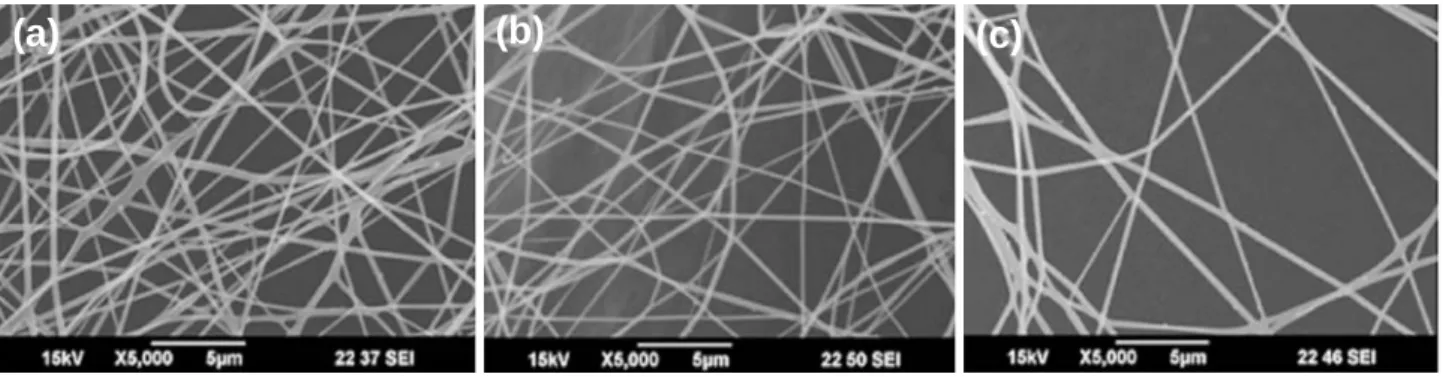
Fig. 2 shows the SEM images of the PVA nanofibers containing silver nanoparticles. Fig. 2(a) is the electrospun nanofibers of the unirradiated PVA/AgNO3solution, Fig.
2(b) is the electrospun nanofibers of the irradiated PVA/ AgNO3solution, and Fig. 2(c) is the irradiated electrospun
nanofibers of the unirradiated PVA/AgNO3solution. The
morphologies of PVA nanofibers were quite similar,
be-cause the amount of silver nitrate was same. The radiation was affect formation of silver nanoparticles but that was not affects the morphology. Fig. 3 and Fig. 4 show the TEM images of the PVA electrospun nanofibers containing silver nanoparticles. Fig. 3 is the TEM images of the PVA nano-fibers electrospun from the PVA aqueous solutions contain-ing 1.7×10⁻²wt% AgNO
3. Fig. 4 is the TEM images of
the PVA nanofibers electrospun from the PVA aqueous solutions containing 1.7 wt% AgNO3. At higher
concen-tration of AgNO3, silver nanoparticles were spontaneously
generated during the electrospinning process (see Fig. 4(a), (d)). But at lower concentration of AgNO3, silver
nano-particles were generated after the irradiation (see Fig. 3). This result showed that the Ag++
ions reduced to Ag nano-particles by the gamma-ray irradiation. Although the PVA nanofibers were irradiated, number of the Ag nanoparticles were not significantly changed (see Fig. 4(d), (f)), indicat-ing that a lot of the Ag++ions were already reduced to Ag
nanoparticles during the electrospinning process and that PVA acted as a stabilizer of the Ag nanoparticles.
Fig. 4(d), (e) shows the TEM images of the PVA nano-fibers electrospun from the PVA/AgNO3solution before
and after being irradiated by gamma-ray at irradiation dose rate of 2 kGy hr-1for 2.5 h. The size of the Ag
nanoparti-cles in the PVA nanofibers of irradiated solution was larger than those of the unirradiated solution. As shown in Fig. 4(d), the Ag nanoparticles were readily generated during the electrospinning process and there was not observed specific changes their number in the PVA nanofibers after the irradiation (see Fig. 4(f)). After the electrospinning of the irradiated solution, the size of the Ag nanoparticles was increased, but their number was decreased. This implies that the Ag nanoparticles in the irradiated PVA/AgNO3
Fig. 2. SEM images of the PVA nanofibers: (a) electrospun nanofibers of the unirradiated polymer solution, (b) electrospun nanofibers of the irradiated polymer solution, (c) irradiated electrospun nanofibers of the unirradiated polymer solution.
solution aggregated to form larger Ag nanoparticles during the electrospinning process. The irradiation process pro-vided fine control over the size of the Ag nanoparticles in the PVA nanofibers by varying the extent of the reduction and ripening process. The Ag nanoparticles produced are stable and comparable in distribution with those produced by other typical methods.
CONCLUSION
We prepared PVA nanofibers containing Ag nanoparti-cles by two methods using γ-irradiation-induced reduction.
First method was electrospinning of the solution containing Ag nanoparticles. Second method was irradiation of elect-rospun nanofibers containing Ag++
ions. At high concen-Fig. 3. TEM images of the PVA nanofibers (AgNO31×10-3 M):
(a), (d) electrospun nanofibers of the unirradiated polymer solution ((a) ×100 K, (d) ×300 K), (b), (e) electrospun
nano-fibers of the irradiated polymer solution ((b) ×100 K, (e) ×300 K), (c), (f) irradiated electrospun nanofibers of the
unirradiated polymer solution ((c) ×100 K, (f) ×300 K).
Fig. 4. TEM images of the PVA nanofibers (AgNO31×10-1 M):
(a), (d) electrospun nanofibers of the unirradiated polymer solution ((a) ×100 K, (d) ×300 K), (b), (e) electrospun
nano-fibers of the irradiated polymer solution((b) ×100 K, (e) ×
300 K), (c), (f) irradiated electrospun nanofibers of the unirradiated polymer solution ((c) ×100 K, (f) ×300 K).
(a)
(b)
(c)
(d)
(e)
(f)
(a)
(b)
(c)
(e)
(f)
(d)
tration of AgNO3, Ag nanoparticles were also
spontane-ously generated during the electrospinning process. When the irradiated PVA/AgNO3solution was electrospun, the
average size of the Ag nanoparticles was increased, but their number was decreased. It was found that this simple irradiation process could provide fine control over the size of the Ag nanoparticles in the PVA nanofibers.
ACKNOWLEDGMENT
This work was supported by a grant from the Korea Science and Engineering Foundation in the Ministry of Edu-cation, Science and Technology (MEST), Republic of Korea.
REFERENCES
Ding B, Kim H, Lee S, Shao C, Lee D, Park S, Kwag G and Choi K. 2002. Preparation and characterization of nano-scaled poly (vinyl alcohol) fibers via electrospinning. J. Polym. Sci., Part B: Polym. Phys. 40:1261-1268.
Gutierrez M and Henglein A. 1993. Physicochemical pro-perties of small metal particles in solution: “microelec-trode” reactions, chemisorption, composite metal part-icles, and the atom-to-metal transition. J. Am. Phys. Chem.
97:5457-5471.
Hirano S, Wakasa Y, Saka A, Yoshizawa S, Oya-Seimiya Y, Hishinuma Y, Nishimura A, Matsumoto A and Kumakura H. 2003. Preparation of Bi-2223 bulk composed with silver-alloy wire. Physica C. 392:458-462.
Hong KH, Park JL, Sul IH, Youk JH and Kang TJ. 2006. Pre-paration of antimicrobial poly (vinyl alcohol) nanofibers containing silver nanoparticles. J. Polym. Sci. Part B: Polym. Phys. 44:2468-2474.
Jiang HQ, Manolache S, Wong. ACL and Denes FS. 2004. Plasma-enhanced deposition of silver nanoparticles onto polymer and metal surfaces for the generation of antimi-crobial characteristics. J. Appl. Polym. Sci. 93:1411-1422. Longenberger L and Miller G. 1995. Formation of metal
part-icles in aqueous solutions by reactions of metal complexes with polymers. J. Phys. Chem. 99:475.
Marignier JL, Belloni J, Delcourt MO and Chevalier JP. 1985. New microaggregates of non noble metals and alloys pre-pared by radiation induced reduction. Nature 317:344-345. Mizukoshi Y, Okisu K, Maeda Y, Yamamoto TA, Oshima R
and Nagata Y. 1997. Sonochemical preparation of bime-tallic nanoparticles of gold/palladium in aqueous solution. J. Am. Phys. Chem. B. 101:7033-7037.
Ohtaki M and Toshima N. 1990. Photoreduction of rhodium (III) ions in water with ultraviolet light aiming to prepare the dispersions of ultrafine particles. Chem. Lett. 4:489-492.
Reetz MT and Helbig W. 1994. Size-selective synthesis of nanostructured transition metal clusters. J. Am. Chem. Soc.
116:7401-7402.
Ren XL and Tang FQ. 2002. Enhancement effect of Ag-Au nanoparticles on glucose biosensor sensitivity. Acta Chim. Sinica 60:393.
Turkevich J, Turkevich, Stevens PL and Hillier J. 1951. A study of the nucleation and growth processes in the synthesis of colloidal gold. Discuss. Faraday Soc. 11:55-75.
Manuscript Received: July 18, 2008 Revision Accepted: August 19, 2008