ABSTRACT
Curcumin is a natural product extracted from Curcuma longa. It has been reported as a potent
antioxidant and anti-inflammatory compound. Previous studies have demonstrated that curcumin suppresses pro-inflammatory cytokine production via inhibition of NF-κB in macrophages. However, its role in adaptive immune cells such as T cells, in vivo, has not
clearly been elucidated. Here, we examined the effects of curcumin in T follicular helper (TFH)
cells and on Ab production during NP-ovalbumin immunization in mice. The results revealed that curcumin administered daily significantly increased CXCR5+B-cell lymphoma 6+ T
FH cells
and CD95+GL-7+ germinal center (GC) B cells in draining lymph nodes. In addition, curcumin
treatment in mice induced total Ab production as well as high affinity IgG1 and IgG2b Ab production. Collectively, these results suggest that curcumin has positive regulatory roles in TFH cell functions and GC responses. Thus, this could be an advantageous supplement to
enhance humoral immunity against infectious diseases and cancer.
Keywords: Curcumin; T lymphocytes; TFH; Immunoglobulins
INTRODUCTION
Curcumin, a chemical product from Curcuma longa, is a diarylheptanoid that has numerous
interference properties in inflammation, cancer, diabetes, and colitis, among others (1-5). Recent studies have emphasized its role in the regulation of cancer via an increase in tumor immunity; however, most of these studies were focused on the anti-inflammatory action of curcumin via inhibition of ROS and inflammatory cytokine production (6-11). This compound is emerging as an anticancer agent because it not only targets tumor cell growth or metastatic functions but also regulates tumor-immunity by increasing Th1 and inhibiting Treg cells (11-13). According to previous reports, curcumin is also known to regulate the NF-κB pathway of various immune cells such as dendritic cells (14), macrophages (15-17), NK cells (18), T cells (19), and B cells (20), suggesting its potent immune modulatory action on proliferative moieties of immune cells.
A very old study demonstrated that dietary curcumin enhances IgG Ab production in rats (21); however, little is known about its cellular mechanism of action on humoral immunity,
Brief Communication
Received: May 27, 2019 Revised: Sep 16, 2019 Accepted: Oct 7, 2019 *Correspondence to Je-Min ChoiDepartment of Life Science, College of Natural Sciences, Hanyang University, Seoul 04763, Korea.
E-mail: [email protected] Copyright © 2019. The Korean Association of Immunologists
This is an Open Access article distributed under the terms of the Creative Commons Attribution Non-Commercial License (https:// creativecommons.org/licenses/by-nc/4.0/) which permits unrestricted non-commercial use, distribution, and reproduction in any medium, provided the original work is properly cited. ORCID iDs Do-Hyun Kim https://orcid.org/0000-0003-0160-3092 Je-Min Choi https://orcid.org/0000-0002-9482-710X Conflict of Interest
The authors declare no potential conflicts of interest.
Abbreviations
Bcl-6, B-cell lymphoma 6; GC, germinal center; i.p., intra-peritoneally; OVA, NP-ovalbumin; TFH, T follicular helper Author Contributions
Conceptualization: Kim DH, Choi JM; Data curation: Kim DH; Project administration: Choi JM; Supervision: Choi JM; Writing - original
Do-Hyun Kim 1,2, Hong-Gyun Lee1,2, Je-Min Choi 1,2,3,*
1Department of Life Science, College of Natural Sciences, Hanyang University, Seoul 04763, Korea 2Research Institute for Natural Sciences, Hanyang University, Seoul 04763, Korea
3Research Institute for Convergence of Basic Sciences, Hanyang University, Seoul 04763, Korea
Curcumin Elevates T
FH
Cells and
Germinal Center B Cell Response for
Antibody Production in Mice
draft: Kim DH, Choi JM; Writing - review &
editing: Lee HG, Choi JM. such as Ab production and isotype class switching. During Ag exposure, B cells can generate high affinity Abs with different isotypes by cross-talk between B and T cells in the germinal
center (GC) (22-24). T follicular helper (TFH) cells, which express the CXCR5 and key
transcription factor B-cell lymphoma 6 (Bcl-6), are localized in the GC in draining lymph nodes (25). TFH cells provide critical costimulatory signals and cytokines to B cell cognates,
resulting in somatic mutation for affinity maturation and isotype-class switch (26-29). Here, we attempted to examine curcumin function in mice to assess whether it is involved in Ab production with regard to the TFH and GC response. We selected intra-peritoneal
administration due to the low stability of curcumin via the oral route; moreover, we opted for NP-ovalbumin (NP-OVA) immunization with the complete adjuvant method to induce a significant amount of TFH and GC B cells in the draining lymph nodes. The results revealed
that curcumin causes in a significant increase in CXCR5+Bcl6+ T
FH and CD95+GL-7+ GC B
cells, with elevated total Ab production as well as high affinity Ag specific IgG1 and IgG2b Ab production.
MATERIALS AND METHODS
Mice
C57BL/6 female mice (6–8 weeks old) were purchased from Orient Bio (Daejeon, South Korea) and maintained at the Hanyang University mouse facilities under pathogen-free conditions with ad libitum feeding. All animal protocols were approved by the Animal Experimentation Ethics Committee of Hanyang University, and the experiments were performed according to the guidelines of the Institutional Animal Care and Use Committee of Hanyang University (2016-0141).
NP-OVA immunization and curcumin administration
NP-OVA (50 μg, Bioresearch Technologies, Novato, CA, USA), diluted in Complete Fruend's Adjuvant (Chondrex, Redmond, WA, USA) was subcutaneously injected into the dorsal region of mice. Seven days after immunization, the mice were sacrificed, and the inguinal lymph node cells were isolated and analyzed by flow cytometry. Curcumin was purchased from Abcam (141921; Cambridge, England) and dissolved in DMSO. Curcumin (200 μg) was injected intra-peritoneally (i.p.) daily following the indicated experimental scheme.
Flow cytometry for T
FHcells and GC B cells
To analyze the TFH cells and GC responses, the inguinal lymph node cells were isolated and
stained with anti-mouse CXCR5-biotin for 30 min at 4°C, followed by anti-mouse CD44-Ag-presenting cell (APC)/Cy7 and CD4-Percp/Cy5.5 and streptavidin-APC staining. After fixation and permeabilization using a Foxp3 Staining Kit (eBioscience, San Diego, CA, USA), intracellular Bcl-6 and Foxp3 was stained using anti-mouse Bcl-6-PE (BD Bioscience, San Jose, CA, USA) and Foxp3-FITC (eBioscience) for 40 min at room temperature. To analyze GC B cells, the isolated single cells were stained with anti-mouse B220-Percp/Cy5.5, GL-7-FITC, and CD95-PE for 30 min at 4°C. Then, the cells were examined using a FACSCanto II system (BD Bioscience); the data were analyzed using FlowJo software (Treestar, Ashland, OR, USA).
ELISA for Ab measurement
The total Ab production in mouse serum was measured using the IgM ELISA kit (eBioscience) and IgG ELISA kit (Abcam). To measure low and high affinity Abs, NP30-conjugated BSA or
NP7-conjugated BSA (Biosearch Technologies, Petaluma, CA, USA) were coated, respectively.
For the isotype Ab analysis, HRP-conjugated goat anti-mouse IgG1, IgG2b, IgG2c, and IgG3 Abs (Southern Biotech, Birmingham, AL, USA) were used to detect the Abs.
Statistical analysis
Data were analyzed using Student's t-test by Prism5 software (GraphPad, San Diego, CA,
USA). The p-values less than 0.05 were considered statistically significant.
RESULTS AND DISCUSSION
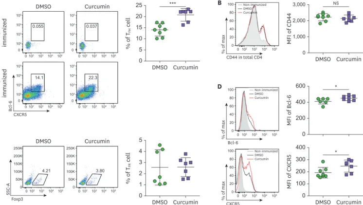
Curcumin administration increases Bcl-6
+CXCR5
+T
FH
cells
To examine curcumin function in the TFH and GC response to a specific Ag, 6-week-old
female C57BL/6 mice were subcutaneously immunized with NP-OVA in complete adjuvant on day 0, followed by daily administration of 200 μg curcumin by i.p. injection until day 6 (Fig. 1). At day 7, inguinal lymph node cells were isolated and analyzed by flow cytometry. As shown in Fig. 2A, NP-OVA immunization, with DMSO vehicle, led to a significant increase in Bcl-6+CXCR5+ T
FH cells compared to non-immunized group. Curcumin treatment significantly
increased Bcl-6+CXCR5+ T
FH cells, which was gated from CD4+CD44+ cells in comparison
with the DMSO vehicle control group. The CD44 expression level of CD4 T cells (Fig. 2B) and percentage of Bcl-6+Foxp3+ T follicular regulatory cells (Fig. 2C) was comparable between
the two groups. The expression level of TFH cell marker molecules such as Bcl-6 or CXCR5
(Fig. 2D) was significantly increased, collectively suggesting that curcumin administration significantly induces TFH cells in the draining lymph nodes during Ag immunization.
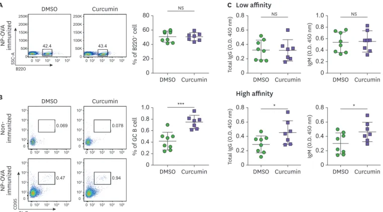
Curcumin administration increases GL-7
+CD95
+GC B cells.
Due to the observed increased proportion of TFH cells and the results of a previous study that
showed enhanced B cell function in sheep RBC immunized mice (27), we hypothesized that there would be elevated levels of GC B cells. The inguinal lymph node cells were analyzed by flow cytometry. With no difference in the proportion of B cells (Fig. 3A), NP-OVA
immunization of the DMSO-treated group led to an increase in the GL-7+CD95+ cells, whereas
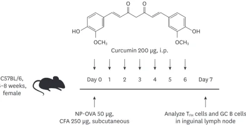
C57BL/6, 6–8 weeks, female NP-OVA 50 µg, CFA 250 µg, subcutaneous Day 0 1 2 3 4 5 6 Day 7
Analyze TFH cells and GC B cells in inguinal lymph node Curcumin 200 µg, i.p.
O O
HO OH
OCH3 OCH3
Figure 1. Experimental scheme of NP-OVA immunization and curcumin administration. Six to eight-week-old female C57BL/6 mice were immunized with NP-OVA in complete adjuvant, and 200 µg of curcumin was i.p. injected daily until day 6. The mice were sacrificed and the draining lymph node cells were analyzed on day 7. CFA, complete freund's adjuvant.
curcumin administration significantly increased the proportion of GC B cells in the draining lymph nodes (Fig. 3B). In addition, there was an increase in the high affinity total IgG and IgM Abs in the serum upon curcumin treatment (Fig. 3C), suggesting that increased TFH cells
by curcumin administration could result in elevated GC B cells and Ab production.
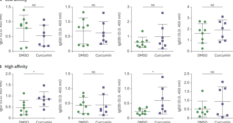
Curcumin administration increases production of high affinity IgG1 and IgG2b Ab
As TFH cells are key players in affinity maturation and isotype class-switching by the
stimulation of GC B cells, we examined various NP-specific isotypes of the Abs in the serum to confirm the consequences of increased TFH and GC responses by curcumin administration.
Fig. 4A and B shows the significant increase in IgG1 and IgG2b Ab production upon curcumin treatment, whereas no significant differences were observed for other isotypes or low affinity Abs. These results suggest that curcumin treatment stimulates high affinity Ab production with isotype class-switching.
Curcumin, one of the most widely studied natural compounds, has diverse biological functions in attenuating chronic inflammatory diseases, including cancer, asthma, inflammatory bowel disease, and rheumatoid arthritis (30,31). These therapeutic functions of curcumin have been mainly associated with the suppression of the production of inflammatory cytokines TNF, IL-1, IL-6, and IL-8 (18,32). However, recent studies have reported that curcumin can directly regulate T cells by inhibiting IL-2 signaling and NF-κB activation (33,34). Moreover, curcumin inhibited Th1 differentiation by blocking
JAK-% of T FH cell 0 10 20 5 15 25 Curcumin DMSO % of T FR cell 0 2 4 1 3 5 Curcumin DMSO MFI of CX CR 5 0 100 300 200 400 Curcumin DMSO MFI of Bcl -6 0 400 200 600 DMSO A C D Non-immuniz ed NP-OV A immuniz ed Curcumin DMSO CXCR5 Bcl -6 Curcumin 0.055 105 104 103 102 102 103 104 105 0 0 105 104 103 102 102 103 104 105 0 0 105 104 103 102 102 103 104 105 0 0 105 104 103 102 102 103 104 105 0 0 250K 200K 150K 100K 102 103 104 105 50K 0 250K 200K 150K 100K 50K 0 0 0102 103 104 105 0.037 14.1 22.3 4.21 3.80 Foxp3 SSC -A 100 80 60 40 102 103 104 105 20 0 0 CXCR5 % of max 100 80 60 40 102 103 104 105 20 0 0 Bcl-6 % of max * * Non-immunized DMSO Curcumin Non-immunized DMSO Curcumin MFI of CD 44 0 2,000 1,000 3,000 Curcumin DMSO NS B 100 80 60 40 102 103 104 105 20 0 0 CD44 in total CD4 % of max *** Non-immunized DMSO Curcumin
Figure 2. Curcumin administration increases Bcl-6+CXCR5+ T
FH cells. (A) Flow cytometric analysis of CD4+CD44+CXCR5+Bcl-6+ TFH cells in the inguinal lymph nodes
from NP-OVA-immunized mice treated with DMSO or curcumin. (B) The expression level of CD44 on CD4+ cells (C) The proportion of CD4+CD44+CXCR5+
Bcl-6+Foxp3+ T
FR cells. (D) The level of Bcl-6 and CXCR5 on CD4+CD44+ cells. Each dot represents an individual sample. Data are presented as the mean±standard
deviation of 2 independent experiments (n=7–8).
MFI, mean fluorescence intensity; SSC-A, standard scrapie cell assay; TFR, T follicular regulatory; NS, not significant.
STAT signal activation (35,36). Here, we demonstrate for the first time, to our knowledge, the positive biological function of curcumin in TFH cell differentiation and GC B cell
formation. We found that curcumin administration in vivo increases Bcl-6+CXCR5+ T FH cells
and GL-7+CD95+ GC B cells, with high affinity Ab production. As curcumin has a direct
effect on the biological functions of T cells, we further examined curcumin treatment in T cell differentiation and found that curcumin appears to specifically inhibit Th1 and Th2 differentiation rather than Th17 (Supplementary Fig. 1). Collectively, these results indicate that curcumin modulates multiple molecular targets and has potent anti-inflammatory activities regulating effector T cell functions (TFH, Th1, and Th2 cells). Direct regulation of
the NF-κB and JAK-STAT signaling pathway by curcumin might be the clue to elucidating the potential mechanisms involved in the generation of TFH cells and GC responses. Furthermore,
considering previous reports that curcumin can enhance B cell function (21,37), dissecting the mechanism underlying the Ab production increasing effect of curcumin should be addressed in further studies.
In conclusion, this study is the first to report that the administration of curcumin increases humoral immunity by Ab production, which is presumably mediated by increased TFH cells in
the draining lymph nodes. Interestingly, curcumin also contributes in the production of high affinity Abs of the IgG1 and IgG2b isotypes during immunization. Although the molecular mechanisms of curcumin's action on the TFH response should be further evaluated in detail,
we believe that curcumin could be an advantageous supplement, to enhance protective immunity via increased Ab production, in the treatment of infectious diseases or cancer.
% of GC B cell 0 0.4 0.8 0.2 0.6 1.0 Curcumin DMSO % of B 220 + cell 0 20 60 40 80 Curcumin DMSO DMSO B A C Low affinity High affinity Non-immuniz ed NP-OV A immuniz ed NP-OV A immuniz ed Curcumin DMSO GL-7 CD 95 Curcumin 0.069 0.078 42.4 43.4 0.47 0.94 105 104 103 102 102 103 104 105 0 0 105 104 103 102 102 103 104 105 0 0 105 104 103 102 102 103 104 105 0 0 105 104 103 102 102 103 104 105 0 0 250K 200K 150K 100K 102 103 104 105 50K 0 250K 200K 150K 100K 50K 0 0 0102 103 104 105 B220 SSC -A *** Total Ig G (O .D . 450 nm) 0 0.4 0.2 0.6 0.8 Curcumin DMSO Total Ig G (O .D . 450 nm) 0 0.2 0.6 0.4 0.8 Curcumin DMSO * IgM (O .D . 450 nm) 0 0.4 0.2 0.6 0.8 Curcumin DMSO IgM (O .D . 450 nm) 0 0.2 0.6 0.4 1.0 0.8 Curcumin DMSO * NS NS NS
Figure 3. Curcumin administration increases GL-7+CD95+ GC B cells. Flow cytometric analysis of (A) B220+ B cell and (B) B220+GL-7+CD95+ GC B cells in the
inguinal lymph nodes. (C) Total IgM and IgG Ab level in the serum of NP-OVA-immunized mice. Each dot represents an individual sample. Data are presented as the mean±standard deviation of 2 independent experiments (n=7–8).
SSC-A, standard scrapie cell assay; NS, not significant. *p<0.05, ***p<0.001.
ACKNOWLEDGEMENTS
This research has been supported by the Ottogi Ham Taiho Foundation.
SUPPLEMENTARY MATERIAL
Supplementary Figure 1
Curcumin treatment suppresses in vitro Th1/2 differentiation. (A) Magnetic-Activated Cell
Sorting-sorted naïve CD4 T cells were cultured in a 2 µg anti-CD3 and anti-CD28 Ab-coated 96-well plate under lineage-specific cytokine skewing conditions. Th1: 0.2 ng/ml IL-12, 50 U/ml IL-2; Th2: 20 ng/ml IL-4, 50 U/ml IL-2; Th17: 20 ng/ml IL-6, 0.5 ng/ml TGFβ, 20 ng/ml IL-1β, and 20 ng/ml IL-23 for 5 days. (A, B) The cells were analyzed by flow cytometry, and cytokine production was measured by ELISA.
Click here to view
REFERENCES
1. Mukhopadhyay A, Basu N, Ghatak N, Gujral PK. Anti-inflammatory and irritant activities of curcumin analogues in rats. Agents Actions 1982;12:508-515.
PUBMED | CROSSREF A IgG 1 (O .D . 450 nm ) 0 0.5 1.0 1.5 Curcumin DMSO Low affinity IgG 2c (O .D . 450 nm ) 0 0.5 1.0 1.5 Curcumin DMSO IgG 2b (O .D . 450 nm ) 0 1 2 3 Curcumin DMSO IgG 3 (O .D . 450 nm ) 0 1 2 4 3 Curcumin DMSO NS NS NS NS B IgG 1 (O .D . 450 nm ) 0 0.5 1.0 1.5 2.0 Curcumin DMSO High affinity IgG 2c (O .D . 450 nm ) 0 0.5 1.0 1.5 Curcumin DMSO IgG 2b (O .D . 450 nm ) 0 0.5 1.0 1.5 Curcumin DMSO IgG 3 (O .D . 450 nm ) 0 0.5 1.0 1.5 2.5 2.0 Curcumin DMSO * NS * NS
Figure 4. Curcumin administration increases the production of high affinity IgG1 and IgG2b Abs. (A) NP30-(low affinity) or (B) NP7-(high affinity) specific Abs
to measure IgG1, IgG2c, IgG2b, and IgG3 in the serum of NP-OVA-immunized mice. Each dot represents an individual mouse. Data are presented as the mean±standard deviation of 2 independent experiments (n=7–8).
NS, not significant. *p<0.05.
2. Huang TS, Lee SC, Lin JK. Suppression of c-Jun/AP-1 activation by an inhibitor of tumor promotion in mouse fibroblast cells. Proc Natl Acad Sci U S A 1991;88:5292-5296.
PUBMED | CROSSREF
3. Brouet I, Ohshima H. Curcumin, an anti-tumour promoter and anti-inflammatory agent, inhibits induction of nitric oxide synthase in activated macrophages. Biochem Biophys Res Commun 1995;206:533-540.
PUBMED | CROSSREF
4. Sidhu GS, Mani H, Gaddipati JP, Singh AK, Seth P, Banaudha KK, Patnaik GK, Maheshwari RK. Curcumin enhances wound healing in streptozotocin induced diabetic rats and genetically diabetic mice. Wound Repair Regen 1999;7:362-374.
PUBMED | CROSSREF
5. Lubbad A, Oriowo MA, Khan I. Curcumin attenuates inflammation through inhibition of TLR-4 receptor in experimental colitis. Mol Cell Biochem 2009;322:127-135.
PUBMED | CROSSREF
6. Mukhopadhyay A, Bueso-Ramos C, Chatterjee D, Pantazis P, Aggarwal BB. Curcumin downregulates cell survival mechanisms in human prostate cancer cell lines. Oncogene 2001;20:7597-7609.
PUBMED | CROSSREF
7. Zhen L, Fan D, Yi X, Cao X, Chen D, Wang L. Curcumin inhibits oral squamous cell carcinoma proliferation and invasion via EGFR signaling pathways. Int J Clin Exp Pathol 2014;7:6438-6446.
PUBMED
8. Zhong Y, Yu W, Feng J, Fan Z, Li J. Curcumin suppresses tumor necrosis factor-α-induced matrix metalloproteinase-2 expression and activity in rat vascular smooth muscle cells via the NF-κB pathway.
Exp Ther Med 2014;7:1653-1658.
PUBMED | CROSSREF
9. Guo LD, Shen YQ, Zhao XH, Guo LJ, Yu ZJ, Wang D, Liu LM, Liu JZ. Curcumin combined with oxaliplatin effectively suppress colorectal carcinoma in vivo through inducing apoptosis. Phytother Res 2015;29:357-365.
PUBMED | CROSSREF
10. Ye M, Zhang J, Zhang J, Miao Q, Yao L, Zhang J. Curcumin promotes apoptosis by activating the p53-miR-192-5p/215-XIAP pathway in non-small cell lung cancer. Cancer Lett 2015;357:196-205.
PUBMED | CROSSREF
11. Xu B, Yu L, Zhao LZ. Curcumin up regulates T helper 1 cells in patients with colon cancer. Am J Transl Res
2017;9:1866-1875. PUBMED
12. Zou JY, Su CH, Luo HH, Lei YY, Zeng B, Zhu HS, Chen ZG. Curcumin converts Foxp3+ regulatory T cells to T helper 1 cells in patients with lung cancer. J Cell Biochem 2018;119:1420-1428.
PUBMED | CROSSREF
13. Zhao GJ, Lu ZQ, Tang LM, Wu ZS, Wang DW, Zheng JY, Qiu QM. Curcumin inhibits suppressive capacity of naturally occurring CD4+CD25+ regulatory T cells in mice in vitro. Int Immunopharmacol 2012;14:99-106.
PUBMED | CROSSREF
14. Kim GY, Kim KH, Lee SH, Yoon MS, Lee HJ, Moon DO, Lee CM, Ahn SC, Park YC, Park YM. Curcumin inhibits immunostimulatory function of dendritic cells: MAPKs and translocation of NF-kappa B as potential targets. J Immunol 2005;174:8116-8124.
PUBMED | CROSSREF
15. Pan Y, Zhang X, Wang Y, Cai L, Ren L, Tang L, Wang J, Zhao Y, Wang Y, Liu Q, et al. Targeting JNK by a new curcumin analog to inhibit NF-kB-mediated expression of cell adhesion molecules attenuates renal macrophage infiltration and injury in diabetic mice. PLoS One 2013;8:e79084.
PUBMED | CROSSREF
16. Chen D, Nie M, Fan MW, Bian Z. Anti-inflammatory activity of curcumin in macrophages stimulated by lipopolysaccharides from Porphyromonas gingivalis. Pharmacology 2008;82:264-269.
PUBMED | CROSSREF
17. Xu Y, Liu L. Curcumin alleviates macrophage activation and lung inflammation induced by influenza virus infection through inhibiting the NF-κB signaling pathway. Influenza Other Respi Viruses 2017;11:457-463.
PUBMED | CROSSREF
18. Jagetia GC, Aggarwal BB. “Spicing up” of the immune system by curcumin. J Clin Immunol 2007;27:19-35.
PUBMED | CROSSREF
19. Kliem C, Merling A, Giaisi M, Köhler R, Krammer PH, Li-Weber M. Curcumin suppresses T cell activation by blocking Ca2+ mobilization and nuclear factor of activated T cells (NFAT) activation. J Biol Chem
2012;287:10200-10209. PUBMED | CROSSREF
20. Han SS, Chung ST, Robertson DA, Ranjan D, Bondada S. Curcumin causes the growth arrest and apoptosis of B cell lymphoma by downregulation of egr-1, c-myc, bcl-XL, NF-kappa B, and p53. Clin Immunol 1999;93:152-161.
PUBMED | CROSSREF
21. South EH, Exon JH, Hendrix K. Dietary curcumin enhances antibody response in rats. Immunopharmacol Immunotoxicol 1997;19:105-119.
PUBMED | CROSSREF
22. Poholek AC, Hansen K, Hernandez SG, Eto D, Chandele A, Weinstein JS, Dong X, Odegard JM, Kaech SM, Dent AL, et al. In vivo regulation of Bcl6 and T follicular helper cell development. J Immunol 2010;185:313-326.
PUBMED | CROSSREF
23. Choi YS, Kageyama R, Eto D, Escobar TC, Johnston RJ, Monticelli L, Lao C, Crotty S. ICOS receptor instructs T follicular helper cell versus effector cell differentiation via induction of the transcriptional repressor Bcl6. Immunity 2011;34:932-946.
PUBMED | CROSSREF
24. Crotty S. T follicular helper cell differentiation, function, and roles in disease. Immunity 2014;41:529-542.
PUBMED | CROSSREF
25. Vinuesa CG, Linterman MA, Yu D, MacLennan IC. Follicular helper T cells. Annu Rev Immunol
2016;34:335-368. PUBMED | CROSSREF
26. Park HJ, Kim DH, Lim SH, Kim WJ, Youn J, Choi YS, Choi JM. Insights into the role of follicular helper T cells in autoimmunity. Immune Netw 2014;14:21-29.
PUBMED | CROSSREF
27. Jeon YH, Choi YS. Follicular helper T (Tfh) cells in autoimmune diseases and allograft rejection. Immune Netw 2016;16:219-232.
PUBMED | CROSSREF
28. Choi SS, Jang E, Oh YK, Jang K, Cho ML, Park SH, Youn J. Aged sanroque mice spontaneously develop Sjögren's syndrome-like disease. Immune Netw 2019;19:e7.
PUBMED | CROSSREF
29. Lee J, Park H, Eom J, Kang SG. MicroRNA-mediated regulation of the development and functions of follicular helper T cells. Immune Netw 2018;18:e7.
PUBMED | CROSSREF
30. Aggarwal BB, Harikumar KB. Potential therapeutic effects of curcumin, the anti-inflammatory agent, against neurodegenerative, cardiovascular, pulmonary, metabolic, autoimmune and neoplastic diseases.
Int J Biochem Cell Biol 2009;41:40-59.
PUBMED | CROSSREF
31. Hatcher H, Planalp R, Cho J, Torti FM, Torti SV. Curcumin: from ancient medicine to current clinical trials. Cell Mol Life Sci 2008;65:1631-1652.
PUBMED | CROSSREF
32. Aggarwal BB, Sung B. Pharmacological basis for the role of curcumin in chronic diseases: an age-old spice with modern targets. Trends Pharmacol Sci 2009;30:85-94.
PUBMED | CROSSREF
33. Forward NA, Conrad DM, Power Coombs MR, Doucette CD, Furlong SJ, Lin TJ, Hoskin DW. Curcumin blocks interleukin (IL)-2 signaling in T-lymphocytes by inhibiting IL-2 synthesis, CD25 expression, and IL-2 receptor signaling. Biochem Biophys Res Commun 2011;407:801-806.
PUBMED | CROSSREF
34. Ranjan D, Chen C, Johnston TD, Jeon H, Nagabhushan M. Curcumin inhibits mitogen stimulated lymphocyte proliferation, NFkappaB activation, and IL-2 signaling. J Surg Res 2004;121:171-177.
PUBMED | CROSSREF
35. Natarajan C, Bright JJ. Curcumin inhibits experimental allergic encephalomyelitis by blocking IL-12 signaling through Janus kinase-STAT pathway in T lymphocytes. J Immunol 2002;168:6506-6513.
PUBMED | CROSSREF
36. Fahey AJ, Adrian Robins R, Constantinescu CS. Curcumin modulation of IFN-beta and IL-12 signalling and cytokine induction in human T cells. J Cell Mol Med 2007;11:1129-1137.
PUBMED | CROSSREF
37. Antony S, Kuttan R, Kuttan G. Immunomodulatory activity of curcumin. Immunol Invest 1999;28:291-303.