Regulation of Feeding Behavior and
Hypothalamic Neuropeptide Gene
Expression by Melanocortin
Analogues
Thesis by
Byung-Jin Kim
Brain Korea 21 Project for Medical Sciences
The Graduate School of Yonsei University
Regulation of Feeding Behavior and
Hypothalamic Neuropeptide Gene
Expression by Melanocortin
Analogues
Directed by Professor Ja-Hyun Baik
A Dissertation submitted to
the Graduate School of Yonsei University
December , 2001
By
Byung-Jin Kim
Brain Korea 21 Project for Medical Sciences
The Graduate School of Yonsei University
A Dissertation for the Degree of Master in
Medical Sciences by Byung-Jin Kim has been
approved by
(Supervisory committee, Chairman)
(Supervisory committee)
(Supervisory committee)
Brain Korea 21 Project for Medical Sciences
The Graduate School of Yonsei University
감사의
글
항상 지도와 관심으로 보아주시는 백자현 선생님께 먼저 감사를 전합니다. 아울러 논문 심사위원으로 수고해주신 임승길 선생님과 장정원 선생님께도 감사를 전합니다. 듬직한 동료이자 선배였던 수현씨, 이은진 선생님 그리고 조상래 선생님께 감사를 전합니다. 그리고 같은 실험실에 들어와 같이 많은 것을 생각하고 느꼈던 명환, 성재, 용년, 일선, 청섭, 미현, 안연희 선생님께 감사를 전합니다. 아울러 한 식구처럼 대해주신 3층과 5층의 많은 선생님들게 그리고 언제나 많은 얘기를 나누었던 약리학교실의 성지영 선생과 민영규 선생님께도 고마운 마음을 전합니다. 아울러 항상 많은 도움을 주시던 4층의 많은 분들에게 감사를 전합니다. 항상 신나는 격려를 해주던 나우누리 경북대 통신동아리 회원들에게 그리고 지난 16년 동안 한결 같은 친구로 남아준 상욱군과 영돈군, 석창군에게 진심어린 감사를 전합니다. 마지막으로 언제나 염원과 사랑으로 지켜봐 주시는 부모님, 사랑스런 두 여동생 윤경과 윤미 그리고 항상 기도로 돌봐주시는 고모님, 그리고 아프신 몸에도 불구하고 항상 염원해주시는 할머니, 모든 나의 가족들에게 감사를 전하면서 이 논문을 마칩니다. 2001년 12월 김 병 진Contents
Abstract ………1
I. Introduction ……… 3
II. Material and Methods ………7
1. Animals preparation, intracerebroventricular
administrations and food-intake measurements ………… 7
2. Brain sample preparation and in situ hybridization ……7
Animal sacrifice and brain section … … … 7
Probe synthesis … … … 8
Hybridization … … … 9
Section emulsion and quantification … … … 1 0
3. RT-PCR and southern Blot Analysis … … … 1 0
III. Results ………14
1. Inhibition of feeding by ICV administration of the
melanocortin analogues and body weight change…………14
2. Effect of melanocortin analogues on the expression of
IV. Discussion ……… 35
V. Conclusion ……… 41
Reference ………43
List of Figures and Tables
Table 1. Amino acid sequence of melanocortin and analogues … 12
Table 2. Primers for PCR amplification and internal primers for
southern blot analysis … … … 13
Figure 1. Inhibition of feeding by ICV administration of several
melanocortin analogues … … … 1 6
Figure 2. Daily body weight change by ICV administration of
melanocortin analogues … … … 1 9
Figure 3. Photogram and graph of in situ hybridization for MCH
expression in ventromedial hypothalamus(VMN) and
lateral hypothalamus(LH). … … … 2 3
Figure 4. Photogram and graph of in situ hybridization for AGRP
expression in arcuate nucleus(ARC). … … … 2 5
Figure 5. Photogram and graph of in situ hybridization for NPY
expression in arcuate nucleus(ARC). … … … 2 7
Figure 6. RT-PCR analysis for hypothalamic mRNA expression
levels of MCH, AGRP and NPY after 3hr of
Figure 7. RT-PCR analysis for hypothalamic mRNA expression
levels of MCH, AGRP and NPY after 1hr of
Abstract
Regulation of Feeding Behavior and
Hypothalamic Neuropeptide Gene Expression
by Melanocortin Analogues
Byung-Jin Kim
Brain Korea 21 Project for Medical Sciences
The Graduate School, Yonsei University
(Directed by Professor Ja-Hyun Baik)
Melanocortins are known to be involved in the inhibition of food-intake and energy metabolism. Intracerebroventricular(I.C.V)
administration of several different analogues of α-MSH , such as
α-MSH, NDP-MSH, α-MSH-ND, [Gln6] α-MSH-ND, [Lys6] α- MSH-ND, which were substituted in the position of His6 with Gln and Lys, and Cyclic 16k-MSH to C57J/BL6 mice showed that significant inhibition of time course food-intake compared to
saline-administerd control. However, truncated form of [Gln6] α-
hybridization and RT-PCR analysis revealed that expression of
MCH was significantly decreased by α-MSH, NDP-MSH, α-MSH-
ND, [Gln6] α-MSH, [Lys6] α-MSH and Cyclic 16k-MSH after 1
and 3hr of administration and expression of AGRP and NPY in
hypothalamus was significantly decreased by α-MSH, [Gln6] α-
MSH, [Lys6] α-MSH and Cyclic 16k-MSH after 3 hr of
administration. Administration of NDP-MSH and α-MSH-ND
induced a biphasic regulation in expression of AGRP and NPY, showing a decrease after 1hr and an increase after 3hr. Our results suggest that MC3R and MC4R melanocortin receptors mediate hypophagic signaling in association with differential regulation of hypothalamic neuropeptide. Identification of potential target genes in this regulation are currently undertaking to understand the signaling mediated by MC3R and MC4R in the feeding circuitry.
Key Words: Melanocortin receptor, Peptide analogues, Food-intake, Gene Expression, Hypothalamus.
Regulation of Feeding Behavior and
Hypothalamic Neuropeptide Gene Expression
by Melanocortin Analogues
(Directed by Professor Ja-Hyun Baik)
Brain Korea 21 Project for Medical Sciences
The Graduate School, Yonsei University
Byung-Jin Kim
I. Introduction
Obesity is now a worldwide public health problem owing to increased risk for type II diabetes, hypertention, hyper-lipidaemia, and certain cancers. In recent years, important investigations have been made in identifying the components that regulate body weight including several genes responsible for animal and human obesity. A key element of physiological system that regulates body weight is the hormone, leptin. Leptin is produced by fat tissue and upon
activation of leptin receptor in the brain, a series of neuronal response is required for food intake and energy balance to be
affected1. Several distinct hypothalamic neuropeptides have
emerged as candidate mediators of adiposity signals in CNS. For
examples, 2MCH(melanin-concentrating hormone), 3 , 4NPY
(neuropeptide Y), 5AGRP (Agouti-related protein) and 6galanin are
known to stimulate food intake while 7mahogany, 8neurotensin,
s o m a t o s t a t i n , c o r t i c o t ro p h i n- r e l e a s i n g f a c t o r ( C R F ) a n d cholecystokinin are known for their anorexigenic effect. Over recent years, much attention has been drawn to the involvement of the melanocortins in control of feeding behavior. Alpha-melanocyte stimulating hormone (MSH) is peptide hormone with 13 amino acids derived through a series of proteolytic cleavages from the precursor peptide pro-opiomelanocortin (POMC). This peptide mediates its effects through G-protein coupled receptors by stimulating adenylate cyclase. So far, at least five subtypes of melanocortin receptors are identified. Melanocortin 1 receptor (MC1R) is expressed in melanocytes and known to be involved in skin pigmentation and the MC2R is the ACTH receptor and specifically expressed in the adrenal gland.
MC5R is expressed in many peripheral tissues and suggested to
regulate hair lipid production and thermal regulation. 9,10 MC3R is
expressed in specific brain regions, ventromedial hypothalamus (VMH) and arcuate nucleus(ARC) whereas the MC4R is expressed more widely across the brain and spinal cord. It has been reported
that activation of MC4R by α-MSH increases energy expenditure
and decreases food intake, genetic disruption of MC4R also caused
obesity in mice11,12. Therefore, MC4R receptor-selective agonists
have been considered as potential candidates for the treatment of
abnormal eating behaviors including obesity and anorexia13,14. It
has been reported recently that genetic disruption of this MC3R in mice caused increase of fat mass and decrease of lean body mass without outstanding increase of food intake and showed distinct feature of obesity as compared to that of MC4R knock-out mice. These reports suggested differential physiological functions of
MC3R in obesity with MC4R 15,16 and that melanocortin 3 and 4
receptor play a important mediator of feeding and energy expenditure in hypothalamus.
17Previously, we analyzed several α-MSH analogues, such as α-
ND, which were substituted in the position of His6 with Gln and Lys, and Cyclic 16k-MSH upon stimulation of MC3R and MC4R using a CRE-mediated reporter gene transcription activity assay.
In our CRE-mediated gene transcription activity assay, α-MSH -
ND was the most efficient α-MSH analogue on MC4R whereas
NDP-MSH was the most efficient for MC3R. Changing the His6
residue of α-MSH -ND to Gln, Lys markedly decreased the
CRE-mediated luciferase activity for MC3R as compared to MC4R. In the present study, we have analyzed the effect of these analogues on
feeding behavior in vivo by 18,19,20 intracerebroventricular(ICV)
administration in mice and 2 1we also assessed regulation of
expression of several neuropeptides involved in the regulation of food-intake, by in situ hybridization and RT -PCR analysis.
II. Materials and Methods
1. Animals preparation, intracerebroventricular administrations and food-intake measurements
Male C57/BL6 mice weighing 20-30g (6-8 weeks old) were maintained in individual metabolic cages with free access for feed and water under 12hr light cycle and 12 hr dark cycle(lights on at 0800 hr) with controlled temperature (21℃-23℃). Before ICV administration, mice were fasted for 24hr. Administration was proceeded with Hamilton syringe (26 gauge needle) and performed
essentially as described21. Peptides(Table.1) was prepared with
concentration 3 nmole in 5µl volume for 1 administration with
same volume of saline control. Administration was performed at the beginning of the light phase and measurement was performed at the end of this period. Food and water intake was measured at 1, 2, 4, 6, 8, 10, 12 and 24hr after each administration.
2. Brain sample preparation and in situ hybridization
Animal sacrifice and brain section
Animals were sacrificed 1hr or 3hr after final administration. Brains were immediately excised after sacrifice and stored at –70℃.
Prepared Brains were mounted in frozen stage and serially sectioned of 10µm slices with a microtome.
Probe synthesis
Antisense NPY was prepared by linearizing the plasmid pBLNPY-1 with Fsp I, which contains 511 bp of the rat NPY gene(provided by Dr.Steven L.Sabol). Antisense MCH was prepared by linearizing the plasmid pGEM4-MCH with Xba I, which contains 700 bp of the rat MCH gene(provided by Dr.Rebecca M. Demo). Antisense galanin was prepared by linearizing the plasmid pBluescript-galanin with Hind III, which contains 700 bp of the rat galanin gene (provided by Dr.Tom Teal). Antisense AGRP is prepared by linearizing the plasmid pBluescript-AGRP with EcoR I , which contains 300 bp of mouse
AGRP gene. [35S] cRNA probes were prepared by transcribing 1µg
of each linearized DNA with T3 polymerase(NPY), T7 polymerase(galanin and AGRP) and SP6 polymerase(MCH) for
1hr 30 min at 37℃ in a reaction containing [35S]CTP(Amersharm
Hybridization
Before hybridization, all sections were fixed with acetone and 37% formaldehyde solution at 4℃. After fixation, sections were acetylated with 0.1M triethanolamine (pH8.0) solution for 5 min and then treated in 50% formamide-1ⅹSSC solution at 60℃ for
10 min. Hybridization was performed for 24hr at 52℃ with 35S-
labeled probes(2.5ⅹ107 cpm/ml with yeast tRNA(50µg/µl) in 50%
formamide, 0.3M NaCl, 1ⅹDenhardt’s solution, 5 mM EDTA, 1mM sodium phosphate buffer, 10% dextran sulfate, 10mM DTT and 20mM Tris). Sections were then washed with 50% formamide-1ⅹSSC solution twice at 55℃ for 1 hr. After rinsing with 2ⅹSSC twice, sections were treated with RNase A(20µg/ml) and RNase T(1 unit/ml) for 30 min at 37℃. Then sections were treated with 50% formamide-2ⅹSSC solution twice at 55℃ for each 1 hr. Sections were desalted in a series of washes with 2ⅹSSC for 15 min at room temperature, 0.1ⅹSSC for 15 min at 50℃ and 0.1ⅹSSC for 30 min at room temperature. Dehydration of sections were performed with ascending cold ethanol (30%, 70% and 100%) and sections were dried at room temperature for 40 min.
Section emulsion and quantification
Dried sections were dipped in emulsion solution (Kodak NTB2). Slides were exposed at 4℃ for 1-7 weeks, developed in Kodak GBX developer and counterstained with 0.01% toluidine blue. Quantification of auto-radiogram on X-ray film (Kodak AR) were measured by using TINA 2.0 program and slides were quantified by using the MCID program.
3. RT-PCR and southern Blot Analysis
Total RNA was prepared from isolated hypothalamus of mice brain using LiCl RNA extraction buffer. First strand cDNAs were generated from total RNA using reverse-transcription with random primer by denaturating at 90℃ for 4 min, annealing at room temperature for 10 min and extending at 42℃ for 50 min. Primers for PCR amplification and internal forward primers for southern blot analysis were generated from cDNA sequences in gene bank(NCBI) or related references (Table.2). PCR amplification for each genes was performed with cycle 94℃ 30sec, 55℃ 30 sec and 72℃ 1 min for 35 cycles. Agarose gels with these PCR products were transferred to nylon transfer membrane (hybond-N+,
Amersharm) and membranes were hybridized by using forward
primers which were labeled with γ32P-ATP(NEN). Co-
amplification of the GAPDH gene was performed to normalize the expression.
III. Results
1. Inhibition of feeding by ICV administration of the melanocortin analogues and body weight change.
Mice were induced to feed by food deprivation for 24 hr
before intracerebroventricular (I.C.V) administration of α-MSH
analogues. Different melanocortin analougues [NDP-MSH, α-
MSH-ND, [Gln6] α-MSH-ND, [Lys6] α-MSH-ND, cyclic16k-MSH
and [Gln6] α-MSH-ND (6-10) : Table 1] were administered and
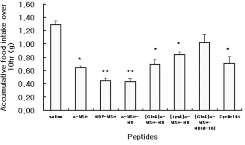
their effect on food intake inhibition were analyzed in vivo. Food- intake was measured at 1, 2, 4, 6, 8, 10, 12 and 24 hr after administration of peptide. Fig 1 showed the time-course food intake over 12hr(A) and 24hr(B) period and accumulative food intake
over 2hr (C) and 10hr (D). NDP-MSH and α-MSH-ND significantly
inhibited food intake by 65-70% as compared to saline administered control over 10 hr period (** P<0.01). Other peptides,
such as α-MSH, [Gln6] α-MSH-ND, [Lys6] α-MSH-ND and
cyclic16k-MSH also significantly inhibited food intake by 36-50% over 10hr period (* P<0.05, n=10) after administration. However,
truncated form of [Gln6] α-MSH-ND showed no significant effect
MSH-ND were the most efficient peptides for inhibition of food
intake and [Gln6] α-MSH-ND, [Lys6] α-MSH-ND and cyclic16k-
MSH also showed significant inhibition on food intake whereas
truncated form of [Gln6] α-MSH-ND did not show significant
effect on food intake inhibition. These data are consistent with our previous results with CRE-luciferase activity assay in vitro, where the activity of these analogues were accessed on the basis of G protein coupling efficiency. These results also suggested the structural importance of the core-sequence of melanocortin analogues(Asp-His-D-Phe-Arg) for binding and activation of MC3R and MC4R .
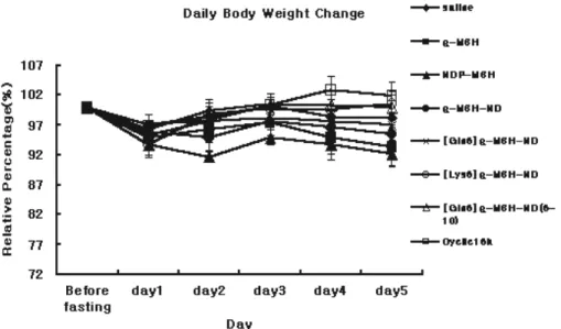
Body weight of each peptide administrated mice were measured at every 24hr after administration(Fig 2). Slight but not statically significant decrease of body weight(7-8%) were showed
by administration of α-MSH and NDP-MSH in 5th days after
administration and other peptides, α-MSH-ND, [Gln6] α-MSH-ND,
[Lys6] α-MSH-ND and cyclic 16k-MSH showed no significant
Fig 1. Inhibition of feeding by ICV administration of several
melanocortin analogues. α-MSH, NDP-MSH, α-MSH-ND, [Gln6] α
-MSH-ND, [Lys6] α-MSH-ND and cyclic16k-MSH(3nmole) produced the
significant food-intake inhibition effect whereas truncated form of [Gln6] α
-MSH-ND(3nmole) had no significant effect on food intake inhibition. (A), time course food intake over 24hr period, (B) time course food-intake over 12hr period. Accumulative food intake over 2hr (C) and 10 hr (D) from start of administration. All value are mean±SEM, n=10 per each group. Data were analyzed by mean of ANOVA followed by Dunnett test for individual comparisons.(*P<0.05, **P<0.01 vs. saline)
Fig 2. Daily body weight change by ICV administration of melanocortin analogues. All values are mean±SEM n=10 per each group. Data were
analyzed by mean of ANOVA followed by Dunnett test for individual comparisons.
2. Effect of melanocortin analogues on the expression of hypothalamic neuropeptides.
Expression of several hypothalamic neuropeptides, such as MCH, AGRP and NPY, after 3hr of administration was analyzed by in situ hybridization and RT-PCR and expression of these genes after 1hr of administration was analyzed by RT-PCR to investigate the regulation of these genes by melanocortin analogues.
Administration of α-MSH induced a decrease of expression of
MCH by 10% (* P<0.05) and 12-17% of MCH expression were
significantly decreased by NDP-MSH, α-MSH-ND [Gln6] α-MSH-
ND, [Lys6] α -MSH-ND and cyclic16k-MSH administration (**
P<0.01). Expression of MCH was significantly decreased in ventromedial hypothalamus (VMH) and lateral hypothalamus (LH) region showing a close association with the food intake inhibition. However, MCH expression was not affected by
administration of truncated form of [Gln6] α-MSH-ND (Fig 3. (A)
and (B)).
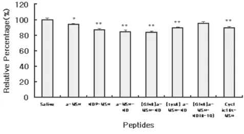
Expression of AGRP was decreased significantly by 13-15% in
brain arcuate nucleus (ARC) by administration of [Gln6] α-MSH-
ND, [Lys6] α-MSH-ND and cyclic 16k-MSH (* P<0.05). However,
administration of NDP-MSH induced a significant increase of AGRP expression by 18% (** P<0.01) and similar effect was
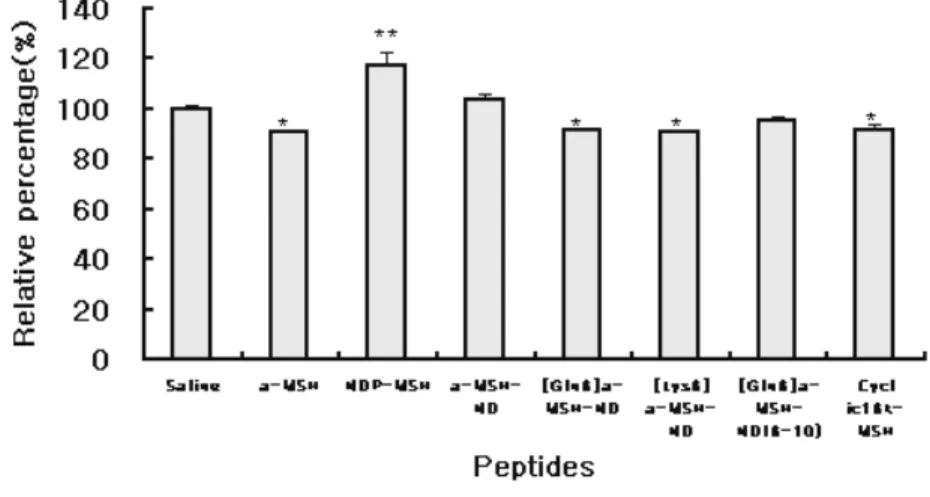
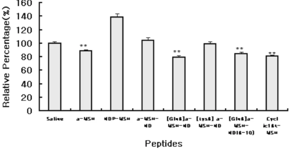
observed in α-MSH-ND administered group. Expression of NPY in
ARC was significantly decreased by 21% by administration of α-
MSH , [Gln6] α-MSH-ND and cyclic16k-MSH but also by the
truncated form of [Gln6] α-MSH-ND administration(** P<0.01).
Whereas, administration of NDP-MSH and α-MSH-ND did not
induce significant change in NPY expression (Fig 5 (A) and (B)).
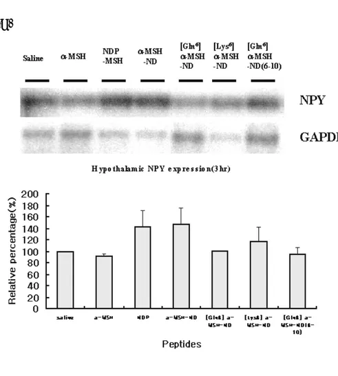
In parallel, expression of hypothalamic mRNA corresponding to neuropeptide, MCH, AGRP and NPY, after 3hr of administration was analyzed by RT-PCR (Fig 6). Expression of
MCH was decreased by MSH, NDP-MSH, MSH-ND [Gln6]
α-MSH-ND, cyclic16k-MSH, and [Gln6] α-MSH-ND(6-10). AGRP
expression was decreased in α-MSH, [Gln6] α-MSH-ND, [Lys6] α-
MSH-ND and cyclic16k-MSH-administered group and increased in
NDP-MSH and α-MSH-ND-administered group. NPY expression
was decreased by α-MSH, [Gln6] α-MSH-ND, [Lys6] α-MSH-ND
and cyclic16k-MSH administration and increased by NDP-MSH
and α-MSH-ND administration. Therefore, RT-PCR analysis
supported our observations made by in situ hybridization .
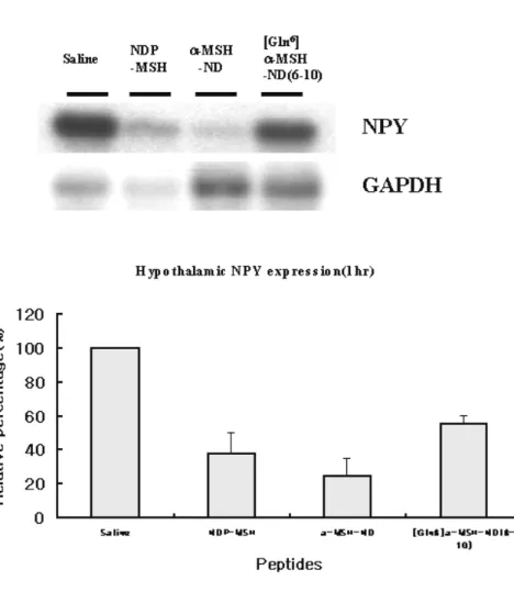
Expression of MCH, AGRP and NPY after 1hr of NDP-MSH
Expression of MCH was decreased by 10-20% by NDP-MSH and α-MSH-ND (Fig 7. A) and expression of AGRP and NPY was decreased by 40-70% after 1hr of administration of NDP-MSH and α-MSH-ND (Fig 7. B, C).
(B )
Fig 3. Photogram and graph of in situ hybridization for MCH expression in ventromedial hypothalamus(VMN) and lateral
hypothalamus(LH). Expression of MCH was decreased significantly with
by administration of melanocortin analogues. All values are mean±SEM. n=3 per individual group. Data were analyzed by mean of ANOVA followed by Dunnett test for individual comparisons.(*P<0.05, **P<0.01 vs. saline)
(B)
Fig 4. Photogram and graph of in situ hybridization for AGRP
expression in arcuate nucleus(ARC). Expression of AGRP was decreased
significantly by a-MSH, [Gln6] a-MSH-ND, [Lys6] a-MSH-ND and cyclic16k-MSH whereas expression was increased significantly by NDP-MSH. All values are mean±SEM. n=3 per individual group. Data were analyzed by mean of ANOVA followed by Dunnett test for individual comparisons.(*P<0.05, **P<0.01 vs saline)
(B)
Fig 5. Photogram and graph of in situ hybridization for NPY expression in arcuate nucleus(ARC). Expression of NPY was decreased significantly
by a-MSH, [Gln6] a-MSH-ND, truncated form of [Gln6] a-MSH-ND and cyclic16k-MSH whereas expression was increased by NDP-MSH. All values are mean±SEM. n=3 per individual group. Data were analyzed by mean of ANOVA followed by Dunnett test for individual comparisons.(*P<0.05, **P<0.01 vs. saline)
(C)
Fig. 6 RT-PCR analysis for hypothalamic mRNA expression levels of MCH, AGRP and NPY after 3hr of administration of melanocortin analogues. Expression of hypothalamic mRNA levels is quantified by
RT-PCR and southern blot anaysis. Expression level of MCH(A), AGRP(B) and NPY(C) was standardized by co-amplification with GAPDH primers
Fig. 7 RT-PCR analysis for hypothalamic mRNA expression levels of MCH, AGRP and NPY after 1hr of administration of melanocortin analogues. Expression of hypothalamic mRNA levels is quantified by
RT-PCR and southern blot anaysis. All expression level of MCH(A), AGRP(B) and NPY(C) was standardized by co-amplification with GAPDH primers
Ⅳ
. Discussion
Several melanocortin receptors were cloned and identified and it has been thought that these melanocortin receptors may be
involved in mediating the diverse effects of melanocortins. Recently, brain specific melanocortin receptor MC3R and MC4R were identified and it has been reported that these two subtypes are important mediators in feeding behavior and energy expenditure.
Many analogues of α-MSH which is endogenous agonist of MC3R
and MC4R have been constructed and analyzed in the view of receptor selectivity and biological activity to evolve as an anti-
obesity medicine.23
We previously reported the differential regulation of cAMP-mediated gene transcription and ligand selectivity by two hypothalamic melanocortin receptor subtypes, MC3R and MC4R
with several melanocortin analogues15. Here, in the present study,
we analyzed the effect of these analogues(Table.1) on food intake in
vivo. Intracerebroventricular administration of different melanocortin analogues to C57/BL6 mice showed that NDP-MSH
and α-MSH-ND were the most efficient peptides in food-intake
inhibition and other peptides, such as α-MSH , [Gln6] α-MSH-ND,
[Lys6] α-MSH-ND and cyclic 16k-MSH, also significantly inhibited
food-intake whereas truncated form of [Gln6] α-MSH-ND had no
our previous results. Indeed, in previous our assay of CRE-
mediated gene transcription activity, α-MSH-ND was the most
efficient α-MSH analogue for MC4R whereas NDP-MSH was the
most efficient for MC3R. However, truncated form of [Gln6] α-
MSH-ND showed low CRE-mediated gene transcription activity upon stimulation of MC3R and MC4R.
It is reported that structure of melanocortin analogue and receptor selectivity is strongly relevant and important. It has
previously been demonstrated that the core sequence of α-MSH
analogues (Asp-His-D-Phe-Arg) is critically important for ligand-receptor interaction and selectivity and we previously reported that
type I β turn structure in α-MSH-ND is important for receptor
binding and ligand selectivity24. Indeed, analogues which contain
core amino-acid sequence or modified core sequence (Asp-Gln-D- Phe-Arg and Asp-Lys-D-Phe-Arg) displayed remarkable efficiency for MC3R and MC4R in our previous report and these peptides also showed the significant inhibition effect on food intake.
However, truncated form of [Gln6] α-MSH-ND(6-10) which is lack
These data strongly indicate that these core-residues are important for receptor binding and activation.
Many orexigenic and anorexigenic genes in CNS are reported with gene expression regulation by leptin and reported that hypothalamus is a major center in the control of food and body mass. However, the interaction and the mechanisms of regulation between hypothalamic neuropeptides are not much defined. In this study, we analyzed gene expression of feeding-related hypothalamic neuropeptides after administration of melanocortin analogues by in
situ hybridization and RT-PCR. First, we analyzed the expression
of MCH in hypothalamus after 1 and 3 hr of melanocortin analogue administration . MCH expression in VMH and LH was significantly decreased in association with food-intake inhibition results by administration of melanocortin analogues. It was previously reported that MCH is a critical regulator of feeding and energy balance which probably acts downstream of leptin ad
melanocortin system2. Recently, it is reported that hypothalamic
expression of MCH, which are normally up-regulated in fasted animals leading to an increase in food intake, are significantly
Thus, MCH seems to play a regulatory role in interaction with several receptors as downstream components of feeding behavior. On the basis of our data, it is expected that MCH play as a mediator for adiposity signal in downstream of melanocortin system.
Expression of AGRP and NPY in hypothalamus was
significantly decreased by α-MSH, [Gln6] α-MSH, [Lys6] α-MSH
and Cyclic 16k-MSH after 3hr of administration. Interestingly, expression of AGRP and NPY was decreased by administration of
NDP-MSH and α-MSH-ND after 1hr and their expression was
increased lately after 3hr. These data demonstrated that NDP-MSH
and α-MSH-ND which showed the strongest effects on food intake
inhibition, induced a biphasic regulation of AGRP and NPY expression. It is possible that this up-regulation might be a feed back regulation upon the stimulation of melanocortin receptors by
NDP-MSH and α-MSH-ND. Taken together, our data suggest that
melanocortin system and AGRP/NPY system are balanced and harmonized for the regulation of feeding and energy expenditure in hypothalamus.
AGRP and NPY are highly expressed in ARC where is co-localized with expression region of melanocortin and its receptors, MC3R and MC4R with high density. Indeed, AGRP is the
endogenous antagonist of MC3R/4R26 and α-MSH and AGRP are
synthesized within adjacent, but distinct subgroup of hypothalamic neurons (POMC and AGRP neuron) that are sensitive to input
from adiposity related signal, leptin27. The AGRP and NPY are
co-expressed in ARC neuron and it is suggested that leptin stimulates POMC neurons and increase signaling of MC3R and MC4R which have an opposite effect on NPY/AGRP neuron. POMC and NPY/AGRP neurons are co-expressed with leptin receptor indicating that ARC is a principal site for transducing adiposity
signal from leptin28. It was also reported that neuropeptide Y
(NPY) mRNA levels was increased in ARC during fasting, whereas
POMC mRNA levels was decreased30. NPY expression was
significantly attenuated by leptin in ARC suggesting that ARC is an important region for expression and regulation of AGRP, NPY and melanocortin system. Thus, our data clearly demonstrated that expression of NPY and AGRP is regulated by melanocortin analogue administration which is mediated by MC3R and MC4R.
Furthermore, in our experiments, regulation of expression for
AGRP and NPY was different by NDP-MSH and α-MSH-ND as
compared to other melanocortin analogues. These data raise a possibility that the specific melanocortin analogues for MC3R or MC4R might induce the distinct gene expression regulation of hypothalamic neuropeptides with differential adiposity signaling mediated by MC3R and MC4R.
In conclusion, our results suggest that the effect of melanocortin analogues on food intake inhibition related to the structure of peptides and this structure is important for physiological role of melanocortin receptor. And we also suggest that hypophagic effect of brain melanocortin receptor MC3R and MC4R is associated with differential regulation on expression of hypothalamic neuropeptides. Identification of potential target genes in this regulation are currently undertaking to understand the signaling mediated by MC3R and MC4R.
V. Conclusion
Intracerebroventricular administration of different melanocortin
analogues to C57/BL6 mice showed that NDP-MSH and α-MSH-
ND were the most efficient peptides in food-intake inhibition and
other peptides, such as α-MSH , [Gln6] α-MSH-ND, [Lys6] α-
MSH-ND and cyclic 16k-MSH, also significantly inhibited food-
intake whereas truncated form of [Gln6] α-MSH-ND had no effect.
Expression of hypothalamic neuropeptides such as MCH, AGRP and NPY after administration of melanocortin analogues was analyzed by in situ hybridization and RT-PCR. MCH expression
was significantly decreased by administration of α-MSH, NDP-
MSH, α-MSH-ND, [Gln6] α-MSH, [Lys6] α-MSH and Cyclic 16k-
MSH after 1 and 3hr. Expression of AGRP and NPY in ARC was
significantly decreased by α-MSH, [Gln6] α-MSH, [Lys6] α-MSH
and Cyclic 16k-MSH after 3hr of administration. Administration of
NDP-MSH and α-MSH-ND induced a biphasic regulation in
expression of AGRP and NPY, showing a decrease after 1hr and an increase after 3hr. Our results suggest that MC3R and MC4R melanocortin receptors mediate hypophagic signaling in association
with differential and harmonized regulation of other hypothalamic neuropeptides.
References
1. Cohen P, Zhao C, Cai X, Montez JM, Rohani SC, Feinstein P, Mombaerts P, Friedman JM. Selective deletion of leptin receptor in neurons leads to obesity. J Clin Invest Oct;108(8):1113-21(2001).
2. Shimada M, Tritos NA, Lowell BB, Flier JS, Maratos-Flier E. Mice lacking melanin concentrating hormone are hypophagic and lean. Nature 396, 670-74(1998).
3. Bi S, Ladenheim EE, Schwartz GJ, Moran TH. A role for NPY overexpression in the dorsomedial hypothalamus in hyperphagia and obesity of OLETF rats. Am J Physiol Regul Integr Comp Physiol 281(1) : R 254-60 (2001).
4. Niimi M, Sato M, Taminato T. Neuropeptide Y in central control of feeding and interactions with orexin and leptin. Endocrine 2, 269-73 (2001)
mouse obesity syndrome and mechanisms of Agouti-induced obesity. Obesity Res 7, 506-13 (1999)
6. Kyrkouli SE, Stanley BG, Seirafi RD, Leibowitz SF. Stimulation of feeding by galanin-anatomical localization and behavioral specificity of this peptide effects in the brain. Peptides 15, 1267 -72(1994)
7. Dinulescu DM, Fan W, Boston BA, McCall K, Lamoreux ML, Moore KJ, Montagno J, Cone RD. Mahogany (mg) stimulates feeding and increases basal metabolic rate independent of its suppression of agouti. Proc Natl Acad Sci U S A 1998 Oct 13;95(21):12707-12.
8. Beck. B Cholecystokinin, neurotensin and corticotrophin- releasing factor-3 important anorexic peptides. Ann Endocrinology 53, 44-9 (1992)
hypothalamic proopiomelanocortin mRNA by leptin in ob/ob mice. Endocrinology 138, 5063 -67(1997)
10. Mizuno TM. Hypothalamic proopiomelanocortin mRNA is reduced by fasting in ob/ob and db/db mice, but it is stimulated by leptin. Diabetes 47, 294 (1998)
11. Huszar D, Lynch CA, Fairchild-Huntress V, Dunmore JH, Fang Q, Berkemeier LR, Gu W, Kesterson RA, Boston BA, Cone RD, Smith FJ, Campfield LA, Burn P, Lee F. Targeted Disruption of the melanocortin 4 receptor results in obesity in mice. Cell 88, 131 (1997)
12. Linda Ste. Marie, Grant I. Miura, Donald J. Marsh, Keith Yagaloff, and Richard D. Palmiter. A metabolic defect promote obesity in mice lacking melanocortin-4 receptors. Proc. Natl Acad. Sci 97, 12339-44 (2000)
13. Schioth HB, Muceniece R, Mutulis F, Prusis P, Lindeberg G, Sharma SD, Hruby VJ, Wikberg JE. Selectivity of
Cyclic[D-Nal7] and [D-Phe7] substituted MSH analogues for melanocortin receptor subtypes. Peptides 18, 1009-13(1997)
14. Lee JH, Lim SK, Huh SH, Lee D, Lee W. Solution structures of the melanocyte-stimulating hormone by two- dimensional NMR spectroscopy and dynamical simulated-annealing caculations. Eur. J. Biochem 257, 31-40 (1998)
15. Lee EJ, Lee SH, Jung JW, Lee WT, Kim BJ, Park KW, Lim SK, Yoon CJ, Baik JH. Differential regulation of cAMP- mediated gene transcription and ligand selectivity by MC3R and MC4R melanocortin receptors. Eur J Biochem 268(3), 582-91(2001)
16. Williams DL, Kaplan JM, Grill HJ. The role of the dorsal vagal complex and the vagus nerve in feeding effects of melanocortin-3/4 receptor stimulation. Endocrinology. 141, 1332-37( 2000).
MA, Dekoning J, Baetscher M, Cone RD. A unique metabolic syndrome causes obesity in the melanocortin-3 receptor-deficient mouse. Endocrinology 141, 3518-21 (2000)
18. Chen AS, Marsh DJ, Trumbauer ME, Frazier EG, Guan XM, Yu H et al. Inactivation of the mouse melanocortin-3 receptor results in increased fat mass and reduced lean body mass. Nature Genetics 26. 97-102 (2000)
19. Abbott CR, Rossi M, Kim M, AlAhmed SH, Taylor GM, Ghatei MA, Smith DM, Bloom SR. Investigation of the melanocyte stimulating hormones on food intake. Lack Of evidence to support a role for the melanocortin-3-receptor. Brain Res. 30, 203-10.(2000)
20. Small CJ, Kim MS, Stanley SA, Mitchell JR, Murphy K, Morgan DG, Ghatei MA, Bloom SR. Effects of chronic central nervous system administration of agouti-related protein in pair-fed animals. Diabetes. 50, 248-54(2001)
21. laursen, S.E and Belknap J.K. Intracerebroventricular injections in mice. Some methodological refinements. J Pharmarcol Methods 16, 355-57 (1986)
22. Christophe Breton, Francoise Presse, Guillaume Hervieu, and Jean-Louis Nahon. Structure and regulation of the mouse melanin-concentrating hormine mRNA and gene. Molecular & Cellular Neuroscience 4, 271-84(1993)
23. Ying-Kui Y, Chris D, Carrie HL and Ira G. Molecular Nasis for the interaction of [Nle4, D-Phe7] melanocyte stimulating
hormone with the human melanocortin 1 receptor(melanocyte
α-MSH receptor) J Bio Chem 272 No 37 23000-10(1997)
24. Li SZ, Lee JH, Lee W, Yoon CJ, Baik JH, Lim SK. Type I beta-turn conformation is important for biological activity of the melanocyte-stimulating hormone analogues. Eur J Biochem 1999 Oct 1;265(1):430-40.
25. Haskell-Luevano C, Cone RD, Monck EK, Wan YP. Related Articles Structure activity studies of the melanocortin-4 receptor by in vitro mutagenesis: identification of agouti-related protein (AGRP), melanocortin agonist and synthetic peptide antagonist interaction determinants. Biochemistry. 2001 May 22;40(20):6164-79
26. Baskin DG, Hahn TM, Schwartz MW. Leptin sensitive neurons in the hypothalamus Horm Metab Res 1999 May;31(5):345-50
27. Yamada M, Miyakawa T, Duttaroy A, Yamanaka A, Moriguchi T, Makita R, Ogawa M, Chou CJ, Xia B, Crawley JN, Felder CC, Deng CX, Wess J. Mice lacking the M3 muscarinic acetylcholine receptor are hypophagic and lean. Nature. 2001 Mar 8;410(6825):207-12
28. Michael A.C, James L.S, Marcelo R, Marcelo G.C, Sabrina D, Tamas L.H, Roger D.C, Malcom J.L Leptin activates
anorexigenic POMC neurons through a neural network in the arcuate nuleus. Nature 411, 480-84 (2001)
29. Ebihara K, Ogawa Y, Katsuura G, Numata Y, Masuzaki H, Satoh N, Tamaki M, Yoshioka T, Hayase M, Matsuoka N, Aizawa-Abe M, Yoshimasa Y, Nakao K. Involvement of agouti-related protein, an endogenous antagonist of hypothalamic melanocortin receptor, in leptin action. Diabetes 1999 Oct;48(10):2028-33
30. Shimizu-Albergine M, Ippolito DL, Beavo JA.Downregulation of fasting-induced cAMP response element-mediated gene induction by leptin in neuropeptide Y neurons of the arcuate nucleus. J Neurosci 2001 Feb 15;21(4):1238-46
29. Forbes S, Bui S, Robinson BR, Hochgeschwender U, Brennan MB. Integrated control of appetite and fat metabolism by the leptin-proopiomelanocortin pathway. Proc Natl Acad Sci U S A 2001 Mar 27;98(7):4233-7
30. Schioth HB, Muceniece R, Mutulis F, Prusis P, Lindeberg G, Sharma SD, Hruby VJ, Wikberg JE. Selectivity of cyclic [D-Nal7] and [D-Phe7] substituted MSH analogues for the melanocortin receptor subtypes. Peptides. 1997;18(7):1009-13.
국문요약
멜라노코틴 유도체에 의한 섭식행동 조절 및
시상하부 신경펩타이드의 조절
멜라노코틴은 시상하부의 멜라노코틴 3 수용체 (MC3R)와 4 (MC4R) 수용체에 결합하여 식욕조절 및 에너지 대사를 조절하는 비만 조절인자로 알려져 있다. 본 연구에서는 멜라노코틴과 그 구조적 변형을 꾀한 NDP-MSH, α−MSH-ND 와, α−MSH-ND 의 6 번째 His6 을 Gln 과 Lys 으로 치환한 [Gln6] α−MSH-ND, [Lys6] α−MSH-ND 그리고 Cyclic16k-MSH 등의 유도체들이 마우스의 섭식에 미치는 영향을 연구하였으며 Intracerebro-ventricular (I.C.V) administration 으로 이 유도체들을 마우스에 주입한 결과 이들에 의해 마우스의 섭식이 감소함을 관찰하였다. 반면 [Gln6] α−MSH-ND 의 절단된 형인 [Gln6] α−MSH-ND (6-10) 에 의해서는 섭식이 크게 영향을 받지 않았다. 이와 함께 멜라노코틴 유도체들의 주입이 시상하부의 비만조절 인자들의 발현조절에 미치는 영향을 In situ hybridization 과 RT-PCR 로 분석하였다. Melanin-concentrating hormone (MCH)은 멜라노코틴유도체의 주입 1 시간 및 3 시간 후에 그 발현이 감소하였다. 또 다른 시상하부 신경 펩타이드인 agouti-related protein (AGRP)과 nueropeptide Y (NPY)의 경우 α−MSH, [Gln6] α−MSH-ND, [Lys6] α−MSH-ND 그리고 Cyclic 16k-MSH 의 주입 3 시간 후에 그 발현이 감소하였다. NDP-MSH 와 α−MSH-ND 의 주입에 의한 AGRP 및 NPY 의 발현은 주입 1 시간 후에 감소하며 3 시간 후에는 증가하였다. 이러한 결과들은 멜라노코틴 3 과 4 수용체의 섭식 저해 작용이 다른 신경 펩타이드 들의 다양한 조절작용과 연관되어 있음을 제시한다. 그러므로 멜라노코틴 및 그 수용체의 작용에 대해 표적이 될 가능성을 가진 유전자를 규명한다면 섭식 및 에너지 대사 조절에 있어서의 멜라노코틴 3, 4 수용체의 신호전달기전의 이해는 물론 비만치료제 개발에 대한 기초가 될 것이다. 핵심되는 말 : 멜라노코틴 수용체, 유도체, 섭식, 유전자발현, 시상하부.