© 2014 The Korean Academy of Medical Sciences.
This is an Open Access article distributed under the terms of the Creative Commons Attribution Non-Commercial License (http://creativecommons.org/licenses/by-nc/3.0) which permits unrestricted non-commercial use, distribution, and reproduction in any medium, provided the original work is properly cited.
pISSN 1011-8934 eISSN 1598-6357
The Significance of Clinical and Laboratory Features in the
Diagnosis of Glycogen Storage Disease Type V: A Case Report
Glycogen storage disease type V (GSD-V) is the most common disorder of muscle glycogenosis with characteristic clinical and laboratory findings. A 32-yr-old woman complained of exercise intolerance and myoglobulinuria since early adolescence. She reported several episodes of second-wind phenomenon. Physical examination did not show any neurological abnormality, including fixed muscle weakness or atrophy. Serum creatine kinase level was 1,161 IU/L at rest. The result of the non-ischemic forearm exercise test was compatible with GSD-V. Mutation analysis identified the compound heterozygous mutations of the PYGM, p.D510fs and p.F710del, which has not yet been reported in Korea. The present case recognizes that detail clinical and laboratory analysis is the first step in the diagnosis of GSD-V.
Keywords: Glycogen Storage Disease Type V; McArdle’s Disease; Phosphorylase, Glycogen, Muscle (PYGM)
Hyung Jun Park,1 Ha Young Shin,2
Yu Na Cho,2 Seung Min Kim,2
and Young-Chul Choi2,3
1Department of Neurology, Mokdong Hospital, Ewha Womans University School of Medicine, Seoul; 2Department of Neurology, Yonsei University College of Medicine, Seoul; 3Rehabilitation Institute of Neuromuscular Disease, Yonsei University College of Medicine, Seoul, Korea
Received: 8 October 2013 Accepted: 8 January 2014 Address for Correspondence: Young-Chul Choi, MD
Department of Neurology, Gangnam Severance Hospital, Yonsei University College of Medicine, 612 Eonju-ro, Gangnam-gu, Seoul 135-720, Korea
Tel: +82.2-2019-3323, Fax: +82.2-3462-5904 E-mail: [email protected]
http://dx.doi.org/10.3346/jkms.2014.29.7.1021 • J Korean Med Sci 2014; 29: 1021-1024
INTRODUCTION
Glycogen storage disease type V (GSD-V), also known as McAr-dle’s disease, is the most common disorder of muscle glyco-genosis (1). GSD-V is caused by alterations in the PYGM which encodes myophosphorylase. Myophosphorylase catalyzes the phosphorolytic cleavage of glycogen to glucose-1-phosphate in skeletal muscle. Therefore, enzyme deficiency results in the de-fective breakdown of glycogen into glucose and glycogen accu-mulation in muscle (2).
GSD-V is inherited in an autosomal recessive pattern, and the prevalence has been estimated at about 1 in 100,000-167,000 (3, 4). Most adult patients present with typical clinical features such as exercise intolerance, episodic myoglobulinuria and the second-wind phenomenon. In addition, the resting serum cre-atine kinase (CK) level, forearm exercise test, and electrophysi-ological studies can contribute to diagnosis. Subsequent muta-tion analysis and myophosphorylase activity assay can be used to confirm the diagnosis.
There has only been one case report of Korean patients with GSD-V (5). However, the previous report did not feature typical clinical presentations of GSD-V such as myoglobulinuria and the second-wind phenomenon. Here, we report a Korean case of GSD-V with typical clinical and laboratory findings.
CASE DESCRIPTION
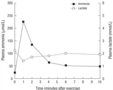
A 32-yr-old woman (Fig. 1A, II-3) was referred to our clinic due to easy fatigability, muscle cramps and myoglobulinuria in June 2013. Her family history was unremarkable. She recalled easy fatigability, muscle cramps and contractures after intense exer-cise during physical education classes since early adolescence. In addition, she had several episodes of dark urine following in-tense exercise such as long-distance running. These symptoms of exercise intolerance had been aggravated one year before. She also reported marked improvement in exercise tolerance after about 20 min, a process termed the ‘second-wind phe-nomenon’; specifically, she had seen improvement in excessive fatigue, breathlessness, and tachycardia. Physical examination did not show fixed muscle weakness and muscle atrophy. All of her sensory modalities and tendon reflexes were intact. Labo-ratory studies revealed a serum creatine kinase (CK) level of 1,161 IU/L (normal < 215 IU/L) at rest. Nerve conduction study (NCS) and needle electromyography study (EMG) did not show any abnormalities at rest. The non-ischemic forearm exercise test showed no increase in venous lactate level with a normal increase in ammonia (Fig. 2). To confirm easy fatigability and weakness after exercise, we performed motor NCS on the ulnar nerve after voluntary maximal contraction for two minutes and 50 Hz nerve stimulations for one second. However, the
ampli-CASE REPORT
Park HJ, et al. • Glycogen Storage Disease Type V with PYGM Mutation
1022 http://jkms.org http://dx.doi.org/10.3346/jkms.2014.29.7.1021
tudes of the compound muscle action potentials did not de-crease after maximal contraction and nerve stimulation. In ad-dition, EMG did not show silent electrical activities during mus-cle cramps. Based on the clinical and laboratory features, she was diagnosed with glycogenoses with exercise-induced weak-ness, especially GSD-V. Therefore, we performed PYGM geno-typing.
Mutational analysis
A DNA sample was extracted from peripheral leukocytes using the Quick Gene blood kit (Fujifilm, Tokyo, Japan). The primer set that covers all 20 exons and their flanking intron regions of the PYGM was designed with Primer 3web v.4.0 (http://prim-er3.wi.mit.edu/) using sequences from the Gen Bank accession number NT_167190.1. All the exons of the PYGM were sequenc-ed using BigDye Terminator v.3.1 (Applisequenc-ed Biosystems, Foster City, CA, USA) followed by polymerase chain reaction. Electro-phoresis and analysis of the reaction mixtures were done with ABI3130xl Genetic Analyzer (Applied Biosystems). A compound heterozygous mutation was identified; one allele had a frame-shift mutation of c.1531delG (p.D510fs) in exon 13 and the other allele had a deletion mutation of c.2128_2130delTTC (p.F710del) in exon 17 (Fig. 1B). These mutations have been previously re-ported to be underlying causes of GSD-V in Japanese patients (6).
DISCUSSION
We described a Korean patient with PYGM mutations who had typical clinical and laboratory findings for identification of GSD-V. Even though GSD-V is the most common disorder of muscle glycogenosis, our patient is only the second case of genetically proven GSD-V in Korea.
Four characteristic clinical features are important for initial suspicion of GSD-V. First, exercise intolerance such as easy fati-gability, muscle cramps and contractures is triggered by static contractions and dynamic exercise. Almost all patients are ad-mitted to the hospital for exercise intolerance. The pathophysi-ology of exercise intolerance is not fully understood, but may be associated with impaired glycolysis (2). A deficient glycogen-dependent adenosine triphosphate (ATP) supply might result in down-regulation of Na+/K+ pumps in myocytes, leading to
loss of membrane excitability (7, 8). The defective breakdown of glycogen into glucose is reproduced by the forearm exercise test. The forearm exercise test showed no increase in lactate level with a normal increase in the ammonia level. The forearm ex-ercise test was originally carried out under ischemic conditions; however, we did not conduct the test under ischemic conditions because the diagnostic value is just as good in non-ischemic conditions and muscle injury can be avoided (9). In addition, several previous studies demonstrated loss of membrane excit-ability as the amplitude of CMAPs decreased after maximal contracture and high frequency repetitive nerve stimulation, and silent electrical activities during muscle cramping (10, 11). However, we did not identify these findings in the patient pre-sented in this case study. It may be associated with benign clini-cal symptoms without fixed muscle weakness.
Second, the serum CK level is elevated in almost all patients Fig. 1. Pedigree and Sequencing chromatograms in Korean patient with PYGM
muta-tions. (A) Pedigree of a family with glycogen storage disease type V (GSD-V). Arrow indicates the proband, whose DNA was used for direct sequencing of PYGM. Filled symbol indicate affected member. (B) Sequencing chromatograms of PYGM mutations. One allele had a frameshift mutation of c.1531delG (p.D510fs) in exon 13 and the other allele had a deletion mutation of c.2128_2130delTTC (p.F710del) in exon 17. c.1531delG (p.D510fs) Antisense sequence c.2128_2130delTTC (p.F710del) 2 1 1 2 3 3 I II III A B
Fig. 2. Non-ischemic forearm exercise test in a patient with GSD-V. The non-ischemic forearm exercise test showed no increase in venous lactate level with a normal in-crease in ammonia.
Time (minutes after exercise)
0 1 2 3 4 5 6 7 8 9 10 Plasma ammonia ( μmol/L) 300 250 200 150 100 50 0 Plasma lacta te (mmol/L) 6 5 4 3 2 1 0 Ammonia Lactate
Park HJ, et al. • Glycogen Storage Disease Type V with PYGM Mutation
http://jkms.org 1023
http://dx.doi.org/10.3346/jkms.2014.29.7.1021
with GSD-V even in the absence of exercise. One recent report indicated that hyperCKemia (> 200 IU/L) was present in 99% of patients and the majority (79%) of patients had a level greater than 1,000 IU/L (4). The elevation of serum CK level at rest is likely associated with muscle damage due to impaired glycoly-sis during exercise. However, the serum CK level at rest did not correlate with clinical severity because exercise-induced mus-cle damage can stimulate musmus-cle repair and adaptive hypertro-phy (12). In addition, one study demonstrated no significant difference in serum CK level at rest between physically active and inactive patients (4).
Third, about half of all patients with GSD-V have experienced one or more episodes of hyperCKemia (several thousand IU/L) or myoglobulinuria after intense exercise. In addition, about 50% of patients with myoglobulinuria develop acute renal fail-ure, which is almost always reversible, but nevertheless requires emergency treatment (13).
Fourth, the second-wind phenomenon, an especially unique feature of GSD-V, differentiates GSD-V from other muscle gly-cogenoses such as muscle phosphofructokinase deficiency (13). This phenomenon is reported by almost patients during the medical interview. If it is not found in the medical history, it is easily reproduced by diagnostic cycle test (14). Therefore, it is the key finding that leads to initial suspicion of GSD-V. This phenomenon is caused by an increased supply of glucose and free fatty acids after a long period of exercise. These changes lead to a switch in metabolic pathways from endogenous glyco-genolysis to oxidative phosphorylation of blood-borne fatty ac-ids (15). However, continuing to exercise also results in severe muscle cramps and myoglobulinuria.
Besides these major clinical features, some patients with GSD-V can also show fixed muscle weakness. Most patients includ-ing the patient in this report did not complain of muscle weak-ness, but approximately one-third of patients develop proximal muscle weakness with aging (2, 16).
Mutation analysis and the myophosphorylase activity assay are used to confirm the diagnosis of GSD-V; however, molecu-lar analysis is more important and more commonly used be-cause muscle biopsy is needed for the enzyme activity assay. At last count in February 2013, 131 different mutations have been reported in the human gene mutation database (http://www. hgmd.org). Genotype has not been correlated with the severity of clinical phenotype in previous studies (17, 18). However, there has been a higher incidence of some genotypes in specific eth-nic groups; for example, p.R50X and p.G205S in the white pop-ulation, p.F710del in the Japanese poppop-ulation, and p.W798R in the Spanish population (13). Likewise, two mutations in our patient have previously been reported in Japan, a neighboring country of Korea (6). In particular, the p.F710del mutation has been identified in 73% of Japanese patients (19).
In conclusion, we identified a compound heterozygous
mu-tation of the PYGM in a Korean patient with typical clinical and laboratory findings. We recognize that detailed clinical and lab-oratory analysis is only the first step in the diagnosis of disease, especially GSD-V.
ORCID
Hyung Jun Park http://orcid.org/0000-0003-4165-8901 Ha Young Shin http://orcid.org/0000-0002-4408-8265 Yu Na Cho http://orcid.org/0000-0003-2865-6012 Seung Min Kim http://orcid.org/0000-0002-4384-9640 Young-Chul Choi http://orcid.org/0000-0001-5525-6861
REFERENCES
1. Vieitez I, Teijeira S, Fernandez JM, San Millan B, Miranda S, Ortolano S, Louis S, Laforet P, Navarro C. Molecular and clinical study of McArdle’s disease in a cohort of 123 European patients: identification of 20 novel mutations. Neuromuscul Disord 2011; 21: 817-23.
2. Dimaur S, Andreu AL, Bruno C, Hadjigeorgiou GM. Myophosphorylase deficiency (glycogenosis type V; McArdle disease). Curr Mol Med 2002; 2: 189-96.
3. Haller RG. Treatment of McArdle disease. Arch Neurol 2000; 57: 923-4. 4. Lucia A, Ruiz JR, Santalla A, Nogales-Gadea G, Rubio JC,
García-Con-suegra I, Cabello A, Pérez M, Teijeira S, Vieitez I, et al. Genotypic and phenotypic features of McArdle disease: insights from the Spanish nation-al registry. J Neurol Neurosurg Psychiatry 2012; 83: 322-8.
5. Sohn EH, Kim HS, Lee AY, Fukuda T, Sugie H, Kim DS. A novel PYGM mutation in a Korean patient with McArdle disease: the role of nonsense-mediated mRNA decay. Neuromuscul Disord 2008; 18: 886-9.
6. Tsujino S, Shanske S, Goto Y, Nonaka I, DiMauro S. Two mutations, one novel and one frequently observed, in Japanese patients with McArdle’s disease. Hum Mol Genet 1994; 3: 1005-6.
7. Haller RG, Clausen T, Vissing J. Reduced levels of skeletal muscle Na+K+ -ATPase in McArdle disease. Neurology 1998; 50: 37-40.
8. Clausen T, Nielsen OB. The Na+,K(+)-pump and muscle contractility. Acta Physiol Scand 1994; 152: 365-73.
9. Kazemi-Esfarjani P, Skomorowska E, Jensen TD, Haller RG, Vissing J. A nonischemic forearm exercise test for McArdle disease. Ann Neurol 2002; 52: 153-9.
10. Dyken ML, Smith DM, Peake RL. An electromyographic diagnostic screen-ing test in McArdle’s disease and a case report. Neurology 1967; 17: 45-50. 11. Lorenzoni PJ, Lange MC, Kay CS, Scola RH, Werneck LC. Motor nerve
conduction study in McArdle disease: case report. Arq Neuropsiquiatr 2005; 63: 874-7.
12. Clarkson PM, Hubal MJ. Exercise-induced muscle damage in humans. Am J Phys Med Rehabil 2002; 81: S52-69.
13. Lucia A, Nogales-Gadea G, Pérez M, Martín MA, Andreu AL, Arenas J. McArdle disease: what do neurologists need to know? Nat Clin Pract Neurol 2008; 4: 568-77.
14. Vissing J, Haller RG. A diagnostic cycle test for McArdle’s disease. Ann Neurol 2003; 54: 539-42.
Park HJ, et al. • Glycogen Storage Disease Type V with PYGM Mutation
1024 http://jkms.org http://dx.doi.org/10.3346/jkms.2014.29.7.1021
second “second wind” in McArdle disease: oxidative mechanisms. Arch Neurol 2002; 59: 1395-402.
16. Wolfe GI, Baker NS, Haller RG, Burns DK, Barohn RJ. McArdle’s disease presenting with asymmetric, late-onset arm weakness. Muscle Nerve 2000; 23: 641-5.
17. Bruno C, Cassandrini D, Martinuzzi A, Toscano A, Moggio M, Morandi L, Servidei S, Mongini T, Angelini C, Musumeci O, et al. McArdle disease: the mutation spectrum of PYGM in a large Italian cohort. Hum Mutat 2006; 27: 718.
18. Martín MA, Rubio JC, Buchbinder J, Fernández-Hojas R, del Hoyo P, Teijeira S, Gámez J, Navarro C, Fernández JM, Cabello A, et al. Molecu-lar heterogeneity of myophosphorylase deficiency (McArdle’s disease): a genotype-phenotype correlation study. Ann Neurol 2001; 50: 574-81. 19. Sugie H, Sugie Y, Ito M, Fukuda T, Nonaka I, Igarashi Y. Genetic analysis
of Japanese patients with myophosphorylase deficiency (McArdle’s dis-ease): single-codon deletion in exon 17 is the predominant mutation. Clin Chim Acta 1995; 236: 81-6.