*Corresponding author. Tel: 82-2-958-6788; Fax: 82-2-958-6919; E-mail: [email protected]
Received 22 September 2008
Keywords: Biomarker, Colorectal cancer, Early detection,
Fecal-oc-cult blood test, Noninvasive marker, Proteomics
Noninvasive molecular biomarkers for the detection of
colorectal cancer
Hye-Jung Kim
1, Myeong-Hee Yu
2, Hoguen Kim
3, Jonghoe Byun
4& Cheolju Lee
1,*
1Life Sciences Division and 2Functional Proteomics Center, Korea Institute of Science and Technology, Seoul 136-791, Korea, 3Department of Pathology, Yonsei University College of Medicine, Seoul 120-752, Korea, 4Department of Molecular Biology, Dankook University, Yongin-si, Gyeonggi-do 448-701, Korea
Colorectal cancer (CRC) is the third most common malignancy in the world. Because CRC develops slowly from removable precancerous lesions, detection of the disease at an early stage during regular health examinations can reduce both the in-cidence and mortality of the disease. Although sigmoidoscopy offers significant improvements in the detection rate of CRC, its diagnostic value is limited by its high costs and inconvenience. Therefore, there is a compelling need for the identification of noninvasive biomarkers that can enable earlier detection of CRC. Accordingly, many validation studies have been con-ducted to evaluate genetic, epigenetic or protein markers that can be detected in the stool or in serum. Currently, the fe-cal-occult blood test is the most widely used method of screen-ing for CRC. However, advances in genomics and proteomics combined with developments in other relevant fields will lead to the discovery of novel non invasive biomarkers whose use-fulness will be tested in larger validation studies. Here, non-invasive molecular biomarkers that are currently used in clin-ical settings and have the potential for use as CRC biomarkers are discussed. [BMB reports 2008; 41(10): 685-692]
Introduction
Colorectal cancer (CRC) is the third most common malignancy in the world. In addition, there are approximately 1,000,000 new cases of CRC and 500,000 deaths associated with CRC each year. Indeed, CRC represents one of the primary causes of cancer deaths in Europe and the USA (1). In Korea, CRC is the fourth leading cause of mortality by cancer, and its in-cidence is increasing (2). CRC is believed to develop slowly via a progressive accumulation of genetic mutations; therefore, the risk of recurrence and subsequent death due to CRC is closely related to the stage of the disease at the time of primary
diagnosis. Recent studies have shown that shifting the de-tection of the disease to an earlier stage via mass screening and intervening at this stage can reduce the risk of death from CRC (3, 4). These findings strongly demonstrate the clinical need for biomarkers for early detection of CRC.
Biomarkers are substances that are used as indicators of a biological state. Accordingly, biomarkers have characteristics that enable them to be objectively measured and evaluated as indicators of normal biological processes, pathogenic proc-esses, or pharmacologic responses to a therapeutic inter-vention (5). One of the key requirements of a test for CRC is that it must allow detection of the disease at earlier stages so that the disease can be cured effectively. Accordingly, such tests should have high sensitivity and specificity while produc-ing a low number of false-negative and false-positive results to prevent subjecting healthy individuals to unnecessary colonoscopies. Sigmoidoscopy offers significant improvements in detection rates for CRC. However, the diagnostic value of sigmoidoscopy is limited with regards to costs, risks, and in-convenience (6, 7). Noninvasive biomarkers can be analyzed relatively easily and economically; therefore, they have the po-tential to greatly enhance screening acceptance. Several non-invasive tests for CRC detection are available, of which the fe-cal-occult blooding test (FOBT) is the most commonly used (8-10). However, this method lacks sensitivity as well as specif-icity for screening an average risk population. As a result, new cancer biomarkers that will further enhance the detection of the disease and trigger follow-up colonoscopies when neces-sary must be developed. In addition to such screening bio-markers, prognostic markers that predict the likely course of the cancer, stratification markers that predict the likely re-sponse to a drug prior to beginning treatment, and efficacy markers that monitor the efficacy of drug treatment may even-tually reduce the mortality rate of CRC.
Recent advances in genomics and proteomics have con-tributed to our understanding of pathways that control the growth, differentiation, and death of cells. Genomic techni-ques such as DNA microarray analysis and proteomic methods such as 2-dimensional electrophoresis and mass spectrometry are now commonly used to evaluate the expression profiles of genes and proteins in cells, tissues, and bodily fluids (11, 12).
Mini Review
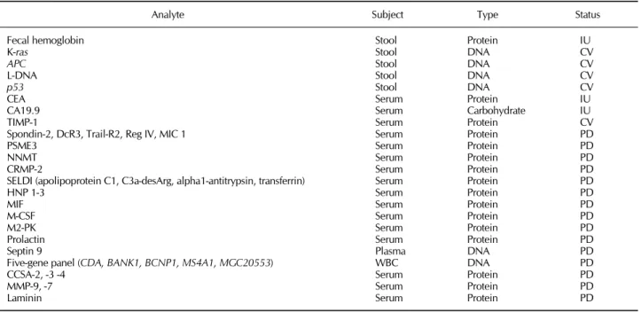
Analyte Subject Type Status Fecal hemoglobin K-ras APC L-DNA p53 CEA CA19.9 TIMP-1
Spondin-2, DcR3, Trail-R2, Reg IV, MIC 1 PSME3
NNMT CRMP-2
SELDI (apolipoprotein C1, C3a-desArg, alpha1-antitrypsin, transferrin) HNP 1-3 MIF M-CSF M2-PK Prolactin Septin 9
Five-gene panel (CDA, BANK1, BCNP1, MS4A1, MGC20553) CCSA-2, -3 -4 MMP-9, -7 Laminin Stool Stool Stool Stool Stool Serum Serum Serum Serum Serum Serum Serum Serum Serum Serum Serum Serum Serum Plasma WBC Serum Serum Serum Protein DNA DNA DNA DNA Protein Carbohydrate Protein Protein Protein Protein Protein Protein Protein Protein Protein Protein Protein DNA DNA Protein Protein Protein IU CV CV CV CV IU IU CV PD PD PD PD PD PD PD PD PD PD PD PD PD PD PD IU, in use; CV, clinical validation; PD, preclinical development
Table 1. Noninvasive molecular biomarkers for the detection of CRC Identification of genes or proteins that are characteristic of the development of cancer can potentially uncover biomarkers that will aid in the diagnosis of CRCs. This review will focus on potential biomarkers that have recently been discovered and may be used in the future to detect CRC using non-invasively-collected samples as well as biomarkers that are currently being used in clinical settings (Table 1).
Fecal markers
Fecal hemoglobinStool-based screening for CRC is simple, inexpensive and the least invasive method of screening available (13). FOBT, which is the most widely used screening modality for CRC, de-tects hemoglobin enzymatically or immunologically (14). Enzymatic FOBT measures the peroxidase-like activity of he-moglobin that originates from any source; therefore, it is sus-ceptible to both colorectal and upper gastrointestinal bleeding. In addition, the ingestion of certain foods (red meats, fruits and vegetables) and medicines (non-steroidal anti-inflammatory drugs) can also yield false-positive results. Immunological FOBT uses antibodies that specifically detect human hemoglo-bin; therefore, it is not impacted by plant peroxidase in the diet. An important limitation of the FOBT is its relatively poor sensitivity at detecting early-stage lesions. In addition, FOBT shows low sensitivity for the detection of both adenomas (~10%) and CRCs (40-85%). Finally, large randomized clin-ical trials have shown that FOBT is not very reliable and that
they reduce cancer mortality by only 30% (3, 15).
Genes and epigenetic markers
Colonocytes, which are shed into the fecal stream, provide in-formative material that can be used to detect genes and epi-genetic markers in feces (16). Unlike fecal blood, which is shed only intermittently, it is believed that colonocytes are shed continuously. Furthermore, the shedding of colonocytes from CRC occurs more frequently than from normal colonic epithelium. Fecal colonocytes are assessed by analyzing DNA mutations for targets such as K-ras, p53, and adenomatous pol-yposis coli (APC), by analyzing epigenetic markers such as mi-crosatellite instability (MSI), or by measuring unfragmented long-form DNA (L-DNA).
K-ras, which encodes a Ras family protein, functions as a guanine nucleotide binding protein that is involved in a signal transduction pathway that includes the phosphatidylinosi-tol-3-kinase and serine/threonine protein kinase B pathways (17). K-ras mutations have been found in 40-50% of sporadic colon cancers and adenomas (18). In addition, studies have demonstrated that K-ras mutations are present in aberrant crypt foci (a suspected pre-cancerous lesion) in 13-95% of all CRC cases (19-21), which suggests that K-ras mutations may be an important early event in tumorogenesis.
p53 encodes a tumor suppressor protein that regulates the ex-pression of genes involved in apoptosis, angiogenesis, the cell cycle and maintenance of the genome (22). Approximately 50% of all human cancers contain mutated p53 genes, and 30-60% of
CRCs have demonstrated mutations in the gene that vary based on the stage, grade and location of the cancer (23) These muta-tions appear to be formed relatively late in the genesis of CRC, and altered p53 has only a modest impact on outcome. Therefore, the relatively low mutation rate of p53 in early-stage tumors limits its sole use in DNA-based detection of CRC. APC protein is another tumor suppressor that assembles on a scaffold protein, axin, with β-catenin and glycogen synthase kinase 3β to coordinate the regulation of β-catenin signaling (24). Inactivation of the APC protein is responsible for both in-herited and sporadic forms of CRC. Like K-ras, mutation of APC appears to be an early-genetic event in the progression from adenoma to carcinoma, which indicates its potential for use as a screening marker. However, unlike K-ras, the muta-tions are distributed throughout the coding region, thereby making it technically difficult and time consuming to detect all of the potential mutations during screening programs for CRC (21, 25, 26).
Microsatellites are stretches of short DNA sequences that contain a motif of 1-5 nucleotides that are tandemly repeated (27). The most common microsatellite in human DNA is a di-nucleotide repeat of cytosine and adenine. These tandem re-peats occur throughout the human genome. MSI occurs when microsatellites undergo a change in length that covers approx-imately 15% of the CRC. MSI tumors show a better prognosis than stage-matched microsatellite stable tumors (28). In spora-dic CRCs, the development of MSI most commonly occurs due to epigenetic silencing of the DNA mismatch repair gene, MLH1 (29). There are several MSI markers, among which BAT26 is probably the most widely used.
Shedding of colonocytes is a normal consequence of ex-foliation (30). Untransformed colonocytes are shed con-tinuously from the colonic mucosa. These cells usually under-go apoptosis, which is characterized by DNA fragmentation and laddering. Conversely, malignant colonocytes shed from CRC tumors have a decreased rate of apoptosis relative to nor-mal colonocytes, which facilitates detection of intact genomic DNA (L-DNA) as a potential stool-based marker. Boynton et al. (31) amplified six genomic fragments of different length from each of four different genetic loci (APC, p53, BRCA1, and BRCA2) using fecal specimens collected from 25 CRC patients and 77 controls. In their study, when a positive L-DNA was de-fined as >18 bands detected from a possible 24 bands (4 loci × 6 fragments), the specificity for CRC detection was 97% and the sensitivity was 57%.
A large population-based study conducted by Imperiale et al. revealed that a fecal DNA panel consisting of 21 mutations (3 in the K-ras gene, 10 in the APC gene, and 8 in the p53 gene; the MSI marker BAT-26; and L-DNA) detects a greater proportion of important colorectal neoplasia than FOBT with-out compromising specificity (32). In addition, the sensitivity of the fecal DNA panel was 52% for invasive caners and 41% for invasive cancers plus adenomas with high-grade dysplasia, whereas that of the FOBT was 13% for the former and 14% for
the latter. In subjects with negative findings on colonoscopy, the DNA panel had a specificity of 94%, whereas FOBT had a specificity of 95%. Taken together, the results of this study clearly demonstrate that the DNA panel has a higher sensi-tivity than FOBT without reduced specificity.
Serum or blood markers
CEACarcinoembryonic antigen (CEA) is a high molecular weight glycoprotein that belongs to the immunoglobulin superfamily. The carboxy-terminal of CEA contains a hydrophobic region that is modified to provide a glycosyl phosphatidylinositol link to the cell membrane. Although its presence can be de-termined in biopsy samples, it is usually identified in serum. This protein has been used for many years as a biomarker of CRCs as well as for other cancers (33). Specifically, high CEA levels are associated with cancer progression, and levels of the marker are expected to fall following cancer surgery (34). However, in the absence of cancer, high CEA levels may also occur in response to other conditions such as hepatitis, in-flammatory bowel disease, pancreatitis, and obstructive pul-monary disease. Furthermore, CEA may not be detected when cancer is in advanced stages. As a result, CEA does not pro-vide sufficient sensitivity and reliability for the early detection of cancer. Indeed, using a cut-off point of 2.5 ng/ml results in the sensitivity of CEA for early CRC (i.e., Dukes’ A and B dis-ease) being only 30-40% with a specificity of 87% (9). Therefore, the NIH does not recommend that CEA be used to screen for early CRC (33). Instead, the potential value of the CEA test lies in its use to measure the course of the progression of cancer as a prognostic marker once it has been diagnosed, with higher CEA levels being indicative of greater disease se-verity and a poorer prognosis.
CA 19-9
Carbohydrate antigen 19-9 (CA 19-9), which is the second most investigated gastrointestinal tumor marker, is known to be a sialylated Lewis-a antigen (35). CA 19-9 was originally de-fined by a monoclonal antibody produced by a hybridoma prepared from the spleen cells of a mouse that had been im-munized with the human colorectal carcinoma cell line, SW 1116. Although CA 19-9 is the best marker available for pan-creatic adenocarcinoma, it is less sensitive than CEA for CRC and provides less information than CEA when used for mon-itoring patients that have already been diagnosed with CRC (36). Other carbohydrate antigens such as CA 50, CA 195, CA 242, CA M26, CA M25, CA M43 and CA 72-4 have also been evaluated extensively (37); however, due to their observed sensitivity, stage dependency and specificity, these antigens are not useful markers for the detection of CRC.
Tissue inhibitor of metalloproteinase type 1
mul-tifunctional glycoprotein that inhibits most matrix metal-loproteinases (MMPs). The total levels of TIMP-1 have been shown to be significantly higher in patients with both colonic and rectal cancers, while healthy blood donors have a low and very narrow range of plasma TIMP-1 levels (38, 39). More im-portantly, TIMP-1 is capable of detecting early stage colon can-cer with a sensitivity and specificity comparable to those ob-tained when detecting late stage cancers. Conversely, patients with colon adenomas, inflammatory bowl diseases or primary breast cancer do not show increased plasma levels of total TIMP-1 (40). Preoperative TIMP-1 levels have been proposed as a stage-independent prognostic marker for CRC in multi-variate statistical analyses in two separate high-powered stud-ies (41, 42). However, the results of these studstud-ies revealed that elevated levels of TIMP-1 were not restricted to advanced stages of CRC alone; therefore, larger studies are required to validate the use of TIMP-1 for early diagnosis and evaluation of the prognosis of CRC.
Five- serum-marker panel (spondin-2, DcR3, Trail-R2, Reg IV, MIC 1)
diaDexus Inc. recently evaluated four serum biomarkers, spon-din-2, tumor necrosis factor receptor superfamily member 6B (DcR3), TRAIL receptor 2 (TRAIL-R2) and Reg IV in 600 serum samples in collaboration with the Mayo Clinic (43). All four markers, as well as a fifth marker, macrophage inhibitory cyto-kine 1 (MIC1), were found to be elevated in patients with CRC when compared to normal controls and patients with benign disease. In addition, this five-serum marker panel offers better sensitivity and specificity than CEA. Accordingly, the company is currently developing biomarker panels that include the 5 markers as diagnostic modalities to improve the detection rate of early stage CRC.
Nicotinamide N-methyltransferase and proteasome activator complex subunit 3
Roche Diagnostics GmbH utilized two-dimensional gel elec-trophoresis and mass spectrometry to analyze 16 matched CRC and adjacent normal tissue samples. Proteins found to be elevated in cancer tissue were then further validated with se-rum samples. Elevated levels of nicotinamide N-methyl-transferase (NNMT) and proteasome activator complex subunit 3 (PSME3), which are not predicted to be secreted, were found in serum from patients with CRC (44, 45). Validation studies using 109 CRC samples, 317 healthy control samples, and 87 samples from patients with benign bowel diseases revealed that the diagnostic accuracy of PSME3 was similar to that of CEA, and that NNMT was better than CEA at detecting CRC. However, the abundance of PSME3 is less stage-dependent than CEA.
Collapsin response mediator protein-2
Although direct analysis of human samples may sometimes be very challenging, discovering candidate biomarkers from
can-cer cell lines and subsequent validation in human samples is plausible (46, 47). By analyzing the secretomes of 21 cancer cell lines derived from 12 cancer types, Wu et al. identified collapsin response mediator protein-2 (CRMP-2) and evaluated it as a potential CRC biomarker in the serums of 201 CRC pa-tients and 210 healthy controls (48). The use of CRMP-2 alone showed better sensitivity but poorer specificity than CEA. However, combined detection using CEA and CRMP-2 pro-duced better sensitivity (77%) and specificity (95%) than de-tection using either of these markers alone (43 and 61% sensi-tivity, respectively; 87 and 65% specificity, respectively). Therefore, CRMP-2 may be a valuable serum marker when used in combination with CEA.
Six SELDI peaks corresponding to apolipoprotein C1, complement C3a-desArg, α1-antitrypsin and transferrin
Surface-enhanced laser desorption/ionization (SELDI) mass spectrometry is a technology that can produce proteomic fin-gerprints from biological samples using a relatively high- throughput platform (49). In this method, samples are applied to proteinchip arrays coated with chromatographic surfaces and then analyzed by mass spectrometry. Martin et al. used SELDI to investigate the serum proteome of 62 CRC patients and 31 noncancer subjects (50). They identified four proteins with diagnostic potential: apolipoprotein C1, complement C3a-desArg (the stable form of C3a anaphylatoxin), α1-anti-trypsin and transferrin. Artificial neural networks trained using only the intensities of six SELDI peaks corresponding to the identified proteins were able to classify the patients enrolled in the study with 95% sensitivity and 91% specificity, while the use of CEA to evaluate the same sample set showed 54% sen-sitivity and 93% specificity.
Habermann et al. demonstrated that C3a-desArg is present at significantly higher levels in serum from patients with color-ectal adenomas and carcinomas than in serum from healthy in-dividuals (51). Specifically, in a blinded validation study (n = 59), the use of C3a-desArg alone predicted the presence of col-orectal malignancy with a sensitivity of 97% and a specificity of 96%. C3a-desArg and other proteins corresponding to SELDI peaks are common serum proteins, and changes in their concentrations most likely reflect epiphenomena rather than secretion by cancer cells.
α-Defensins
Two independent approaches using SELDI-TOF to analyze the protein profiles of colon cancer serum and protein profiles of CRC tumors demonstrated that human neutrophil peptides (HNP)-1, HNP-2 and HNP-3, also known as α-defensin-1, α-defensin-2, and α-defensin-3, are up-regulated in patients with CRC (52, 53). Indeed, the HNP1-3 level in the serum of 48 CRC patients and 42 normal controls was capable of identi-fying CRC with a sensitivity of 69% and a specificity of 100%. However, a larger study is required to refine and validate the diagnostic accuracy of these findings.
Macrophage migration inhibitory factor
Based on an observation that the gene expression level of mac-rophage migration inhibitory factor (MIF) is elevated in CRC tissues, our group has evaluated the use of the protein as a po-tential biomarker for CRC. In an analysis of serum samples of 129 patients with colon cancer and 53 healthy control sub-jects, the serum MIF level was found to be significantly in-creased in patients with CRC (54). Although the specificity of MIF is not as high as that of CEA (90.6% vs. 100.0%), MIF is more sensitive during early cancer detection (47.3% vs. 29.5%), which suggests that MIF may be used as a diagnostic marker in CRC.
Macrophage-colony stimulating factor
The serum levels of both macrophage-colony stimulating fac-tor (M-CSF) and granulocyte-colony stimulating facfac-tor are sig-nificantly higher in CRC patients than in healthy subjects (55, 56). In addition, serum levels of M-CSF are more associated with lymph node metastasis than CEA and CA 19-9, which suggests that serum M-CSF elevation in CRC patients might help predict the risk of lymph node metastasis of this tumor. Finally, M-CSF appears to offer additional information to that presented by classic prognostic factors.
Prolactin
Prolactin, which is a hormone with multiple biological actions that is synthesized by the anterior pituitary gland, is elevated in patients with CRC. A study that evaluated 47 CRC patients and 51 healthy controls revealed that prolactin can predict CRC with a sensitivity and specificity of 77% and 98%, re-spectively (57).
M2-pyruvate kinase
M2-pyruvate kinase is an isoform of glycolytic enzyme pyruvate kinase. Although the protein is a cytosolic enzyme, it is li-berated into circulation via a mechanism that is not yet known. However, it has been suggested that M2-pyruvate kinase is re-leased into circulation from dying cancer cells. Therefore, M2-pyruvate may be a useful marker for the detection of CRC. In addition, two independent studies revealed that the use of M2-pyruvate kinase for the detection of CRC has a sensitivity of 48-58% and a specificity of 90-95%. Furthermore, when com-bined with CEA, the sensitivity of M2-pyruvate increases with-out decreasing the specificity (58, 59).
Methylated septin-9 DNA
The assessment of epigenetic events is one of the most promis-ing means of identifypromis-ing biomarker candidates for the early de-tection of cancer. DNA methylation in which cytosines within the palindromic dinucleotide 5'-CpG-3' sequence are methy-lated shapes the chromatin structure of DNA according to its functional state (60). The cancer genome is frequently charac-terized by hypermethylation of specific genes. Therefore,
Epigenomics AG has developed a blood-test for CRC that is based on methylation of SEPT9, NGFR and TMEEF2 (61). To evaluate this test, free-floating DNA was extracted from plasma samples of 133 CRC patients and 179 healthy controls in the same age range, and the methylation levels were then meas-ured using restriction enzyme-based qPCR. The biomarker with the highest performance was found to be SEPT9, which was capable of detecting CRC with a specificity and sensitivity of 95% and 52%, respectively, when a cutoff of 0.011 μg/L of methylated SEPT9 DNA was used.
Five gene markers in whole blood
Gene expression patterns in the peripheral blood reflect changes that occur within the cells and tissues of the body (62). Han et al. extracted total RNA from the white blood cells of peripheral blood and identified differentially regulated genes using a microarray (63). Specifically, they used a panel comprised of five genes including B-cell scaffold protein with ankyrin repeats 1 (BANK1), B-cell novel protein 1 (BCNP1), cytidine deaminase (CDS), FERM domain containing 3 (MGC20553), and membrane-spanning 4-domains, subfamily A, member 1 (MS4A1) to detect CRC and found that this test had a sensitivity of 88-94% and a specificity of 64-77%.
Other markers
Three proteins, colon cancer-specific antigen (CCSA)-2, CCSA-3 and CCSA-4, have shown promise as markers for the detection of CRC. Getzenberg et al. identified several nuclear matrix proteins from CRC cell lines and tested some of them to determine if they would be useful as cancer biomarkers (64). Using a cutoff value of 2 μg/ml for CCSA-3, both CRC and ad-vanced adenoma were detected with 89% sensitivity and 82% specificity. In addition, the sensitivity and specificity was 85% and 91%, respectively, when CCSA-4 was used with a cutoff value of 0.3 μg/ml. Finally, the use of CCSA-2 at a cutoff of 10.8 μg/ml had an overall specificity of 78% and sensitivity of 97% when used to separate individuals with advanced ad-enomas and CRC from normal, hyperplastic, and nonadvanced adenoma populations (65). Although the initial studies have had promising outcomes, the molecular identities of the three proteins have not yet been publically disclosed.
Remodeling of the extracellular matrix is important in the development of cancers, and several extracellular matrix pro-teins that can be liberated into circulation have been evaluated as potential biomarkers. The results of these evaluations have revealed that the serum levels of MMP9 and MMP7 depend on the presence of colorectal malignancy (66, 67). In addition, it has been suggested that serum laminin and MMP7 can be used as independent prognostic markers of CRC (67, 68).
Conclusion and perspectives
FOBT is currently the only screening modality for CRC. DNA-based fecal markers are promising but are not widely
used in clinical settings. In addition, a lack of sensitivity and specificity preclude the use of all existing serum markers for the early detection of CRC. CEA is used to monitor therapy in advanced CRC, and the preoperative level of CEA is used to provide prognostic information. However, there is insufficient evidence for routine use of other classic serum markers such as carbohydrate antigens and TIMP-1. Therefore, large-scale validation studies are required to evaluate the potential for the use of biomarkers that have recently been discovered through omics technology.
Genomics, proteomics, and a combination of both of these methods play major roles in the discovery of biomarkers. As with many other methods, each approach has its advantages and disadvantages. However, in light of the fact that many im-portant functions of proteins require post-translational mod-ification or their interaction with other protein(s), proteomics will continue to play a dominant role in the biomarker field. Clinical proteomics as a principal tool for the detection of dif-ferentially expressed proteins in samples from CRC patients versus healthy donors is required to significantly increase the repertoire of candidate biomarkers. In addition, steps should be taken to design adequate clinical trials and build a business model that can be used for the commercialization of newly discovered biomarkers. These efforts will hasten the translation of proteomic discoveries into clinical practice in the not-so-dis-tant future.
Acknowledgements
This study was supported by grants from the Functional Proteomics Center of the 21st Century Frontier R&D Program funded by the Korean Ministry of Education, Science and Technology, Republic of Korea.
REFERENCES
1. Bingham, S. and Riboli, E. (2004) Diet and cancer--the European Prospective Investigation into Cancer and Nutrition. Nat. Rev. Cancer 4, 206-215.
2. Sung, J. J., Lau, J. Y., Goh, K. L., Leung, W. K. and Asia Pacific Working Group on Colorectal Cancer (2005) Increasing incidence of colorectal cancer in Asia: im-plications for screening. Lancet Oncol. 6, 871-876. 3. Hardcastle, J. D., Chamberlain, J. O., Robinson, M. H.,
Moss, S. M., Amar, S. S., Balfour, T. W., James, P. D. and Mangham, C. M. (1996) Randomised controlled trial of faecal-occult-blood screening for colorectal cancer. Lancet
348, 1472-1477.
4. Kronborg, O., Fenger, C., Olsen, J., Jorgensen, O. D. and Sondergaard, O. (1996) Randomised study of screening for colorectal cancer with faecal-occult-blood test. Lancet
348, 1467-1471.
5. Rubenstein, K. (2007) Disease-related biomarkers: their potential in patient screening, prognosis, and strat-ification, Insight Pharma Reports, Needham, MA, USA 6. Geenen, J. E., Schmitt, M. G., Jr., Wu, W. C. and Hogan,
W. J. (1975) Major complications of coloscopy: bleeding
and perforation. Am. J. Dig. Dis. 20, 231-235.
7. Winawer, S., Fletcher, R., Rex, D., Bond, J., Burt, R., Ferrucci, J., Ganiats, T., Levin, T., Woolf, S., Johnson, D., Kirk, L., Litin, S. and Simmang, C. (2003) Colorectal can-cer screening and surveillance: clinical guidelines and ra-tionale-Update based on new evidence. Gastroenterology
124, 544-560.
8. Booth, R. A. (2007) Minimally invasive biomarkers for de-tection and staging of colorectal cancer. Cancer Lett. 249, 87-96.
9. Duffy, M. J., van Dalen, A., Haglund, C., Hansson, L., Klapdor, R., Lamerz, R., Nilsson, O., Sturgeon, C. and Topolcan, O. (2003) Clinical utility of biochemical mark-ers in colorectal cancer: European Group on Tumour Markers (EGTM) guidelines. Eur. J. Cancer 39, 718-727. 10. Habermann, J. K., Bader, F. G., Franke, C., Zimmermann,
K., Gemoll, T., Fritzsche, B., Ried, T., Auer, G., Bruch, H. P. and Roblick, U. J. (2008) From the genome to the pro-teome-biomarkers in colorectal cancer. Langenbecks Arch. Surg. 393, 93-104.
11. Kim, S. Y. and Hahn, W. C. (2007) Cancer genomics: in-tegrating form and function. Carcinogenesis 28, 1387- 1392.
12. Faca, V., Krasnoselsky, A. and Hanash, S. (2007) Innovative proteomic approaches for cancer biomarker discovery. BioTechniques 43, 279, 281-273, 285.
13. Duffy, M. J., van Dalen, A., Haglund, C., Hansson, L., Holinski-Feder, E., Klapdor, R., Lamerz, R., Peltomaki, P., Sturgeon, C. and Topolcan, O. (2007) Tumour markers in colorectal cancer: European Group on Tumour Markers (EGTM) guidelines for clinical use. Eur. J. Cancer 43, 1348-1360.
14. Huang, C. S., Lal, S. K. and Farraye, F. A. (2005) Colorectal cancer screening in average risk individuals. Cancer Causes Control 16, 171-188.
15. Mandel, J. S., Bond, J. H., Church, T. R., Snover, D. C., Bradley, G. M., Schuman, L. M. and Ederer, F. (1993) Reducing mortality from colorectal cancer by screening for fecal occult blood. Minnesota Colon Cancer Control Study. N. Engl. J. Med. 328, 1365-1371.
16. Loktionov, A., O'Neill, I. K., Silvester, K. R., Cummings, J. H., Middleton, S. J. and Miller, R. (1998) Quantitation of DNA from exfoliated colonocytes isolated from human stool surface as a novel noninvasive screening test for col-orectal cancer. Clin. Cancer Res. 4, 337-342.
17. Shaw, R. J. and Cantley, L. C. (2006) Ras, PI(3)K and mTOR signalling controls tumour cell growth. Nature
441, 424-430.
18. Fearon, E. R. and Vogelstein, B. (1990) A genetic model for colorectal tumorigenesis. Cell 61, 759-767.
19. Losi, L., Roncucci, L., di Gregorio, C., de Leon, M. P. and Benhattar, J. (1996) K-ras and p53 mutations in human colorectal aberrant crypt foci. J. Pathol. 178, 259-263. 20. Shivapurkar, N., Huang, L., Ruggeri, B., Swalsky, P. A.,
Bakker, A., Finkelstein, S., Frost, A. and Silverberg, S. (1997) K-ras and p53 mutations in aberrant crypt foci and colonic tumors from colon cancer patients. Cancer Lett.
115, 39-46.
21. Smith, A. J., Stern, H. S., Penner, M., Hay, K., Mitri, A., Bapat, B. V. and Gallinger, S. (1994) Somatic APC and
K-ras codon 12 mutations in aberrant crypt foci from hu-man colons. Cancer Res. 54, 5527-5530.
22. Mills, A. A. (2005) p53: link to the past, bridge to the future. Genes Dev. 19, 2091-2099.
23. Iacopetta, B. (2003) TP53 mutation in colorectal cancer. Hum. Mutat. 21, 271-276.
24. Hart, M. J., de los Santos, R., Albert, I. N., Rubinfeld, B. and Polakis, P. (1998) Downregulation of beta-catenin by human Axin and its association with the APC tumor sup-pressor, beta-catenin and GSK3 beta. Curr. Biol. 8, 573-581.
25. Srivastava, S., Verma, M. and Henson, D. E. (2001) Biomarkers for early detection of colon cancer. Clin. Cancer Res. 7, 1118-1126.
26. Ahlquist, D. A., Skoletsky, J. E., Boynton, K. A., Harrington, J. J., Mahoney, D. W., Pierceall, W. E., Thibodeau, S. N. and Shuber, A. P. (2000) Colorectal can-cer screening by detection of altered human DNA in stool: feasibility of a multitarget assay panel. Gastroenterology 119, 1219-1227.
27. Dietmaier, W., Wallinger, S., Bocker, T., Kullmann, F., Fishel, R. and Ruschoff, J. (1997) Diagnostic microsatellite instability: definition and correlation with mismatch repair protein expression. Cancer Res. 57, 4749-4756.
28. Ribic, C. M., Sargent, D. J., Moore, M. J., Thibodeau, S. N., French, A. J., Goldberg, R. M., Hamilton, S. R., Laurent-Puig, P., Gryfe, R., Shepherd, L. E., Tu, D., Redston, M. and Gallinger, S. (2003) Tumor micro-satellite-instability status as a predictor of benefit from flu-orouracil-based adjuvant chemotherapy for colon cancer. N. Engl. J. Med. 349, 247-257.
29. Esteller, M., Levine, R., Baylin, S. B., Ellenson, L. H. and Herman, J. G. (1998) MLH1 promoter hypermethylation is associated with the microsatellite instability phenotype in sporadic endometrial carcinomas. Oncogene 17, 2413- 2417.
30. Albaugh, G. P., Iyengar, V., Lohani, A., Malayeri, M., Bala, S. and Nair, P. P. (1992) Isolation of exfoliated co-lonic epithelial cells, a novel, non-invasive approach to the study of cellular markers. Int. J. Cancer 52, 347-350. 31. Boynton, K. A., Summerhayes, I. C., Ahlquist, D. A. and
Shuber, A. P. (2003) DNA integrity as a potential marker for stool-based detection of colorectal cancer. Clin. Chem.
49, 1058-1065.
32. Imperiale, T. F., Ransohoff, D. F., Itzkowitz, S. H., Turnbull, B. A. and Ross, M. E. (2004) Fecal DNA versus fecal occult blood for colorectal-cancer screening in an average-risk population. N. Engl. J. Med. 351, 2704-2714. 33. Bates, J. (2008) Cancer biomarkers: adoption is driving
growth, Insight Pharma Reports, Needham, MA, USA 34. Duffy, M. J. (2001) Carcinoembryonic antigen as a marker
for colorectal cancer: is it clinically useful? Clin. Chem.
47, 624-630.
35. Magnani, J. L., Nilsson, B., Brockhaus, M., Zopf, D., Steplewski, Z., Koprowski, H. and Ginsburg, V. (1982) A monoclonal antibody-defined antigen associated with gas-trointestinal cancer is a ganglioside containing sialylated lacto-N-fucopentaose II. J. Biol. Chem. 257, 14365-14369. 36. Duffy, M. J. (1998) CA 19-9 as a marker for gastro-intestinal cancers: a review. Ann. Clin. Biochem. 35 (Pt
3), 364-370.
37. Hundt, S., Haug, U. and Brenner, H. (2007) Blood mark-ers for early detection of colorectal cancer: a systematic review. Cancer Epidemiol. Biomarkers Prev. 16, 1935- 1953.
38. Holten-Andersen, M. N., Murphy, G., Nielsen, H. J., Pedersen, A. N., Christensen, I. J., Hoyer-Hansen, G., Brunner, N. and Stephens, R. W. (1999) Quantitation of TIMP-1 in plasma of healthy blood donors and patients with advanced cancer. Br. J. Cancer 80, 495-503.
39. Sorensen, N. M., Schrohl, A. S., Jensen, V., Christensen, I. J., Nielsen, H. J. and Brunner, N. (2008) Comparative studies of tissue inhibitor of metalloproteinases-1 in plas-ma, serum and tumour tissue extracts from patients with primary colorectal cancer. Scand. J. Gastroenterol. 43, 186-191.
40. Holten-Andersen, M. N., Fenger, C., Nielsen, H. J., Rasmussen, A. S., Christensen, I. J., Brunner, N. and Kronborg, O. (2004) Plasma TIMP-1 in patients with color-ectal adenomas: a prospective study. Eur. J. Cancer 40, 2159-2164.
41. Holten-Andersen, M., Christensen, I. J., Nilbert, M., Bendahl, P. O., Nielsen, H. J., Brunner, N. and Fernebro, E. (2004) Association between preoperative plasma levels of tissue inhibitor of metalloproteinases 1 and rectal can-cer patient survival. a validation study. Eur. J. Cancan-cer 40, 64-72.
42. Holten-Andersen, M. N., Stephens, R. W., Nielsen, H. J., Murphy, G., Christensen, I. J., Stetler-Stevenson, W. and Brunner, N. (2000) High preoperative plasma tissue in-hibitor of metalloproteinase-1 levels are associated with short survival of patients with colorectal cancer. Clin. Cancer Res. 6, 4292-4299.
43. diaDexus Inc. (2007) Clinical application of diaDexus di-agnostics in colorectal cancer http://www.diadexus.com/ products/research/oncoogy_diagnostics.php
44. Roessler, M., Rollinger, W., Palme, S., Hagmann, M. L., Berndt, P., Engel, A. M., Schneidinger, B., Pfeffer, M., Andres, H., Karl, J., Bodenmuller, H., Ruschoff, J., Henkel, T., Rohr, G., Rossol, S., Rosch, W., Langen, H., Zolg, W. and Tacke, M. (2005) Identification of nicotinamide N-methyltransferase as a novel serum tumor marker for colorectal cancer. Clin. Cancer Res. 11, 6550-6557.
45. Roessler, M., Rollinger, W., Mantovani-Endl, L., Hagmann, M. L., Palme, S., Berndt, P., Engel, A. M., Pfeffer, M., Karl, J., Bodenmuller, H., Ruschoff, J., Henkel, T., Rohr, G., Rossol, S., Rosch, W., Langen, H., Zolg, W. and Tacke, M. (2006) Identification of PSME3 as a novel serum tumor marker for colorectal cancer by combining two-dimensional polyacrylamide gel electrophoresis with a strictly mass spectrometry-based approach for data analysis. Mol. Cell. Proteomics 5, 2092-2101.
46. Volmer, M. W., Stuhler, K., Zapatka, M., Schoneck, A., Klein-Scory, S., Schmiegel, W., Meyer, H. E. and Schwarte-Waldhoff, I. (2005) Differential proteome analy-sis of conditioned media to detect Smad4 regulated se-creted biomarkers in colon cancer. Proteomics 5, 2587- 2601.
47. Chang, J. W., Kang, U.-B., Kim, D. H., Yi, J. K., Lee, J. W., Noh, D.-Y., Lee, C. and Yu, M.-H. (2008) Identification of
circulating endorepellin LG3 fragment: Potential use as a serological biomarker for breast cancer. Proteomics Clin. Appl. 2, 23-32.
48. Wu, C. C., Chen, H. C., Chen, S. J., Liu, H. P., Hsieh, Y. Y., Yu, C. J., Tang, R., Hsieh, L. L., Yu, J. S. and Chang, Y. S. (2008) Identification of collapsin response mediator protein-2 as a potential marker of colorectal carcinoma by comparative analysis of cancer cell secretomes. Proteomics 8, 316-332.
49. Engwegen, J. Y., Gast, M. C., Schellens, J. H. and Beijnen, J. H. (2006) Clinical proteomics: searching for better tu-mour markers with SELDI-TOF mass spectrometry. Trends Pharmacol. Sci. 27, 251-259.
50. Ward, D. G., Suggett, N., Cheng, Y., Wei, W., Johnson, H., Billingham, L. J., Ismail, T., Wakelam, M. J., Johnson, P. J. and Martin, A. (2006) Identification of serum bio-markers for colon cancer by proteomic analysis. Br. J. Cancer 94, 1898-1905.
51. Habermann, J. K., Roblick, U. J., Luke, B. T., Prieto, D. A., Finlay, W. J., Podust, V. N., Roman, J. M., Oevermann, E., Schiedeck, T., Homann, N., Duchrow, M., Conrads, T. P., Veenstra, T. D., Burt, S. K., Bruch, H. P., Auer, G. and Ried, T. (2006) Increased serum levels of complement C3a anaphylatoxin indicate the presence of colorectal tumors. Gastroenterology 131, 1020-1029; quiz 1284. 52. Albrethsen, J., Bogebo, R., Gammeltoft, S., Olsen, J.,
Winther, B. and Raskov, H. (2005) Upregulated ex-pression of human neutrophil peptides 1, 2 and 3 (HNP 1-3) in colon cancer serum and tumours: a biomarker study. BMC Cancer 5, 8.
53. Melle, C., Ernst, G., Schimmel, B., Bleul, A., Thieme, H., Kaufmann, R., Mothes, H., Settmacher, U., Claussen, U., Halbhuber, K. J. and Von Eggeling, F. (2005) Discovery and identification of alpha-defensins as low abundant, tu-mor-derived serum markers in colorectal cancer. Gastroenterology 129, 66-73.
54. Lee, H., Rhee, H., Kang, H. J., Kim, H. S., Min, B. S., Kim, N. K. and Kim, H. (2008) Macrophage migration in-hibitory factor may be used as an early diagnostic marker in colorectal carcinomas. Am. J. Clin. Pathol. 129, 772- 779.
55. Mroczko, B., Groblewska, M., Wereszczynska- Siemiatkowska, U., Okulczyk, B., Kedra, B., Laszewicz, W., Dabrowski, A. and Szmitkowski, M. (2007) Serum macrophage-colony stimulating factor levels in colorectal cancer patients correlate with lymph node metastasis and poor prognosis. Clin. Chim. Acta 380, 208-212.
56. Mroczko, B., Groblewska, M., Wereszczynska- Siemiatkowska, U., Kedra, B., Konopko, M. and Szmitkowski, M. (2006) The diagnostic value of G-CSF measurement in the sera of colorectal cancer and ad-enoma patients. Clin. Chim. Acta 371, 143-147.
57. Soroush, A. R., Zadeh, H. M., Moemeni, M., Shakiba, B. and Elmi, S. (2004) Plasma prolactin in patients with col-orectal cancer. BMC Cancer 4, 97.
58. Schneider, J., Bitterlich, N. and Schulze, G. (2005) Improved sensitivity in the diagnosis of gastro-intestinal tu-mors by fuzzy logic-based tumor marker profiles includ-ing the tumor M2-PK. Anticancer Res. 25, 1507-1515. 59. Zhang, B., Chen, J. Y., Chen, D. D., Wang, G. B. and
Shen, P. (2004) Tumor type M2 pyruvate kinase ex-pression in gastric cancer, colorectal cancer and controls. World J. Gastroenterol. 10, 1643-1646.
60. Zhu, J. and Yao, X. (2007) Use of DNA methylation for cancer detection and molecular classification. J. Biochem. Mol. Biol. 40, 135-141.
61. Lofton-Day, C., Model, F., Devos, T., Tetzner, R., Distler, J., Schuster, M., Song, X., Lesche, R., Liebenberg, V., Ebert, M., Molnar, B., Grutzmann, R., Pilarsky, C. and Sledziewski, A. (2008) DNA methylation biomarkers for blood-based colorectal cancer screening. Clin. Chem. 54, 414-423.
62. Liew, C. C., Ma, J., Tang, H. C., Zheng, R. and Dempsey, A. A. (2006) The peripheral blood transcriptome dynam-ically reflects system wide biology: a potential diagnostic tool. J. Lab. Clin. Med. 147, 126-132.
63. Han, M., Liew, C. T., Zhang, H. W., Chao, S., Zheng, R., Yip, K. T., Song, Z. Y., Li, H. M., Geng, X. P., Zhu, L. X., Lin, J. J., Marshall, K. W. and Liew, C. C. (2008) Novel blood-based, five-gene biomarker set for the detection of colorectal cancer. Clin. Cancer Res. 14, 455-460.
64. Brunagel, G., Vietmeier, B. N., Bauer, A. J., Schoen, R. E. and Getzenberg, R. H. (2002) Identification of nuclear ma-trix protein alterations associated with human colon cancer. Cancer Res. 62, 2437-2442.
65. Leman, E. S., Schoen, R. E., Magheli, A., Sokoll, L. J., Chan, D. W. and Getzenberg, R. H. (2008) Evaluation of colon cancer-specific antigen 2 as a potential serum mark-er for colorectal cancmark-er. Clin. Cancmark-er Res. 14, 1349-1354. 66. Hurst, N. G., Stocken, D. D., Wilson, S., Keh, C.,
Wakelam, M. J. and Ismail, T. (2007) Elevated serum ma-trix metalloproteinase 9 (MMP-9) concentration predicts the presence of colorectal neoplasia in symptomatic patients. Br. J. Cancer 97, 971-977.
67. Maurel, J., Nadal, C., Garcia-Albeniz, X., Gallego, R., Carcereny, E., Almendro, V., Marmol, M., Gallardo, E., Maria Auge, J., Longaron, R., Martinez-Fernandez, A., Molina, R., Castells, A. and Gascon, P. (2007) Serum ma-trix metalloproteinase 7 levels identifies poor prognosis advanced colorectal cancer patients. Int. J. Cancer 121, 1066-1071.
68. Saito, N. and Kameoka, S. (2005) Serum laminin is an in-dependent prognostic factor in colorectal cancer. Int. J. Colorectal Dis. 20, 238-244.