대한마취과학회지 2008; 54: S 40~2
□ 영문논문 □ Korean J Anesthesiol Vol. 54, No. 3, March, 2008
S 40
Transtracheal High-frequency Jet Ventilation using a Two-lumen
Central Venous Catheter for Laryngomicrosurgery
− A case report −
1
Department of Anesthesiology and Pain Medicine, and 2Anesthesia and Pain Research Institute, Yonsei University College of Medicine, Seoul, Korea
Seung-Ho Choi, M.D.
1,2, Sun-Joon Bai, M.D.
1,2, Sung-Jin Lee, M.D.
1,2, Hyung-Seok Lee, M.D.
1,
Yang-Sik Shin, M.D.
1,2, and Ki-Young Lee, M.D.
1,2Received:September 6, 2007
Corresponding to:Ki-Young Lee, Department of Anesthesiology and Pain Medicine and Anesthesia, and Pain Research Institute, Yonsei University College of Medicine, 260, Seongsan-ro, Sinchon- dong, Seodaemun-gu, Seoul 120-752, Korea. Tel: 82-2-2228- 2421, Fax: 82-2-312-7185, E-mail: kylee504@yumc.yonsei.ac.kr
A 33 year old female patient was scheduled for laser laryngomicrosurgery to remove a polyp arising from the posterior one third of the vocal cord. A double lumen central venous catheter was inserted through the cricothyroid membrane and transtracheal high frequency jet ventilation was performed via the distal lumen. The proximal lumen was connected to a capnography monitor, enabling breath by breath monitoring of PETCO2. The surgery was successfully completed, and the patient was discharged from the post
anesthesia care unit (PACU) three hours after surgery without any complication. (Korean J Anesthesiol 2008; 54: S 40~2)
Key Words: central venous catheter, laryngomicrosurgery, transtracheal jet ventilation.
Transtracheal jet ventilation can be used for endoscopic laryn-geal surgery and in patients with acute airway problems.1-5) Laryngoscopy and endotracheal intubation are not needed, and no apparatus is placed in the upper airway. We report a case of transtracheal high-frequency jet ventilation (HFJV) using a two-lumen central venous catheter for laser laryngomicro-surgery (LMS) to remove a polyp arising from the posterior one third of the vocal cord.
CASE REPORT
A 33-year-old female patient (weight 54 kg, height 167 cm) was scheduled for laser LMS to remove a vocal cord polyp. The method of ventilation during anesthesia was discussed with the surgeon. We decided to perform transtracheal HFJV using a two-lumen central venous catheter because the polyp arose from the posterior one third of the vocal cord, thus an orotracheal intubation or catheter insertion would interfere surgical access
to the lesion site.
Anesthesia was induced with an intravenous administration of glycopyrrolate 0.004 mg/kg, alfentanil 10μg/kg, propofol 1.5 mg/kg and atracurium 0.5 mg/kg with monitoring electro-cardiography, noninvasive blood pressure, and pulse oximetry. A laryngeal mask airway (LMA) was inserted, and 3% sevoflurane in oxygen was administered to the patient for maintenance of anesthesia.
After confirming that the patient was ventilated appropriately at the extended neck position, a 7 Fr. 20-cm two-lumen central venous catheter (Arrow International Inc., Reading, PA, U.S.A.), containing a 14G distal lumen and an 18G proximal lumen, was inserted into the trachea caudad via the cricothyroid membrane using the Seldinger method (Fig. 1). Both lumens of the catheter were checked for easy aspiration of air, and a capnograph trace was attached to ensure that all lumens were within the tracheal lumen. The catheter was anchored to the neck skin by using a TegadermTM (3M Health Care, St. Paul, MN, U.S.A.). Then, 2 mg of midazolam and 10μg/kg of additional alfentanil were administered intra-venously, and the LMA was removed. The surgeon confirmed that the catheter tip was positioned about 2 cm above the carina via direct bronchoscopy.
Seung-Ho Choi, et al:Transtracheal Jet Ventilation with Central Venous Catheter
S 41 Fig. 1. Transtracheal high-frequency jet ventilation using a two-lumen central venous catheter.
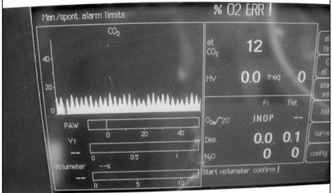
Fig. 2. Capnography monitoring with the proximal lumen of a two-lumen central venous catheter during jet ventilation.
EV jet ventilator (Metran Co., LTD., Saitama, Japan). The proximal lumen was connected to a capnograph. The COMPOS β-EV was set to deliver jet ventilation at a driving pressure of 2.5 bars at a frequency of 120 breaths/min. The upper limit of end-expiratory pressure was set at 0.5 kPa. An inspira-tory:expiratory ratio of 1:2 was used, and the FiO2 was set
at 0.3. The capnography trace was checked intermittently by using three-way stopcock. The capnography trace during jet ventilation is shown in Fig. 2.
A carbon-coated spatula was used by the surgeon for pro-tection of the central venous catheter from the laser beam during the surgery. In 10 minutes of operation time, the vocal polyp was removed, bleeding was controlled, and 1 ml of arterial blood was sampled for gas analysis from the left radial artery.
The SpO2 was continuously maintained at 100% during the
operation. The arterial blood gas analysis was as follows: pH 7.399, PaCO2 40.6 mmHg, PaO2 132.0 mmHg, SaO2 99.9%.
Any complication, including barotrauma, bleeding, or hypoxia, was not seen during the surgery.
At the end of the surgery, jet ventilation was discontinued, and 0.2 mg of glycopyrolate and 10 mg of pyridostigmine were administered intravenously to the patient to reverse the muscle relaxation. The spontaneous respiration of the patient was recovered with ventilatory support via a face mask with 100% O2. The central venous catheter was removed, and the
puncture site upon the cricothyroid membrane was covered with a disposable bandage after sterilization. The patient was transported to the post-anesthesia care unit (PACU).
The patient was discharged from the PACU three hours after surgery and had no postoperative complications.
DISCUSSION
Transtracheal jet ventilation is a useful emergency measure when it is impossible to secure the airway and provide ventilation by conventional means.2,4) It is also indicated in patients undergoing elective surgery in whom conventional or fiberoptic intubation is not feasible.
The use of a central venous catheter for jet ventilation was first described in children in whom the catheters were placed through the larynx.6) Two major complications with trans-tracheal jet ventilation are barotrauma and carbon dioxide accumulation. These complications can be reduced by securing enough gas outlet to prevent sealing of an airway and monitoring PETCO2 to detect carbon dioxide accumulation,
which may be enabled by using a two-lumen central venous catheter for transtracheal jet ventilation.
The central venous catheter is easy to insert into the trachea via the cricothyroid membrane, and the two lumens of the catheter can be checked for patency by aspirating air and tracing a capnogram to ensure that all lumen openings are within the trachea. Additionally, the surgeon can confirm the proper intratracheal position of the catheter tip by a direct bronchoscopy.
A potential problem with this technique is trauma due to movement of the catheter tip with each jet breath. This can be minimized by keeping a short length of the catheter within the trachea and by using the lowest possible driving pressure. Another problem is mucus plugging of the proximal lumen,
Korean J Anesthesiol:Vol. 54. No. 3, 2008
S 42 but this was not found in our practice.
Continuous monitoring of the airway pressure has been advocated to minimize the risk of barotrauma and pneu-mothorax.7) Maintenance of a patent expiratory pathway is crucial in preventing hyperinflation and barotrauma because the only gas outlet should not be sealed. Although the airway pressure could not be monitored in this case due to insufficient preparation of a connecting tube, we believe that our method can ensure more safety in jet ventilation by supplying another gas outlet to prevent barotrauma due to sealing.
The capnograph has previously been used to monitor the efficiency of high-frequency jet ventilation.8-10) Also, monitoring of the airway pressure via the catheter can be continued until the patient is breathing spontaneously. There was no problem in ventilation in our case and the gas exchange profile was adequate with minimal cardiovascular changes.
In this case, the choice of ventilation method was somewhat difficult, since transtracheal HFJV was more invasive than orotracheal HFJV. Therefore, we discussed the method of ventilation during anesthesia with the surgeon. And, we chose transtracheal HFJV because the polyp arose from the posterior one third of the vocal cord, thus an orotracheal intubation or catheter insertion would interfere surgical approach to the lesion site. After completion of the surgery, the surgeon was greatly satisfied with our transtracheal jet ventilation method, which enabled a better surgical condition with less vibration in the surgical area and a good surgical field.
We conclude that transtracheal jet ventilation with a two-lumen central venous catheter is a simple and safe method which enables anesthesiologists to monitor PETCO2 without
interruption of ventilation and gives a better surgical condition
to surgeons.
REFERENCES
1. Gulleth Y, Spiro J: Percutaneous transtracheal jet ventilation in head and neck surgery. Arch Otolaryngol Head Neck Surg 2005; 131: 886-90.
2. Gerig HJ, Schnider T, Heidegger T: Prophylactic percutaneous transtracheal catheterisation in the management of patients with anticipated difficult airways: a case series. Anaesthesia 2005; 60: 801-5.
3. Bourgain JL, Desruennes E, Fischler M, Ravussin P: Transtracheal high frequency jet ventilation for endoscopic airway surgery: a multicentre study. Br J Anaesth 2001; 87: 870-5.
4. Chandradeva K, Palin C, Ghosh SM, Pinches SC: Percutaneous transtracheal jet ventilation as a guide to tracheal intubation in severe upper airway obstruction from supraglottic oedema. Br J Anaesth 2005; 94: 683-6.
5. Miyawaki J, Shono S, Katori K, Sakuragi T, Higa K: Subglottic jet ventilation for pediatric microlaryngosurgery: a case report. J Clin Anesth 2003; 15: 363-5.
6. Dhara SS, Butler PJ: High frequency jet ventilation for microlaryngeal laser surgery. An improved technique. Anaesthesia 1992; 47: 421-4.
7. Bourgain JL, Desruennes E, Cosset MF, Mamelle G, Belaiche S, Truffa-Bachi J: Measurement of end expiratory pressure during transtracheal high frequency jet ventilation for laryngoscopy. Br J Anaesth 1990; 65: 737-43.
8. Gottschalk A, Mirza N, Weinstein GS, Edwards MW: Capnography during jet ventilation for laryngoscopy. Anesth Analg 1997; 85: 155-9.
9. Sehati S, Young JD, Sykes MK, McLeod CN: Monitoring of end-tidal carbon dioxide partial pressure during high frequency jet ventilation. Br J Anaesth 1989; 63(7 Suppl 1): 47-52.
10. Kil HK, Kim WO, Choi HS, Nam YT: Monitoring of PETCO2
during high frequency jet ventilation for laryngomicrosurgery. Yonsei Med J 2002; 43: 20-4.