저작자표시-비영리-변경금지 2.0 대한민국 이용자는 아래의 조건을 따르는 경우에 한하여 자유롭게 l 이 저작물을 복제, 배포, 전송, 전시, 공연 및 방송할 수 있습니다. 다음과 같은 조건을 따라야 합니다: l 귀하는, 이 저작물의 재이용이나 배포의 경우, 이 저작물에 적용된 이용허락조건 을 명확하게 나타내어야 합니다. l 저작권자로부터 별도의 허가를 받으면 이러한 조건들은 적용되지 않습니다. 저작권법에 따른 이용자의 권리는 위의 내용에 의하여 영향을 받지 않습니다. 이것은 이용허락규약(Legal Code)을 이해하기 쉽게 요약한 것입니다. Disclaimer 저작자표시. 귀하는 원저작자를 표시하여야 합니다. 비영리. 귀하는 이 저작물을 영리 목적으로 이용할 수 없습니다. 변경금지. 귀하는 이 저작물을 개작, 변형 또는 가공할 수 없습니다.
Master’s Thesis of Science
in Food Science and Biotechnology
Structure of flavoprotein RclA from
food-borne pathogens, and its
molecular mechanism contributing to
hypochlorous acid resistance
식품 병원균 유래 플라빈단백질 RclA의 구조와
차아염소산 저항성에 기여하는 분자 기전
February 2020
Food Science and Biotechnology Major
Department of Agricultural Biotechnology
The Graduate School
Seoul National University
석사학위논문
Structure of flavoprotein RclA from
food-borne pathogens, and its molecular
mechanism contributing to hypochlorous
acid resistance
지도교수 하 남 출
이 논문을 석사학위논문으로 제출함
2020년 2월
서울대학교 대학원
농생명공학부 식품생명공학전공
백 영 진
백영진의 석사학위논문을 인준함
2020년 2월
위원장
최 영 진 (인)
부위원장
하 남 출 (인)
위원
유 상 열 (인)
i
Abstract
Hypochlorous acid (HOCl) is a powerful sanitizer and has been used to control food-borne pathogens in various food sanitizing industries. The immune system generates HOCl to kill microorganisms via oxidative burst, and the toxicity of HOCl is amplified in the phagosome by importing Cu2+. In Escherichia coli and Salmonella, the transcriptional regulator RclR recognizes HOCl stress and induces the RclA, B, and C proteins to defend against stress. However, the structure and biochemical roles of Rcl proteins remain to be elucidated. In this study, the role of the flavoprotein disulfide reductase (FDR) RclA in the survival of Salmonella in macrophage phagosomes was examined. To understand the molecular mechanism of RclA, the crystal structures of RclA from E. coli were determined. The homodimeric RclA was similar to typical FDRs, exhibiting two conserved cysteine residues near the flavin ring of the cofactor flavin adenine dinucleotide (FAD). Cu2+ accelerated the oxidation of NADH by RclA, leading to a lowering of the oxygen level in the solution. Cu2+ itself was reported to be reduced to become Cu+ during the NADH oxidation by RclA. Mutation of the conserved cysteine residues lowered the specificity to Cu2+ or substantially increased the production of superoxide anion in the absence of Cu2+ compared to the RclA wild type. The lowered oxygen level by RclA would contribute to the inhibition of oxidative bursts in phagosomes. The reduced Cu+ can be
exported by Cu+-specific efflux pumps and would inhibit the toxic radical production by HOCl-Cu2+ reaction. This study provides the molecular basis of bacterial survival in HOCl stress from our immune systems. The HOCl resistance mechanism of RclA further gives an insight into controlling food-borne pathogens with HOCl-based sanitizers. Mimicking the phagosome environment by the combined treatment of HOCl with some inhibitors targeting RclA can lower the dose of HOCl treatment with the increased bactericidal effect.
Keywords: hypochlorous acid, flavoprotein disulfide reductase, copper, oxygen, crystal structure
iii
Contents
Abstract ... i
Contents ... iii
List of Figures ... v
List of table ... vii
I. Introduction ... 1
II. Experimental Procedures ... 4
2.1. Plasmid construction ... 4
2.2. Site directed mutagenesis ... 4
2.3. Purification of RclA wild type and mutants ... 5
2.3.1. Overexpression ... 5
2.3.2. Affinity chromatography ... 6
2.3.3. Ion-exchange chromatography ... 6
2.3.4. Size exclusion chromatography (SEC) and concentration ... 6
2.4. Crystallization of RclA wild type and C43S ... 7
2.5. Data collection and structural determination ... 8
2.6. Enzyme kinetic assays ... 9
2.7. NADH oxidation assay under low O2 conditions ... 10
2.8. Detection of DO in water ... 10
III. Results ... 12
3.1. Overexpression and Purification of RclA wild type ... 12
3.2. Crystallization, structural determination and overall structure of EcRclA wild type ... 18
3.3. Structural comparison to group I FDRs ... 23
3.4. Structural comparison to the group II FDR MerA ... 26
3.5. Cu2+ and Hg2+ accelerate the oxidation of NADH in RclA ... 29
3.6. O2 is required for NADH oxidation in the presence of Cu2+ ... 32
3.7. Cu2+ promotes O 2 consumption without producing superoxide anion ... 35
3.8. Overexpression and purification of RclA C43S and C48S ... 36
3.9. Roles of the two cysteine residues in the CXXXXC motif ... 39
3.10. Structural determination of RclA wild type and RclA C43S in complex with Cu2+ ... 45
3.11. Multiple binding of Cu2+ to RclA ... 47
3.12. Cu2+ binding at the active site ... 50
IV. Discussions ... 54
V. References ... 61
v
List of Figures
Figure 1. Sequence analyses and crystal structure of EcRclA ... 13 Figure 2. Full sequence alignment of EcRclA and representative FDR
proteins with the secondary structure elements... 14 Figure 3. Purification profile of RclA wild type. ... 17 Figure 4. Crystals and X-ray diffraction image of RclA wild type. ... 19 Figure 5. The structure of the asymmetric unit of RclA in the ribbon
representations. ... 21 Figure 6. The overall structure of EcRclA. ... 22 Figure 7. The structure of the active site of EcRclA. ... 24 Figure 8. Structural superposition of EcRclA with the group I FDR proteins.
... 25 Figure 9. Overall superposition between RclA and MerA, which are in
dimeric forms. ... 27 Figure 10. Active site comparison with MerA. ... 28 Figure 11. The NADH oxidation rates by EcRclA in the presence of various
substrates ... 30 Figure 12. O2 depletion and consumption during NADH oxidation by
EcRclA ... 34 Figure 13. SDS-PAGE purification profile of RclA C43S. ... 37 Figure 14. SDS-PAGE purification profile of RclA C48S. ... 38
Figure 15. The location and the surrounding environment of Cys396 of EcRclA. ... 41 Figure 16. The activity of EcRclA mutants C43S and C48S compared to
wild type ... 42 Figure 17. Superoxide anion production during NADH oxidation by RclA
wild type and mutants in the presence of Cu2+. ... 44 Figure 18. Crystals and X-ray diffraction data of RclA wild type and RclA
C43S in complex with Cu2+. ... 46 Figure 19. The overall structure of EcRclA wild type in complex with Cu2+
... 48 Figure 20. Overall structure comparison of EcRclA wild type with the
EcRclA wild type-Cu2+ complex. ... 49 Figure 21. The extra electron density maps at the active site in Cu2+-soaked
structures. ... 52 Figure 22. A proposed reaction order of O2 and Cu2+ reduction by EcRclA.
... 59 Figure 23. The proposed biological role of RclA in the bacterial cell ... 60
vii
List of Table
I. Introduction
Hypochlorous acid (HOCl) is a well-known antibacterial agent, and it has been commonly used as a sanitizer in food industries to control food-borne pathogens (1-3). HOCl is also used in the innate immune systems of higher eukaryotes (4-6). To kill invading bacteria, macrophages and neutrophils in the immune system employ myeloperoxidase to generate HOCl from H2O2, which is produced by the oxidative burst from molecular oxygen by NADPH oxidase (7). HOCl directly oxidizes amino acids containing amines and sulfurs and can cleave DNA once it is converted into a deadly toxic hydroxyl radical in the presence of iron or copper ions via Fenton(-like) reaction (8-11). HypT, RclR, and NemR are HOCl-responsive transcriptional factors in bacteria that increase bacterial survival under HOCl stress (12-14). HypT, belonging to the LysR-type transcriptional regulators, employs a HOCl-sensitive methionine residue to repress genes related to the import of iron to the cytosol in Escherichia coli and Salmonella enterica serovar Typhimurium. HypT plays a critical role in the survival of Salmonella in macrophages (15). NemR of E. coli is a redox-regulated transcriptional repressor that utilizes the oxidation status of HOCl-sensitive cysteine residues. NemR expresses two enzymes, GloA and NemA, to detoxify methylglyoxal and reactive electrophiles (14).
2
and senses HOCl with highly conserved cysteine residues (12,16). RclA, RclB, and RclC proteins were identified as RclR target genes in both E. coli and Salmonella. In E. coli, RclR, RclA, RclB, and RclC contribute to the HOCl-resistance of the bacterium. Deletion of any one of these genes decreases the survival of E. coli under HOCl stress (12). RclA putatively belongs to the flavoprotein disulfide reductase (FDR) family. RclB is a small uncharacterized periplasmic protein that has the signal sequence for secretion. RclC is a multipass inner membrane protein with homology to quinone-binding proteins. However, little is known about the biochemical functions of RclA, RclB, and RclC.
A recent study suggested that the FDR RclA is widely conserved among bacteria that colonize epithelial surfaces, and contributes to host colonization in environments with HOCl-mediated oxidative stress (17). FDRs usually form homodimers and use non-covalently bound flavin adenine dinucleotide (FAD) and a non-flavin redox center, catalyzing NAD(P)H-dependent reduction of a variety of substrates, such as sulfur-containing peptides, metal ions, H2O2, and O2 (18). Like other FAD-dependent reductases, the reducing power of the bound FAD is supplied from the cytosolic NAD(P)H pool. FDRs can be divided into three subgroups depending on the non-flavin redox centers (18). Very recently, Derke et al. reported that RclA from E. coli exhibits Cu2+ reductase activity, reducing Cu2+ to Cu+ to cope with HOCl stress (17). Here, the crystal structures of RclA
from E. coli were determined, containing FAD at its active site. Based on the structures and the subsequent biochemical analyses, the functions of RclA in terms of HOCl resistance in bacteria were investigated.
The HOCl resistance mechanism of RclA gave insights into controlling food-borne pathogens with HOCl-based sanitizers in food processing. Inhibiting RclA of food-borne pathogens This work presents the combination method of HOCl with inhibitor inactivating RclA, to decrease the dose of HOCl and amplifying the bactericidal effect.
4
II. Experimental Procedures
2.1. Plasmid construction
To overexpress the full-length (1–441) RclA wild type, the gene encoding the protein was amplified by polymerase chain reaction (PCR) using the following two primers. The underlined sequences represent the EcoRI and HindIII restriction sites, respectively.
(Forward: 5’-GGCGAATTCATGAATAAATATCAGGCAG-3’) (Reverse: 5’-GCCAAGCTTTTATTTGACTAATGAAAATAG-3’)
The amplified product and the expression vector pProEX-HTa (Invitrogen, USA) were digested with EcoRI and HindIII restriction enzymes. The digested PCR product was inserted into the digested expression vector pProEX-HTa by DNA ligase. The resulting plasmid encodes the hexahistidine tag and the TEV protease cleavage site at the N-terminus of the protein. The plasmids encoding RclA wild type protein were transformed into the
Escherichia coli C43 (DE3) strain by the heat-shock method. The
recombinant plasmid was confirmed by DNA sequencing.
2.2. Site-directed mutagenesis
To produce the RclA C43S and RclA C48S, a single mutation of cysteine to serine was performed using the Quick Change XL Site-directed Mutagenesis
Kit. The complementary mutagenic primers were designed using the QuikChange® Primer Design Program (19) and used to PCR reactions. After amplification, the PCR mixture was incubated with DpnI at 37℃ for 2 h. For overexpression of the recombinant DNA, each plasmid encoding RclA mutant was transformed into E. coli BL21 (DE3) competent cell by the heat-shock method. The sequence of the mutant plasmid was confirmed by DNA sequencing.
2.3. Purification of RclA wild type and mutants
2.3.1. Overexpression
For expression of the recombinant proteins, a single colony of E. coli C43 (DE3) and E. coli BL21 (DE3) was inoculated into 25 mL LB broth including 100 µg/mL ampicillin (Duchefa, Netherlands) and incubated at 37℃ for 14 h. Subsequently, the cells were cultured in 1.5 L Luria Broth medium containing 100 µg/mL ampicillin at 37°C until an OD600 of 0.7 was measured, and the protein expression was induced with 0.5 mM isopropyl-β-D-thiogalactopyranoside (IPTG) at 30°C for 6 h. The cells were harvested by centrifugation at 3850 g for 7 min. After harvest, cells were resuspended in lysis buffer containing 20 mM Tris-HCl (pH 8.0), 150 mM NaCl, and 2 mM 2-mercaptoethanol. The suspensions were disrupted using a continuous-type French press (Constant Systems Limited, United Kingdom) at 23 kpsi. The
6
crude lysate was centrifugated at 19,000 g for 30 min at 4°C and the pellet containing cell debris was discarded.
2.3.2. Affinity chromatography
The supernatant was loaded onto Ni2+-NTA agarose resin (GE Healthcare) and rolled in a column for 45 min at 4°C. After washing the column with ~300 mL lysis buffer supplemented with 20 mM imidazole, the RclA wild-type protein was eluted with lysis buffer supplemented with 250 mM imidazole. The eluate was treated with recombinant TEV protease by incubation at room temperature for 12 h to cleave the hexahistidine tag.
2.3.3. Ion-exchange chromatography
The cleaved proteins were diluted four-fold with 20 mM Tris-HCl (pH 8.0) buffer containing 2 mM 2-mercaptoethanol and loaded onto a HiTrap Q column (GE Healthcare, USA). A linear gradient of increasing NaCl concentration was applied to the column. The eluted fractions containing the protein were pooled and concentrated.
2.3.4. Size exclusion chromatography (SEC) and concentration The concentrated proteins were subjected to size-exclusion chromatography using a HiLoad 26/600 Superdex 200 pg column (GE Healthcare) equilibrated with 20 mM Tris-HCl (pH 8.0) buffer containing 150 mM NaCl and
2-mercaptoethanol. The purified protein was concentrated up to 13 mg/mL using Vivaspin 20 (Sartorius, Germany) and stored at –80°C. Protein concentrations were determined based on FAD content using a previously determined ε456 of 11.3 mM–1 cm–1.
2.4. Crystallization of RclA wild type and C43S
RclA proteins (wild type, C43S) were crystallized at 16°C using the hanging drop vapor diffusion method after mixing 1 µL protein solution and 1 µL precipitation solution. The RclA wild-type protein was first crystallized in a precipitation solution containing 0.2 M ammonium sulfate, 0.1 M Bis-Tris HCl (pH 5.8), 25% (w/v) PEG 3350, and 2 mM TCEP. Subsequently, the crystals were disrupted for microseeding and diluted with precipitation solution containing 0.2 M ammonium sulfate, 0.1 M Bis-Tris HCl (pH 5.8), 13% (w/v) PEG 3350, and 2 mM TCEP. Microseeding was performed by mixing 1 µL protein and 1 µL microseeds diluted with precipitation solution. The RclA wild-type crystals were briefly incubated in precipitation solution supplemented with 20% (v/v) glycerol as a cryoprotectant, and flash-frozen in liquid ethane at –88.5°C, and subsequently transferred into liquid nitrogen to collect the dataset. For the Cu2+-soaked RclA wild-type structure, the crystals were incubated in a reservoir solution supplemented with 5 mM CuCl2 for 15 h. The crystals were incubated in precipitation solution supplemented with 30% (v/v) glycerol and 5 mM CuCl2 for 6 min, and
flash-8
frozen in liquid nitrogen to collect the dataset. RclA C43S was crystallized in a solution containing 0.05 mM ammonium sulfate, 0.05 mM Bis-Tris HCl (pH 5.8), and 30% pentaerythritol ethoxylate (15/4_EO/OH). Microseeding was performed using disrupted RclA C43S crystals, and was diluted with 0.05 mM ammonium sulfate, 0.05 mM Bis-Tris HCl (pH 5.8), 25% pentaerythritol ethoxylate (15/4_EO/OH). For Cu2+-soaked RclA C43S structure, the RclA C43S crystals were incubated in a reservoir solution supplemented with 5 mM CuCl2 for 15 h. After incubation of the crystals in reservoir solution supplemented with 30% (v/v) glycerol and 5 mM CuCl2 for 30 s, crystals were flash-frozen in a nitrogen stream at –173°C.
2.5. Data collection and structural determination
All diffraction datasets were collected at the Pohang Accelerator Laboratory Beamline 5C (20) and were processed using the HKL-2000 package (21,22). The structure of the RclA wild type was determined using the molecular replacement method with MOLREP in the CCP4 package (22), using a model built by the Galaxy web server (23,24) as a search model. The initial model of the RclA wild type was used to solve structures of RclA wild type in complex with Cu2+ and RclA (C43S) in complex with Cu2+ by the molecular replacement method. All of the structures were built using COOT and refined using PHENIX refine software (25,26). The detailed statistics are shown in Table 1.
2.6. Enzyme kinetic assays
All assays were performed using UV-Vis spectrophotometer, GeneQuant 1300 (GE Healthcare) or Thermo Scientific Multiscan Go (Thermo Fisher Scientific). The RclA activity was assayed by adding 2 µM protein (as measured based on flavin content) in 10 mM MES buffer (pH 7.0) containing 100 mM NaCl, 0.2 mM NADH, and 0.2 mM putative substrate. Initial rates of NADH oxidation by RclA with various metal ions were monitored by the loss of absorbance at 340 nm due to NADH oxidation (∆ε340 = 6.2 mM–1 cm– 1); loss of absorbance was monitored for 10 min and the oxidation profile was expressed in a time-dependent manner as the mean ± standard deviation (SD) of three independent experiments. Control assays lacking the substrate or enzyme were routinely included as references. NADH oxidation by RclA mutants C43S and C48S were recorded as previously described for the RclA wild type assay.
For the examination of enzyme dependency on Cu2+ concentration, NADH oxidation was measured at 340 nm with various concentrations of CuCl2. Before the assay, the RclA wild-type protein was desalted with 10 mM MES buffer (pH 7.0) supplemented with 100 mM NaCl to remove any remaining 2-mercaptoethanol from the RclA size exclusion chromatography buffer. The activity assays were initiated by adding 2 µM desalted RclA wild-type protein to 10 mM MES buffer (pH 7.0) containing 100 mM NaCl, 0.2 mM NADH, and 0, 0.025, 0.05, 0.075, 0.1, 0.2, or 0.3 mM CuCl2. Initial rates
10
were monitored by the loss of absorbance at 340 nm due to NADH oxidation (∆ε340 = 6.2 mM–1 cm–1) and expressed as the mean ± SD of four independent experiments.
2.7. NADH oxidation assay under low O
2conditions
The dissolved oxygen (DO) was removed by nitrogen purging. The buffer with 10 mM MES (pH 7.0), 100 mM NaCl, 0.2 mM NADH, and with or without 0.1 mM CuCl2 was contained in 1 mL cuvette and sealed with parafilm. The solution was purged with N2 for 5 min. After nitrogen purging, 2 µM of RclA was added. Initial rates of NADH oxidation by RclA were monitored by the loss of absorbance at 340 nm due to NADH oxidation (∆ε340 = 6.2 mM–1 cm–1) was monitored using a GeneQuant 1300 UV-Vis spectrophotometer (GE Healthcare) and was expressed in time-dependent manner as the mean ± standard deviation (SD) of three independent experiments. A control assay in which the solution was not purged with N2 was included for comparison.
2.8. Detection of DO in water
DO concentrations were measured using the Gravity Analog Dissolved Oxygen Sensor (SKU:SEN0237; DFRobot) with an Arduino Nano board and the Arduino IDE program (27). The programming code was obtained from the DFRobot website (https://wiki.dfrobot.com/Gravity_Analog_Dissolved_
Oxygen_Sensor_SKU_SEN0237). The reaction volume was 5 mL, containing 10 mM MES (pH 7.0) buffer with 100 mM NaCl, 0.2 mM NADH, and 1 μM RclA, with or without 0.2 mM CuCl2. The solution was stirred gently with a 1 cm magnetic stir bar to distribute DO evenly throughout the entire solution. After pre-incubation for 5 min with stirring for equilibrium, the RclA protein was added and the DO concentration was recorded for 10 min. The specific oxidation rate was measured within the initial 1 min and is expressed as the mean ± SD of three independent experiments.
2.9. Measurement of O
2•‾ formation by cytochrome C assay
Superoxide anion (O2•‾) formation was determined by monitoring the reduction of cytochrome C, which was isolated from the equine heart (Sigma Aldrich). The reaction was performed with 200 µL buffer containing 10 mM MES buffer (pH 7.0), 100 mM NaCl, 200 µM NADH, 100 µM CuCl2, 2 µM RclA, and 50 µM cytochrome C. The increase in the absorbance at 550 nm due to cytochrome C reduction was recorded at 37°C for 10 min using a UV-Vis spectrophotometer (Thermo Scientific Multiscan Go). The amount of O2•‾ released was calculated using an extinction coefficient of 21 mM–1 cm–1 and expressed as the mean ± SD of three independent experiments.12
III. Results
3.1. Overexpression and Purification of RclA wild type
RclA is commonly found in Salmonella and E. coli. Since HOCl is produced in macrophage phagosomes as a major bactericidal agent, the function of RclA in resistance to HOCl suggests that RclA plays a role in the survival of the bacteria. Previous unpublished data from the Molecular Food Microbiology Lab of Seoul National University showed that RclA is involved in the pathogenesis of Salmonella by promoting survival in macrophage vacuoles containing sublethal doses of HOCl.
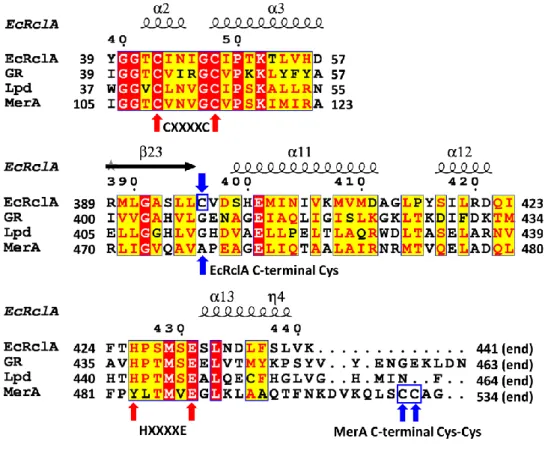
Sequence comparison suggested that RclA belongs to the group I FDRs due to the presence of two important motifs; CXXXXC for the redox-active disulfide as a non-flavin redox center, and the essential HXXXXE which interacts with and promotes the disulfide formation of the CXXXXC motif with its histidine residue (18) (Fig. 1 and Fig. 2).
To gain mechanistic insights on RclA from its 3D structures, the full-length RclA proteins (441 amino acids) of E. coli was overexpressed and purified through affinity chromatography, anion exchange chromatography, and size exclusion chromatography. (Fig. 3) Each step was monitored by SDS-PAGE.
Figure 1. Sequence analyses and crystal structure of EcRclA
Sequence alignment of FDR proteins with the secondary structure elements. For clarity, the regions containing the signature motifs are displayed. The full sequence alignment is presented in Figure 2. The cysteine residues in the CXXXXC motifs and histidine and glutamate residues in the HXXXXE motif are indicated by red arrows. The C-terminal Cys in EcRclA and the C-terminal Cys-Cys motif in MerA are indicated by blue arrows and blue square.
14
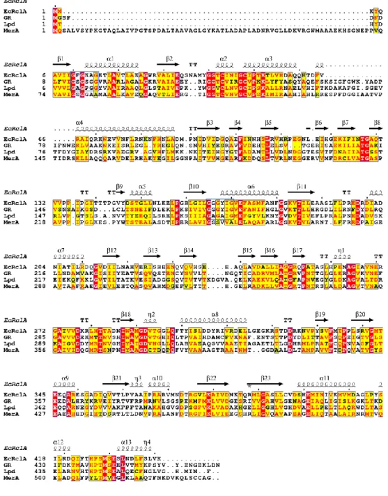
Figure 2. Full sequence alignment of EcRclA and representative FDR proteins with the secondary structure elements.
GR; glutathione-disulfide reductase from Bartonella henselae, (GenBank: WP_011180567), Lpd; dihydrolipoyl dehydrogenase from mycobacterium multispecies (GenBank: WP_003402301.1), MerA; mercuric reductase from
Pseudomonas aeruginosa (GenBank: WP_023980594.1). The sequence
alignment was performed using the T-Coffee server (28) and the ESPript server (29).
Figure 3. Purification profile of RclA wild type.
A. The SDS-PAGE analysis of Ni-NTA affinity chromatography B. The SDS-PAGE analysis of His-tag cleavage using TEV protease.
C. UV and conductivity diagram of the Q-column elution during the anion exchange chromatography.
D. The SDS-PAGE analysis of anion exchange chromatography
E. UV diagram of the superdex column elution during the size exclusion chromatography.
18
3.2. Crystallization, structural determination and overall
structure of EcRclA wild type
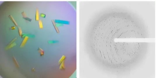
The purified RclA wild type was concentrated up to 13 mg/mL for the crystallization. The crystals of RclA wild type were obtained in the precipitant solution containing 0.2 M ammonium sulfate, 0.1 M Bis-Tris HCl (pH 6.5), 25% (w/v) PEG 3350. Optimizing the precipitant solution to 0.2 M ammonium sulfate, 0.1 M Bis-Tris HCl (pH 5.8), 13% (w/v) PEG 3350, and 2 mM TCEP improved the quality of RclA crystals (Fig. 4).
The crystal structure of RclA from E. coli (EcRclA) was successfully determined at 2.9 Å resolution (Fig. 4 and Table 1). The crystal of RclA belongs to the space group P21, with unit cell dimensions of a=72.4 Å,
b=189.4 Å, and c=95.4 Å. The asymmetric unit contained four protomers
consisting of two homodimers (Fig. 5). Each protomer contained a tightly bound FAD molecule in its active site (Fig. 6).
Figure 4. Crystals (left) and X-ray diffraction image (right) of RclA wild type.
20
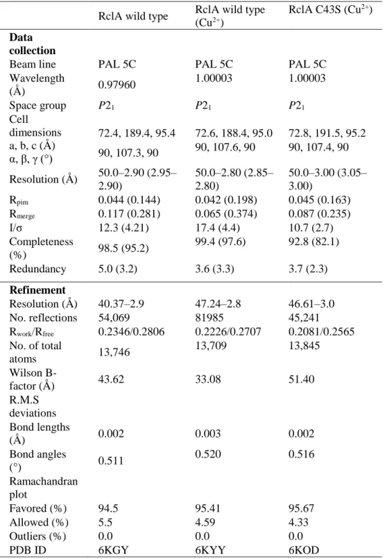
Table 1. Data collection and refinement statistics
RclA wild type RclA wild type
(Cu2+)
RclA C43S (Cu2+)
Data collection
Beam line PAL 5C PAL 5C PAL 5C
Wavelength (Å ) 0.97960 1.00003 1.00003 Space group P21 P21 P21 Cell dimensions a, b, c (Å ) α, β, γ (°) 72.4, 189.4, 95.4 72.6, 188.4, 95.0 72.8, 191.5, 95.2 90, 107.3, 90 90, 107.6, 90 90, 107.4, 90 Resolution (Å ) 50.0–2.90 (2.95– 2.90) 50.0–2.80 (2.85– 2.80) 50.0–3.00 (3.05– 3.00) Rpim 0.044 (0.144) 0.042 (0.198) 0.045 (0.163) Rmerge 0.117 (0.281) 0.065 (0.374) 0.087 (0.235) I/σ 12.3 (4.21) 17.4 (4.4) 10.7 (2.7) Completeness (%) 98.5 (95.2) 99.4 (97.6) 92.8 (82.1) Redundancy 5.0 (3.2) 3.6 (3.3) 3.7 (2.3) Refinement Resolution (Å ) 40.37–2.9 47.24–2.8 46.61–3.0 No. reflections 54,069 81985 45,241 Rwork/Rfree 0.2346/0.2806 0.2226/0.2707 0.2081/0.2565 No. of total atoms 13,746 13,709 13,845 Wilson B-factor (Å ) 43.62 33.08 51.40 R.M.S deviations Bond lengths (Å ) 0.002 0.003 0.002 Bond angles (°) 0.511 0.520 0.516 Ramachandran plot Favored (%) 94.5 95.41 95.67 Allowed (%) 5.5 4.59 4.33 Outliers (%) 0.0 0.0 0.0
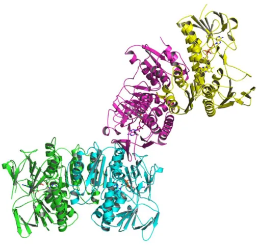
Figure 5. The structure of the asymmetric unit of RclA in the ribbon representations.
The asymmetric unit consists of four protomers, which are colored differently (green, cyan, magenta, and yellow). The bound FAD molecules are shown in the stick representations.
22
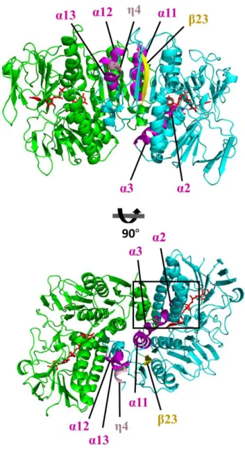
Figure 6. The overall structure of EcRclA.
The subunits are depicted by the ribbon representations in cyan (subunit A) or green (subunit B). Secondary structure elements shown in Figure 2A are indicated with a different color in subunit A. α2, α3, α12, α13 are in magenta. β23 is in yellow, and η4 is in pink. The bound FAD molecules are shown in the red stick representations. The boxed region is enlarged in Figure 7.
3.3. Structural comparison to group I FDRs
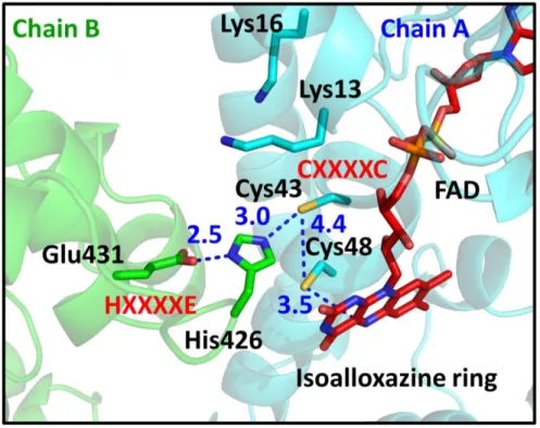
The overall arrangement of the CXXXXC and HXXXXE motifs in the active sites is similar to that of group I FDR family proteins (Fig. 7 and Fig. 8). The two cysteines in the CXXXXC motif are in the reduced form and the distance between the sulfur atoms of the two cysteine residues is ~4.4 Å in the crystal structure. The two cysteine residues are near the isoalloxazine ring of FAD, and Cys48 is closer to FAD than to Cys43 (the distance between the Sγ of Cys48 and the C4a atom of flavin is 3.5 Å). The HXXXXE motif from the other subunit in the dimer is involved in the interaction with the CXXXXC motif and the bound FAD (Fig. 8). His426 and Glu431 are paired with a distance of 2.5 Å, and His426 makes a polar interaction with Cys43 (Fig. 7). Remarkably, two lysine residues (Lys13 and Lys16), which are unique to RclA proteins, were found near Cys43 at the active site. Because group I FDR proteins reduce substrates via the disulfide formation of the CXXXXC motif together with the HXXXXE motif, our findings suggest that RclA shares the enzymatic mechanism of group I FDR proteins.
24
Figure 7. The structure of the active site of EcRclA.
The active site of RclA. The box is enlarged from Figure 6. Each subunit is depicted by the ribbon representation in green or cyan. The bound FAD molecules are shown in the red stick representations. Each residue and FAD are labeled and indicated in the stick representations with the ribbon diagram in the background. The key interactions between the two cysteines of CXXXXC, His426, and Glu431 of HXXXXE, and Cys48 and the C4a atom of FAD are shown in blue dotted lines. Distances are in Å (Blue).
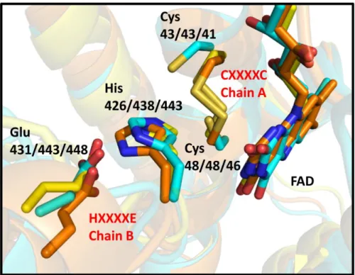
Figure 8. Structural superposition of EcRclA (cyan) with the group I FDR proteins.
Glutathione reductase (orange, PDB ID: 3O0H) and lipoamide dehydrogenase (yellow, PDB ID: 4M52) were ranked as top entries from the DALI server structural analyses, following MerA. The conserved CXXXXC and HXXXXE motifs are labeled in red. Each residue depicted in stick representation is labeled with residue numbers of three proteins (first RclA, second 3O0H, third 4M52).
26
3.4. Structural comparison to the group II FDR MerA
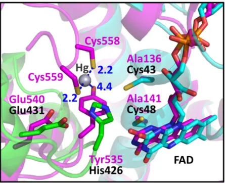
I searched for the closest protein to RclA in terms of structure using the DALI server (30). Mercuric reductase (MerA) (PDB code: 4K7Z), a group II FDR, was ranked as the top entry (Z-score: 43.6). Like EcRclA reduces Cu2+ to Cu+, MerA converts Hg2+ to Hg0 (18). MerA contains the CXXXXC motif, similar to group I FDR proteins, and tyrosine replaces the histidine residue in the HXXXXE motif (Fig. 1). The structural superposition of EcRclA onto MerA revealed a strong structural similarity between the matched regions (sequence identity: 30%, RMSD: 1.522 between 320 Cα atoms, residues 5–433 of EcRclA), except for the Cys-Cys motif for metal ion binding (Fig. 9 and Fig. 10). The C-terminal end loop containing the Cys-Cys motif was folded into the active site near the CXXXXC motif and the tyrosine residue in the MerA structure (Figs. 1 and 10). The Cys-Cys motif and the tyrosine residue interact with a mercuric ion at the active site (31).
Figure 9. Overall superposition between RclA (green and cyan) and MerA (magenta, PDB ID: 4K7Z), which are in dimeric forms.
The boxed region is enlarged in Figure 10. Sequence identity: 30%, RMSD: 1.522 between 320 Cα atoms, residues 5–433 of EcRclA
28
Figure 10. Active site comparison with MerA.
The box is enlarged from Figure 9. RclA (green, cyan) is aligned with MerA (magenta, PDB ID:4K7Z) (32). Each residue aligned in the active site is labeled in a different color (black for RclA and magenta for MerA). His426 in RclA is substituted with Tyr535 in MerA. Mercury was bound to the two C-terminal cysteines of MerA, Cys558, and Cys559. The interactions between the mercury, cysteine, and histidine residues are depicted in dashed lines. The two cysteine residues in the CXXXXC motif of MerA were substituted with alanine to determine the Hg2+-bound structure (32). Distances are in Å (Blue).
3.5. Cu
2+and Hg
2+accelerate the oxidation of NADH in RclA
To elucidate the mechanism of RclA, further biochemical analyses were performed. Initially, the basal level of the NADH oxidase activity of RclA was observed (Fig. 11A and B), which is found in many FDR enzymes (33-35). Previous studies of other FDRs suggested that the basal activity of RclA resulted from oxidation by the molecular oxygen in the buffer (33,35-37). Next, RclA was tested whether it exhibits the MerA-like activity in which NADH oxidase activity is increased by adding metal ion (38,39). The NADH oxidase activity of RclA was measured in the presence of diverse metal ions, such as Hg2+, Cu2+, Fe3+, Mn2+, Mg2+, and Zn2+ (Fig. 11A). Notably, Hg2+ and Cu2+ increased oxidation of NADH by approximately three-fold compared to the basal activity without the metal ions, and Ag+ resulted in an approximately two-fold increase. However, Fe2+, Mg2+, Zn2+, and Mn2+ did not affect the NADH oxidase activity. Cu2+ accelerated the NADH oxidation rate in a dose-dependent manner until reaching 0.3 mM CuCl2 (Fig. 11B and C).To identify other substrates, the NADH oxidation activity was tested with oxidized glutathione (GSSG), lipoic acid, cystine, and HOCl, which could be putative substrates of RclA. No increase was observed in the NADH oxidation rate by RclA with these substances (Fig. 11D). Cu2+ was used in the following analyses because Hg2+ and Ag+ are not relevant in host immune responses and Cu2+ is employed in phagosomes of host innate immune systems.
30
Figure 11. The NADH oxidation rates by EcRclA in the presence of various substrates
A. NADH oxidation rates depending on various transition metal ions. NADH oxidation rates by RclA were measured over time spectrophotometrically (n = 3, mean ± SD). The reaction was initiated by injecting 200 µM NADH into a buffer containing 2 µM RclA with or without 100 µM of metal ions. A control experiment was performed without metal ion. The horizontal line indicates the reference NADH oxidation rate of control.
B. NADH oxidation profile with different concentrations of Cu2+. The reaction was assayed with 200 µM NADH and marked concentrations of Cu -Cl2 (from 0 mM to 0.3 mM). The amounts of the NADH oxidation were measured spectrophotometrically (n = 3, mean ± SD).
C. Initial NADH oxidation rates calculated from the NADH oxidation profile in Figure 4B. The reaction was assayed with 200 µM NADH and marked concentrations of Cu2+. The NADH oxidation rates were measured over time spectrophotometrically (n = 3, mean ± SD).
D. NADH oxidation rate with various substances. The reaction was initiated by injecting 200 µM NADH into a buffer containing 2 µM RclA and 200 µM of various materials. A control experiment was performed without material. NADH oxidation rates were measured over time spectrophotometrically (n = 3, mean ± SD)
32
3.6. O
2is required for NADH oxidation in the presence of Cu
2+ Although Derke et al. reported the reduction of Cu2+ with the reducing power of NADH (17), the redox stoichiometry was not completely accounted for. Only 25% of the reducing power from NADH was used in the Cu2+ reduction to produce Cu+ (Fig. 4B in (17)). Given that molecular oxygen dissolved in the reaction buffer is the oxidant candidate, molecular oxygen was investigated as the second oxidant in the RclA-mediated reaction.Because a significant amount of O2 is dissolved in ambient conditions, the NADH oxidase activity of RclA was investigated under oxygen-depleted conditions by thoroughly purging the buffer with nitrogen gas (Fig. 12A). The NADH oxidation rate of EcRclA was compared in the presence and absence of Cu2+. The depletion of dissolved oxygen almost completely shut down the NADH oxidation by RclA regardless of the presence of Cu2+. Stoichiometric analyses of this reaction accounted for 50% of the NADH oxidation in the presence of Cu2+. Thus, our findings suggest that the presence of Cu2+ and molecular oxygen are interlinked in the oxidation of NADH by RclA, and O2 is required for Cu2+-mediated NADH oxidation by RclA.
34
Figure 12. O2 depletion and consumption during NADH oxidation by
EcRclA
A. NADH oxidation by RclA in O2-depleted conditions. The oxidation rates were measured with 200 µM NADH, 2 µM EcRclA wild type, and with or without 100 µM CuCl2 over time spectrophotometrically (n = 3, mean ± SD). O2 depletion was performed via N2 purging.
B. O2 consumption during the NADH oxidation reaction by RclA. Left, O2 consumption profile during the NADH oxidation reaction. The reaction was initiated by the addition of 200 µM NADH into the pre-incubated reaction solution containing 200 µM Cu2+ and 2 µM RclA. The O
2 concentrations in the buffer were measured over time with the Arduino dissolved oxygen sensor. Data are the averages of three independent experiments. Right, O2 consumption rates during the NADH oxidation reaction with RclA and Cu2+. The O2 consumption rates were measured over time (n = 3, mean ± SD). C. Superoxide anion production during NADH oxidation by EcRclA wild type. Superoxide anion production was measured based on the amount of reduced cytochrome c at 550 nm (line), and NADH oxidation was measured at 340 nm (dotted line) over time spectrophotometrically. The reaction was performed in the presence or absence of 100 µM Cu2+ in buffer containing 200 µM NADH and 2 µM EcRclA wild type. Data are the averages of three independent experiments.
3.7. Cu
2+promotes O
2
consumption without producing
superoxide anion
To determine whether O2 was used as the oxidant during the RclA reaction, the amount of oxygen dissolved in the reaction buffer was measured with an oxygen probe during oxidation of NADH by RclA (Fig. 12B). The specific O2 consumption rate in the presence of 200 µM Cu2+ was about four times faster than that in the absence of Cu2+ (Fig. 12B), as observed in the NADH oxidation rates in the presence and absence of Cu2+ (Fig. 11C). These results indicate that Cu2+ catalyzes the reduction of oxygen during the oxidation of NADH. About 50 µM oxygen was consumed by the time 200 µM NADH was used completely in the reaction (Fig. 12B and 11B), indicating that four NADH molecules were required for one oxygen molecule in this reaction of RclA.
Many FDR enzymes produce superoxide anion as the basal or side activity by a reaction between the bound FADH2 and molecular oxygen (36). Therefore, the amount of superoxide anion produced in the RclA-mediated reaction without Cu2+ was measured using cytochrome C, which is rapidly reduced by superoxide anion with an increase in light absorbance at 550 nm. Like other FDR enzymes, EcRclA without Cu2+ produced superoxide anion (Fig. 12C). Similar experiments were performed in the presence of Cu2+. Production of superoxide anion was not observed in the reaction although more oxygen was consumed that in the absence of Cu2+ (Fig. 12C). These
36
findings indicate that Cu2+ promotes the reaction between oxygen and FADH 2 at the active site of RclA while suppressing the production of superoxide anion.
3.8. Overexpression and purification of RclA C43S and C48S
To investigate the roles of the two conserved cysteine residues (Cys43 and Cys48) in the CXXXXC motif and the additional cysteine residue (Cys396) at the C-terminus, mutant EcRclA proteins C43S, C48S, and C396S were produced. RclA C43S and C48S were overexpressed and purified through affinity chromatography, anion exchange chromatography, and size exclusion chromatography (Figs. 13 and 14). Each step was monitored by SDS-PAGE.
Figure 13. SDS-PAGE purification profile of RclA C43S.
A. The SDS-PAGE analysis of Ni-NTA affinity chromatography of RclA C43S. B. The SDS-PAGE analysis of His-tag cleavage using TEV protease of RclA C43S. C. The SDS-PAGE analysis of anion exchange chromatography of RclA C43S. D. The SDS-PAGE analysis of size exclusion chromatography of RclA C43S.
38
Figure 14. SDS-PAGE purification profile of RclA C48S.
A. The SDS-PAGE analysis of Ni-NTA affinity chromatography of RclA C48S. B. The SDS-PAGE analysis of His-tag cleavage using TEV protease of RclA C48S. C. The SDS-PAGE analysis of anion exchange chromatography of RclA C48S. D. The SDS-PAGE analysis of size exclusion chromatography of RclA C48S.
3.9. Roles of the two cysteine residues in the CXXXXC motif
To investigate the roles of the two conserved cysteine residues (Cys43 and Cys48) in the CXXXXC motif and the additional cysteine residue (Cys396) at the C-terminus, EcRclA C43S, C48S, and C396S were produced, and then tested their activities in the presence or absence of Cu2+. The mutation at Cys396 did not affect the tested RclA properties (data not shown), which is consistent with the structural analyses. The cysteine residue was buried and surrounded by two methionine residues in the hydrophobic region in the crystal structure (Fig. 15). Because the cysteine residue is far from the active site, it is unlikely that Cys396 is involved in the catalysis.
However, analyses of the point mutation at Cys43 or Cys48 in the CXXXXC motif yielded interesting results (Fig. 16A). When Cys48 was changed to a serine, the NADH oxidase activity was highly increased regardless of the presence of Cu2+, compared to those of wild-type enzyme (Fig. 16A and B). These observations indicate that the Cu2+-dependent NADH oxidase activity of RclA is controlled by Cys48. In terms of production of superoxide anion, C48S showed the increased basal superoxide production rate in the absence of Cu2+, indicating that Cys48 may contribute to the suppression of the basal superoxide production by RclA (Fig. 17).
The RclA C43S mutant showed a dramatic increase in NADH oxidase activity in the presence of Cu2+ compared to the wild type (Fig. 16A). However, the mutation at Cys43 increased the NADH oxidase activity in the
40
presence of other metal ions as well. Ag+ accelerated the oxidation rate of NADH as much as Cu2+, and Zn2+ and Hg2+ also further increased the NADH oxidase activity compared to that of wild type enzyme (Fig. 16C). Thus our findings suggest that Cys43 is involved in the metal specificity and further in the binding of Cu2+ at the active site.
Figure 15. The location and the surrounding environment of Cys396 of EcRclA.
A. The overall structure of EcRclA in the near-top view. Each subunit is in a different color (cyan or green). The bound FADs are in the stick representations in red, which indicates the active sites. Cys396 and residues near Cys396 are shown in stick representation in the boxed region.
B. Cys396 and its surrounding residues, which are largely hydrophobic. The box is enlarged from the boxed region of A. The residues are represented in the stick model. The Cys396 residue is in red.
42
Figure 16. The activity of EcRclA mutants C43S and C48S compared to wild type
A. The initial NADH oxidation rate of RclA wild type (WT) and mutants. The reaction was performed with 200 µM NADH and 2 µM EcRclA, with or without 100 µM Cu2+. Data are expressed as the mean ± SD of three independent experiments (µmole NADH ∙min–1∙mg–1 RclA).
and mutants in the absence of Cu2+. The reaction buffer contained 200 µM NADH, 2 µM EcRclA, and 50 µM cytochrome c. Superoxide anion production was measured using the amount of reduced cytochrome c at 550 nm (line); NADH oxidation was measured at 340 nm (dotted line) over time spectrophotometrically at the same time points. Data are the averages of three independent experiments. Each mutant is shown in a different color.
C. NADH oxidation rate comparison between RclA C43S and wild type with various metal ions. The reaction was performed with 200 µM NADH and 2 µM RclA, and 100 µM of metal ions. A control assay was performed without metal ion. Data are expressed as the mean ± SD of three independent experiments (µmole NADH ∙min–1∙mg–1 RclA).
44
Figure 17. Superoxide anion production during NADH oxidation by RclA wild type and mutants in the presence of Cu2+.
The reaction buffer contained 200 µM NADH, 100 µM CuCl2, 5 µM EcRclA, and 50 µM cytochrome C. Superoxide anion production was measured according to the amount of reduced cytochrome C at 550 nm (line), and NADH oxidation was measured at 340 nm (dotted line) at the same time points. Data are the averages of three independent experiments.
3.10. Structural determination of RclA wild type and RclA
C43S in complex with Cu
2+To analyze how Cu2+ affects the activity of RclA at the molecular level, crystal structures of EcRclA in complex with Cu2+ were determined in the presence of 5 mM CuCl2. To prevent the formation of the disulfide bond in the CXXXXC motif by Cu2+, the complex structure of the EcRclA C43S mutant was further determined (see below). (Fig. 18)
46
Figure 18. Crystals and X-ray diffraction data of RclA wild type and RclA C43S in complex with Cu2+.
A Crystals of RclA wild type. B, X-ray diffraction data of RclA wild type with 5 mM CuCl2. C, Crystals of RclA C43S. D, X-ray diffraction data of RclA C43S with 5 mM CuCl2.
3.11. Multiple binding of Cu
2+to RclA
RclA is a histidine-rich protein containing 16 histidine residues (15 surface-exposed histidine residues and His426 at the active site). Strong Cu2+ densities were observed near all of the histidine residues except for His426 in both complex structures, indicating that Cu2+ is bound to almost all of the surface-exposed histidine residues of RclA (Fig. 19). However, no apparent structural changes were observed in the complex structures despite the multiple Cu2+ binding at the histidine residues (Fig. 20). It is noteworthy that surface-exposed histidine residues are abundant in RclA. Because almost all of the histidine residues of RclA can bind to Cu2+ (Fig. 19), RclA could lower the amount of free Cu2+ available to participate in the Fenton reaction with HOCl to produce highly toxic hydroxyl radicals.
48
Figure 19. The overall structure of EcRclA wild type in complex with Cu2+
Each subunit is colored differently (cyan, green). Cu2+ is shown in the sphere (brown). Histidine residues and the bound FAD are shown in stick representation.
Figure 20. Overall structure comparison of EcRclA wild type with the EcRclA wild type-Cu2+ complex.
EcRclA wild type without Cu2+ is in grey; EcRclA wild type-Cu2+ complex is in green. The RMSD between 776 atoms of each dimer was 0.347
50
3.12. Cu
2+binding at the active site
Strong electron density maps near the cysteine residues at the active site also appeared in Cu2+-soaked crystals of the wild type RclA protein. The maps were suggested as heavy-atom sites in the MR-SAD map containing the anomalous signals from Cu2+ by the Phenix program suites. Thus, it is very likely that the strong electron density maps represent the Cu2+ binding (Fig. 21A). The Cu2+ density maps were in between the sulfur atoms of Cys43 and Cys48 in the CXXXXC motif. Moreover, the densities were not found in the C43S mutant structure, indicating the importance of Cys43 for the binding of Cu2+ (Fig. 21B). Combined with the biochemical results with the C43S mutant RclA, the binding between the two cysteine residues is essential in the selectivity to Cu2+ over the other metal ions.
Regarding Cys48, the Cys48 residue is located between the Cu2+ and the FAD molecule. This structure suggests that Cys48 prevents the direct contact between Cu2+ and FAD, and allows the electron flows from FAD to Cu2+ via Cys48. This structural arrangement of Cu2+-Cys48-FAD is well supported by our biochemical results that Cys48 contributed to the prevention of basal production of superoxide anion from oxygen.
Two lysine residues (Lys13 and Lys16), which are unique to RclA proteins, were located at the entrance to the active site. To examine the role of the lysine residues, the mutant protein K13A/K16A was produced and compared the Km and Vmax values for Cu2+ with those of wild type protein.
The mutant protein showed both Km and Vmax values decreased by ~1.3 fold with a similar Vmax/Km value (Fig. 21C). These biochemical results show that the two lysine residues provide the positively-charged environment to limit the prolonged staying of metal ions, promoting the turn-over of the reaction cycles. Alternatively or concomitantly, this positively-charged environment would attract or capture negatively-charged reaction intermediates for the subsequent reactions.
52
Figure 21. The extra electron density maps at the active site in Cu2+
-soaked structures.
A. Binding of Cu2+ at the active site of RclA wild type. Each subunit is colored differently (cyan, green). Cu2+ is shown in the sphere (brown). The electron density map has been contoured at 1.5 σ (sand). Anomalous difference map of the bound Cu2+ has been contoured at 3.0 σ (magenta). Distances are in Å (blue dash)
magenta). The electron density map has been contoured at 1.5 σ (sand). C. The biochemical role of Lys13 and Lys16. Initial velocities of the NADH oxidation by RclA wild type and RclA K13A/K16A were measured for Km and Vmax. The reaction was assayed with 2 µM RclA, 200 µM NADH, and 0, 0.6, 0.15, 0.3, 0.6, 1.5, 3, or 6 mM Cu2+. NADH oxidation rates were measured over time spectrophotometrically (n = 3, mean ± SD). Vmax or Km value was calculated by fitting the data with a Michaelis-Menten equation using an enzyme kinetics tool of the SigmaPlot Version 14.0 (Systat Software, San Jose, CA)
54
IV. Discussions
RclA from E. coli was reported to increase resistance to HOCl treatment (12). This study using Salmonella strains extends the role of RclA in bacterial survival in macrophage phagosomes. To understand the roles of RclA at the molecular level, high-resolution structures of RclA from E. coli exhibiting the typical dimeric features of FDR family proteins were elucidated. The structure of EcRclA showed intermingled properties of group I and II FDRs. Although the overall structure of EcRclA was the most similar to the group II FDR MerA, the key motifs in the active site were more closely related to group I FDRs. While this manuscript was in preparation, Derke et al. reported that EcRclA is a Cu2+ reductase that converts Cu2+ into Cu+ using the reducing power of NADH to cope with HOCl stress in Drosophila (17). Consistently, Cu2+ boosted the NADH oxidation activity in a dose-dependent manner, and Cu2+ was bound to the active site. Furthermore, the O
2 consumption activity of RclA in the presence of Cu2+ was discovered with a limited production of superoxide anion.
Furthermore, the O2 reduction is required for the Cu2+ reduction activity of EcRclA and vice versa, indicating that the NADH oxidation is coupled with the reduction of O2 dissolved in the buffer. Furthermore, neither the reduced portions of Cu2+ nor O
2 was able to account for the released electrons from NADH fully. These findings indicate that the reduction of Cu2+
and O2 should be simultaneously considered a reaction cycle. Because O2 is the starting substance to produce HOCl via oxidative burst in immune cells, the reduced level of O2 in the phagosomes would lower the level of HOCl. To decrease the level of HOCl would be an important role of RclA for bacterial survival in immune cells.
Despite the structural and chemical similarities to MerA mediating a two-electron transfer to a metal ion, a previous study (17) presented evidence for a one-electron transfer to Cu2+ producing Cu+. However, it seems odd that Cu+ is more active than Cu2+ in Fenton-like reactions to produce hydroxyl radicals from HOCl. It is believed that more sophisticated reaction networks with other proteins would be involved.
FDRs are involved in many different chemical reactions using the bound FAD and various electron acceptors, such as disulfide, O2, or metal ions. In the group I FDR glutathione reductase, electrons from NADPH are transferred to the disulfide of the oxidized glutathione using the CXXXXC motif as the key intermediate. However, a small portion of electrons flows into oxygen to generate superoxide anion or H2O2 as a side reaction of FDRs (35). Furthermore, the reduction of glutathione is strongly inhibited by Cu2+ (40), indicating that Cu2+ is bound at the active site, and thus alters the electron flow in the reaction mechanism as a potential regulator. In the cases of MerA and RclA, the electrons flow into Hg2+ and Cu2+, respectively, as the final acceptor, which is the primary reaction of these enzymes. Thus our
56
findings suggest that binding of metal ions at the active site alters the pathway of electrons from FDRs toward the bound metal ions.
Our structural and mutational studies provide hints on the reaction mechanism of RclA (Fig. 8). This study observed the Cu2+ binding in the presumably oxidized FAD-bound structure, presenting the FAD-Cys48-Cu2+ -Cys43 network as the critical electron flow route. The reaction would be triggered when the electrons are provided to FAD by NADH in the presence of O2. The electrons released from FADH2 are first transferred to Cys48 and then transferred to the bound Cu2+ with the Cys43. This Cys48-mediated electron transfer to Cu2+ is consistent with the result that the Cu2+ dependency in NADH oxidation disappeared with increased levels of superoxide anion production in the C48S mutant. It is proposed that the Cys48 prevents the direct single electron transfer from FADH2 to O2 by absorbing electron from FADH2 leading to suppression of superoxide anion production, and transfer the received electrons to Cu2+.
The Cu2+-bound structure, together with the results of the C43S mutation, demonstrated that Cys43 is directly involved in the binding of Cu2+. This C-terminal cysteine residue (Cys43) in the CXXXXC motif would affect the specificity of various metal ions, as the C43S mutant showed different NADH oxidation rates with metal ions compared to the wild type. Since the sulfur atom of Cys43 and the imidazole ring of His426 are within a distance to make a hydrogen bond, the His426-Glu431 pair in the HXXXXE motif
would deprotonate Cys43, promoting binding with the Cu2+. Furthermore, the His-Glu pair could also transfer protons to proton or electron acceptors during the catalysis. To reduce oxygen, the electron transfer should accompany the proton transfer. Thus this proton transfer mediated by this HXXXXE motif would also be important for the catalysis.
Because of the complexity of the reactions, multiple rounds might be required to complete the reaction. Regarding the two lysine residues at the active site, the negatively-charged reaction intermediates such as superoxide anion might be captured by the lysine residues. The oxygen might eventually be transformed into water molecules upon this Cu2+-mediated reaction through the multiple rounds of electron and proton transfers. However, further studies are required to elucidate the electron transfer reactions.
Reactions of RclA, the Cu+-specific CopA in the inner membrane, the Cu+ oxidase CueO in the periplasm, and the Cu2+- specific CusABC efflux pump can be combined in the cytoplasm. (Fig. 23). RclA converts Cu2+ into Cu+ with the consumption of O
2 by NADH. The reduced Cu+ can be exported by Cu+ efflux pump (41) or transported into the periplasm by Cu+ specific copper-exporting P-type ATPase CopA (42). The Cu+ oxidase CueO oxidizes Cu+ to Cu2+ by using O
2 in the periplasm (43), or the transported Cu+ can go out of the cell by the Cus system from the periplasm (41). Thus the net reaction of the four proteins is that Cu2+ is transported from the cytoplasm to the periplasm or out of the cell, and the oxygen in the cytosol and the
58
periplasm are depleted. Since Cu2+ in the cytoplasm could mediate the Fenton-like reaction with HOCl, the lowering level of Cu2+ would be beneficial for bacterial survival. Furthermore, the O2 consumption activity of RclA in the presence of Cu2+ with a limited production of superoxide anion is important. Molecular oxygen is the starting substance to produce HOCl via oxidative bursts in the immune cells. Thus the reduced level of oxygen in the phagosomes capturing the bacteria would lower the level of HOCl, which would be an important role of RclA for the bacteria to survive in the immune cells.
The HOCl resistance mechanism of RclA gave insights into controlling food-borne pathogens with HOCl-based sanitizers in food processing. The dose of HOCl treatment in food sanitizing needs to be controlled, as a high concentration of HOCl can damage the sanitized food and there can be residual chlorine and side products of HOCl. The HOCl treatment with the RclA inactivation by its inhibitor can lower the dose of HOCl treatment with increased antimicrobial effect.
In conclusion, a high-resolution structure of RclA was used to investigate its biochemical roles and functions in the HOCl resistance of bacteria. Although further study is required to elucidate the molecular mechanism, our study provides a molecular basis for how bacteria can cope with our immune system, which may help in the creation of strategies to control pathogenic bacteria.
Figure 22. A proposed reaction order of O2 and Cu2+ reduction by EcRclA. Red arrows represent electron flow. Blue arrows represent proton flow. Residues, substrates, and products of EcRclA are presented in black. The bound FAD in EcRclA is presented in a black box. Interactions between two amino acid residues or between an amino acid residue and Cu2+ ion are shown as dashed lines.
60
Figure 23. The proposed biological role of RclA in the bacterial cell
OM is the bacterial outer membrane, IM is the bacterial inner membrane, CopA is a bacterial p-type ATPase copper transporter (PDB code: 3j08), CusABC is a modeled structure of Cu2+- specific efflux pump from (41), CueO is a multicopper oxidase from E. coli (PDB code: 3OD3).
V. References
1. Leyer, G. J., and Johnson, E. A. (1997) Acid adaptation sensitizes Salmonella typhimurium to hypochlorous acid. Appl. Environ.
Microbiol. 63, 461-467
2. Jaquette, C. B., Beuchat, L. R., and Mahon, B. E. (1996) Efficacy of chlorine and heat treatment in killing Salmonella stanley inoculated onto alfalfa seeds and growth and survival of the pathogen during sprouting and storage. Appl. Environ. Microbiol. 62, 2212-2215 3. Kotula, K. L., Kotula, A. W., Rose, B. E., Pierson, C. J., and Camp,
M. (1997) Reduction of aqueous chlorine by organic material. Journal
of Food Protection 60, 276-282
4. Weiss, S. J., and LoBuglio, A. F. (1982) Phagocyte-generated oxygen metabolites and cellular injury. Lab Invest 47, 5-18
5. Ha, E. M., Oh, C. T., Bae, Y. S., and Lee, W. J. (2005) A direct role for dual oxidase in Drosophila gut immunity. Science 310, 847-850 6. Winterbourn, C. C., and Kettle, A. J. (2013) Redox reactions and
microbial killing in the neutrophil phagosome. Antioxid Redox Signal 18, 642-660
7. Hampton, M. B., Kettle, A. J., and Winterbourn, C. C. (1998) Inside the neutrophil phagosome: oxidants, myeloperoxidase, and bacterial killing. Blood 92, 3007-3017
62
8. Hawkins, C. L., Pattison, D. I., and Davies, M. J. (2003) Hypochlorite-induced oxidation of amino acids, peptides and proteins. Amino Acids 25, 259-274
9. Davies, M. J. (2005) The oxidative environment and protein damage.
Biochim Biophys Acta 1703, 93-109
10. Gray, M. J., Wholey, W. Y., and Jakob, U. (2013) Bacterial responses to reactive chlorine species. Annu Rev Microbiol 67, 141-160
11. Folkes, L. K., Candeias, L. P., and Wardman, P. (1995) Kinetics and mechanisms of hypochlorous acid reactions. Arch Biochem Biophys 323, 120-126
12. Parker, B. W., Schwessinger, E. A., Jakob, U., and Gray, M. J. (2013) The RclR protein is a reactive chlorine-specific transcription factor in Escherichia coli. The Journal of biological chemistry 288, 32574-32584
13. Gebendorfer, K. M., Drazic, A., Le, Y., Gundlach, J., Bepperling, A., Kastenmüller, A., Ganzinger, K. A., Braun, N., Franzmann, T. M., and Winter, J. (2012) Identification of a hypochlorite-specific transcription factor from Escherichia coli. Journal of Biological
Chemistry 287, 6892-6903
14. Gray, M. J., Wholey, W.-Y., Parker, B. W., Kim, M., and Jakob, U. (2013) NemR is a bleach-sensing transcription factor. Journal of
15. Jo, I., Kim, D., No, T., Hong, S., Ahn, J., Ryu, S., and Ha, N.-C. (2019) Structural basis for HOCl recognition and regulation mechanisms of HypT, a hypochlorite-specific transcriptional regulator. Proceedings
of the National Academy of Sciences 116, 3740-3745
16. Martin, R. G., and Rosner, J. L. (2001) The AraC transcriptional activators. Current opinion in microbiology 4, 132-137
17. Derke, R. M., Barron, A. J., Chaple, I. F., Lapi, S. E., Broderick, N. A., and Gray, M. J. (2019) RclA is a thermostable copper (II) reductase required for reactive chlorine resistance and host colonization in Escherichia coli. bioRxiv, 690669
18. Argyrou, A., and Blanchard, J. S. (2004) Flavoprotein disulfide reductases: advances in chemistry and function. Progress in nucleic
acid research and molecular biology 78, 89-142
19. Novoradovsky, A., Zhang, V., Ghosh, M., Hogrefe, H., Sorge, J. A., and Gaasterland, T. (2005) Computational principles of primer design for site directed mutagenesis. in Technical Proceedings of 2005 NSTI
Nanotechnology Conference and Trade Show
20. Park, S.-Y., Ha, S.-C., and Kim, Y.-G. (2017) The protein crystallography beamlines at the pohang light source II. Biodesign 5, 30-34
21. Otwinowski, Z., and Minor, W. (1997) Processing of X-ray diffraction data collected in oscillation mode. in Methods in enzymology, Elsevier.
64 pp 307-326
22. Winn, M. D., Ballard, C. C., Cowtan, K. D., Dodson, E. J., Emsley, P., Evans, P. R., Keegan, R. M., Krissinel, E. B., Leslie, A. G., and McCoy, A. (2011) Overview of the CCP4 suite and current developments. Acta Crystallographica Section D: Biological
Crystallography 67, 235-242
23. Shin, W.-H., Lee, G. R., Heo, L., Lee, H., and Seok, C. (2014) Prediction of protein structure and interaction by GALAXY protein modeling programs. Bio Design 2, 1-11
24. Ko, J., Park, H., Heo, L., and Seok, C. (2012) GalaxyWEB server for protein structure prediction and refinement. Nucleic acids research 40, W294-W297
25. Emsley, P., and Cowtan, K. (2004) Coot: model-building tools for molecular graphics. Acta Crystallographica Section D: Biological
Crystallography 60, 2126-2132
26. Afonine, P. V., Mustyakimov, M., Grosse-Kunstleve, R. W., Moriarty, N. W., Langan, P., and Adams, P. D. (2010) Joint X-ray and neutron refinement with phenix. refine. Acta Crystallographica Section D:
Biological Crystallography 66, 1153-1163
27. Banzi, M., and Shiloh, M. (2014) Getting started with Arduino: the
open source electronics prototyping platform, Maker Media, Inc.
A., Chang, J.-M., Taly, J.-F., and Notredame, C. (2011) T-Coffee: a web server for the multiple sequence alignment of protein and RNA sequences using structural information and homology extension.
Nucleic Acids Research 39, W13-W17
29. Robert, X., and Gouet, P. (2014) Deciphering key features in protein structures with the new ENDscript server. Nucleic Acids Research 42, W320-W324
30. Holm, L. (2019) Benchmarking Fold Detection by DaliLite v. 5.
Bioinformatics
31. Rennex, D., Pickett, M., and Bradley, M. (1994) In vivo and in vitro effects of mutagenesis of active site tyrosine residues of mercuric reductase. FEBS letters 355, 220-222
32. Lian, P., Guo, H.-B., Riccardi, D., Dong, A., Parks, J. M., Xu, Q., Pai, E. F., Miller, S. M., Wei, D.-Q., and Smith, J. C. (2014) X-ray structure of a Hg2+ complex of mercuric reductase (MerA) and quantum mechanical/molecular mechanical study of Hg2+ transfer between the C-terminal and buried catalytic site cysteine pairs. Biochemistry 53, 7211-7222
33. Angiulli, G., Lantella, A., Forte, E., Angelucci, F., Colotti, G., Ilari, A., and Malatesta, F. (2015) Leishmania infantum trypanothione reductase is a promiscuous enzyme carrying an NADPH: O2 oxidoreductase activity shared by glutathione reductase. Biochimica