THESIS OF MASTER DEGREE
Accumulation of mutations in the
precore and the basal core promoter
enhances the progression of the
chronic liver disease in patients
with hepatitis B virus infection
Graduate School, Cheju national university
Department of Medicine
XiuJi Cui
B형 간염 바이러스의 Precore와 Basal
Core Promoter의 변이의 축적과 만성
간질환의 진행
지도교수 송 병 철
최 수 길
이 논문을 의학 석사학위 논문으로 제출함
2004년 12월
최수길의 의학 석사논문을 인준함
심사위원장
㊞위 원
㊞위 원
㊞제주대학교 대학원
2004 년 12월
Accumulation of mutations in the precore and
the basal core promoter enhances the
progression of the chronic liver disease in
patients with hepatitis B virus infection
XiuJi Cui
(Supervised by professor Byung-Cheol Song)
A thesis submitted in partial fulfillment of the requirements for the degree of Master of Medicine
Written under the supervision of Professor Byung-Cheol Song and approved by
Chairperson of committee:
Department of Medicine
Graduate School, Cheju national university
December 2004ABSTRACT
Despite the pathogenic role of the basal core promoter (BCP) and the precore mutations in chronic hepatitis B virus (HBV) infection, their role in the progression of liver disease is still controversial. We analyzed whether the accumulation of these mutations might enhance the progression of HBV-related liver disease. Forty consecutive patients at each clinical status were analyzed. Clinical statuses were as follows: HBeAg-positive asymptomatic carrier (HBeAg+ ASC) (defined as HBeAg+, anti-HBe-, HBV DNA+ by hybridization, normal transaminase); inactive HBsAg carrier; chronic hepatitis B; liver cirrhosis. The genotype and the BCP /precore regions were determined by PCR using genotype specific primers and direct sequencing, respectively. All patients excepted 1 were infected with genotype C of HBV. The A to T mutation at nucleotide 1762 and/or G to A mutation at nucleotide 1764 were found in 30% in HBeAg+ ASC, 65.7% in inactive HBsAg carrier, 95% in chronic hepatitis B, and 90% in liver cirrhosis (p < 0.001). The prevalence of the G to A mutation at nucleotide 1896 was 5% in HBeAg+ ASC, 22.5% in inactive HBsAg carrier, 32.5% in chronic hepatitis B, and 50% in liver cirrhosis, respectively (p<0.001). The T to C/A mutation at nucleotide 1753 in the BCP and G to A mutation at nucleotide1899 in the precore were more frequent in liver cirrhosis than in the other clinical statuses (p < 0.05). In conclusion, sequential accumulations of mutations in the BCP/precore have an important role in the progression of HBV-related liver disease.
Keywords: hepatitis B virus basal core promoter mutation genotype precore mutation chronic hepatitis B
CONTENTS
ABSTRACT...i
LIST OF TABLES...iii
LIST OF FIGURES...iv
I. INTRODUCTION...1
II. PATIENTS AND METHODS...5
1. Patients...5
2. Serologic testing...6
3. Genotyping of HBV...6
4. Sequencing of precore and basal core promoter...6
5. Statistical analyses...8
III. RESULTS...9
1.HBV genotype...9
2.Prevalence of precore/BCP mutations in relation to clinical outcomes...9
3.Prevalence of G1896A mutation in precore and A1762T/G1764A mutations in BCP in relation to HBeAg status...11
IV. DISCUSSION...12
V. REFERENCES...26
VI. ABSTRACT IN KOREAN...35
LIST OF TABLES
1.Table 1. Primer sequences for HBV genotyping used in this study...17 2.Table 2. Characteristics of patients and prevalence(%) of precore/core promoter mutations in relation to clinical outcomes...18 3.Table 3. Characteristics of patients and prevalence(%) of precore/core promoter mutations in relation to HBeAg status...20
4.Table 4. Prevalence(%) of other mutations in relation to clinical outcomes...21
5.Table 5. Prevalence(%) of A1762T/G1764A mutations without G1896A mutation in precore in relation to HBeAg status...22
LIST OF FIGURES
1. Figure 1. Comparison of the frequency of A1762T/G1764A mutations in relation to clinical outcomes...23 2. Figure 2. Comparison of the frequency of the G1896A mutation in relation to clinical outcomes...24 3. Figure 3. Comparison of frequency of the T1753C/A mutation in relation to clinical outcomes...25
I. INTRODUCTION
Hepatitis B virus (HBV) infection is a global health problem and has infected more than 2 billions of people worldwide (Lavanchy et al,. 2004). HBV belongs to hepadnaviridae family, which are hepatotropic DNA virus. HBV has a partially double-stranded 3.2kb genome and replicates via reverse transcription of an RNA intermediate(Delius et al., 1983; Summers et al., 1982). Because HBV polymerase, which serves as reverse transcriptase, lacks of proofreading function, mutations in HBV genomic DNA occur during DNA replication (Locarnini et al., 2003). The most frequently occurring HBV mutations include A to T mutation at nucleotide 1762 and G to A at nucleotide 1764 (A1762T/G1764A) in the basal core promoter (BCP), which usually occurs together (Okamoto et al., 1994), and G to A mutation at nucleotide 1896 (G1896A) in the precore region (Brunetto et al., 1989; Brunetto et al., 1990). The core promoter region of HBV was mapped between nucleotide 1613 and1849 (Yuh et al., 1992; Lo et al., 1994), and consists of the upper regulatory region, which contains both positive and negative cis-acting elements, and the BCP, which contains four TATA-like boxes to control the transcription of precore RNA and pregenomic RNA (Yuh et al., 1992; Chen et al., 1995). The A1762T/G1764A mutation located at the second TATA-like box in the BCP (nt1742-nt1849) region often present in chronic HBV infected patients (Okamoto et al., 1994; Sato et al., 1995). In addition,it was reported that the A1762T/G1764A double mutations in the BCP suppressed the HBeAg synthesis and enhanced the viral replication (Buckwold et al., 1996; Buckwold et al., 1997; Moriyama et al., 1996). Recently, Sato et al. (1995) reported that the A1762T/G1764A mutations in the BCP were associated with fulminant hepatitis. However, subsequent studies (Laskus et al., 1995; Sterneck et al., 1996; Liu et al., 2004) have denied this. In addition, some studies reported that the A1762T/G1764A mutation in the BCP were
associated with the progression of the liver disease (Lindh et al., 1999; Shindo et al., 1999). But other studies reported that there was no relation between the A1762T/G1764A mutation and liver disease (Chun et al., 2000). Therefore, the association between the A1762T/G1764A mutation in the BCP and clinical outcomes is still in controversy.
Another common mutation in the natural course of chronic HBV infection is the precore stop (G1896A) mutation(Carman et al., 1989; Brunetto et al., 1991), which creates a premature stop codon to prevent the production of HBeAg. HBeAg, known as an accessory protein of HBV, is not essential for viral replication (Chang et al., 1987; Chen et al., 1992), but has been used clinically as a marker of viral replication.
The G1896A mutation in the precore was originally hypothesized to a cause of fulminant hepatitis B (Carman et al., 1989; Carman et al., 1991; Omata et al., 1991; Liang et al., 1991) or severe chronic hepatitis (Brunetto et al., 1989; Brunetto et al., 1990; Carman et al., 1989; Okamoto 1990), suggesting that the precore stop codon mutation may be more pathogenic. However, subsequent studies showed that the G1896A mutation in the precore was not associated with fulminant hepatitis (Laskus et al., 1993) and it also can be found in asymptomatic carriers (Tur-Kaspa et al., 1992; Akarca et al., 1994).
Precore stop codon mutation (G1896A) was considered that it was associated with more severe liver disease because of its high prevalence in patients with liver cirrhosis and hepatocellular carcinoma (Ikeda et al., 1998).However, the precore stop codon mutation had also been detected frequently in either HBeAg-positive patients or HBeAg-negative asymptomatic carriers (Okamoto et al., 1994). So, it is still in debate on the relationship between precore stop mutation and severity of liver disease. Many studies suggested that mutation at nucleotide1896 was associated with specific genotype of HBV. Because this mutation was restricted to HBV genotype with T at nucleotide1858, which makes base pair with G at
nucleotide 1896 in stem-loop structure (Tong et al., 1993). If HBV genome has T at nucleotide 1858, such as genotype B and C, G at nucleotide 1896 is easily changed by A for stabilizing the stem-loop structure. (Lok et al., 1994; Li et al., 1993)
However, the reason why the previous studies showed different results in the role of these mutations is not clear. Recently, it has been suggested that HBV genotype has clinical implication in terms of progression of liver disease and genetic mutations.
HBV genotypes are divided into 8 genotypes (A-H), based on intergroup divergence of 8% or greater of the entire genome sequence (Okamoto et al., 1988; Norder et al., 1994; Stuyver et al., 2000; Arauz-Ruiz et al., 2002) and show characteristic geographic distribution (Norder et al., 1994; Lindh et al., 1997; Lee et al., 2001; Ding et al., 2001; Orito et al., 2001: Cho et al., 2001) genotype A is predominantly found in North America, northwest Europe, India, and central Africa; genotypes B and C are mostly found in Asia; genotype D is commonly found in the Mediterranean area and India; genotype E is predominantly found in Africa; genotype F is found in American natives, Polynesia, and Central and South America; and genotype G has been reported in the USA and France. And recently, reported genotype H is found in Central America. (Arauz-Ruiz et al., 2002)
Some studies found that HBV genotype B is associated with a higher rate of spontaneous HBeAg seroconversion (defined as undetectable HBeAg/HBV DNA, and acquisition of anti-HBe) compared with genotype C (Chu et al., 2002; Sugauchi et al., 2002; Sumi et al., 2003). Patients with genotype C and D showed poor response to interferon- therapy compared with patients with genotype B and A, respectively (Kao et al., 2000; Wai et al., 2002; Tharkur et al., 2002), but also, higher relapse rate after lamivudine-induced HBeAg loss (defined as undetectable HBeAg/HBV DNA) (Chien et al., 2003). In addition, prevalence of HBV genotype C and D is more frequent in patients with liver cirrhosis and hepatocellular carcinoma compared with genotype B and A,
respectively (Kao et al., 2000, Sugauchi et al., 2002), indicating that HBV genotypes themselves may have clinical relevance.
Many previous studies analyzed the associations between the HBV genotypes and the precore/BCP mutations. It has been reported that the precore mutation was more frequently found in genotype B compared with genotype C .In contrast, the BCP mutations were more frequently found in genotype C compared with genotype B. (Lindh et al., 1999 Orito et al., 2001). Therefore, the different distribution of HBV genotype in the previous studies might act as confounding factors in the role of the precore/BCP in the progression of the liver disease. Therefore, it needs to study the role of the precore/BCP mutation in the progression of liver disease in adjusting the HBV genotype.
Previously, it has been reported that A1762T/G1764A mutations in the BCP was found around 90% of the patients with chronic liver disease regardless of clinical outcomes of chronic liver disease and HBeAg status in Korea, in which most of the patients were infected with genotype C of HBV (Lee et al., 2001). This finding suggests that A1762T/G1764A mutations by themselves did not have any clinical relevance in the progression of chronic liver disease. Therefore, we assume that another mutation or accumulation of genetic mutation might be associated with the progression of chronic liver disease. Therefore, the aim of this study was to determine the frequency of the precore/BCP mutations in various manifestations of chronic liver disease in the natural course of HBV infection from HBeAg-positive healthy carrier to liver cirrhosis, which is an end result of liver damage. Thereby, we were to evaluate the role of genetic mutations in the progression of chronic liver disease associated with HBV infection.
II. PATIENTS AND METHODS
1. Patients
The study patients with chronic HBV infection were enrolled between January 2002 and December 2003 at Cheju National University Hospital. This study included newly diagnosed patients as well as returning patients attending for follow-up visits. They were followed at 3- to 6- month intervals. At every visit, physical examinations, tests for serum biochemistry, -fetoprotein and ultrasonography were performed. Computed tomography or magnetic resonance imaging and liver biopsy were performed, if clinically needed.
For the analyse of HBV genotypes and mutations in various clinical statuses of chronic HBV infection, we classified the chronic liver diseases into four categories on the basis of laboratory tests, radiological tests, and liver biopsy in some cases. The definitions of chronic liver diseases were as follows: HBeAg-positive asymptomatic carrier, which is usually encountered in the period of immune tolerance phase, was defined as detectable HBsAg, HBeAg and HBV DNA (by hybridization) in serum, undetectable anti-HBeAg, and normal serum alanine amino transferase (ALT); inactive HBsAg carrier was defined as detectable HBsAg and anti-HBe in serum, undetectable HBeAg, undetectable levels of HBV DNA by hybridization, normal serum ALT, and without any evidence of liver cirrhosis and hepatocellular carcinoma; chronic hepatitis B was defined as persistent elevation or fluctuation of serum ALT over 6 months without any evidence of any other etiology of chronic liver disease; liver cirrhosis was diagnosed on the following criteria: having clinically relevant portal hypertension (esophageal varices and/or ascites, splenomegaly with platelet count < 100,000/mm3) (Bruix et al., 1996) and imaging features suggestive of liver cirrhosis on ultrasonography.
consecutive patients were enrolled in each chronic liver disease. Patients were excluded if they had any of the followings: acute hepatitis B, concomitant hepatitis C or D virus infection, any history of antiviral therapy, history of immunosuppressive therapy, and history of heavy alcohol drinking.
For the analysis of genetic mutations and HBV genotypes, residual serum samples were used with written informed consent.
2. Serologic testing
HBsAg, HBeAg and anti-HBe were assayed using commercially available enzyme immunoassay kits from Abbott Laboratories (Wiesbaden, Germany). HBV DNA levels were measured by the Digene Hybrid Capture assay (Digene Corporation, Gaithersburg, MD. USA).
3. Genotyping of HBV
Nucleic acids were extracted from 200㎕ serum that had been stored at 80℃ using a High Pure Viral Nucleic Acid Kit (Roche, Penzberg, Germany). HBV DNA was amplified by nested polymerase chain reaction (PCR) using the universal primers for the outer primers, followed by two different mixtures containing genotype-specific inner primers (Table 1) as described earlier by Naito et al. (2001), which were designed on the basis of the conserved nature of nucleotide sequences in regions of the pre-S1 through S genes, and were evaluated on the basis of the differences in the sizes of the genotype-specific bands. We undertook all necessary precautions to prevent cross-contamination, and negative controls were included at each step.
4. Sequencing of precore and basal core promoter
To detect the mutations in precore and basal core promoter region of HBV, we did bi-directional sequencing with ABI PRISM BigDye Terminator v3.1 Cycle Sequencing Kit (Applied Biosystems, Foster, USA) using an automatic sequencing machine-ABI PRISM 3100 Genetic Analyzer.
(HITACHI, Tokyo, Japan). Before sequencing, we extracted HBV DNA from serum, amplified HBV DNA with nested PCR. And the templates purified from the second PCR products by QIAquick Gel Extraction Kit (Qiagen GmbH, Hilden, Germany) were used in sequencing.
In brief, HBV DNA was extracted from 200㎕ of serum using High Pure Viral Nucleic Acid Kit (Roche, Penzberg, Germany), and serum samples had been stored at -80℃ before using. HBV DNA was amplified by nested polymerase chain reaction (PCR). And the primers used in this study were as follows: the external primers were P1(5'-CATAAGAGGACTCTTGGACT-3', positions 1653-1672) and P2(5'-GGAAAGAAATCAGAAGGCA-3', positions 1974-1956); internal primers were P3(5'-GGACTCTTGGACTCTCAGCAA -3', positions 1660-1680) and P4(5'-TCCACAGAAGCTCCAAATTCTT T-3', positions 1941-1919).
The first PCR was carried out in a tube containing 50 ㎕ which was composed of the following components: 0.2 μM concentration of each of the external primer, 0.2 mM concentration of each of the four dNTP, 1.5 mM MgCl2, Five microliters of 10×PCR buffer (Promega, Madison, USA), and 10
㎕ of solution extracted from serum. The first round PCR was programmed to the first incubate the samples at 94℃ for 5min, followed by 40 cycles at 94℃ for 30sec, at 54℃ for 30sec, and then at 72℃ for 45sec, with a 10 minute extension step at 72℃. The second PCR was programmed same to the first PCR except annealing temperature (60℃) and the amounts of templates (2 ㎕ of the first PCR products). Five microliters of the second PCR products were analyzed by electrophoresis in a 1% agarose gel stained with ethidium bromide and visualized with an ultraviolet translluminator. And the size of PCR products was estimated according to the migration pattern of a 100bp DNA ladder (Promega). Then, the second PCR products were purified from 1% of Agarose gel using QIAquick Gel Extraction Kit (Qiagen GmbH). Two microliters of gel-extracted products were used as sequencing templates. All necessary precautions to prevent cross-contamination were performed, and
negative controls were included in each assay.
5. Statistical analyses
The results were expressed as mean SD. or percentage. The differences among the categorical variables were analyzed by Fisher's exact test or chi-square test. For the continuous variables, Student's t-test was used. A p value of less than 0.05 (two-tailed) was considered to be statistically significant. And the analysis software was Statistical Package for Social Science (SPSS) version 11.5.
III. RESULTS
The demographic and laboratory data of the 160 patients (102 male, 58 female) enrolled in this study are summarized in Table 2. The mean age of the study groups is 26.4±10.3, 34.6±8.5, 35.2±14.2, and 54.2±9.9 years, respectively (p< 0.005). Female was more prevalent in inactive HBsAg carrier, in contrast, male was more prevalent in other spectrums of chronic liver disease (p< 0.005)
To study the prevalence of the precore and the BCP mutations according to the HBeAg status, we also classified the patients into HBeAg-positive group and HBeAg-negative group. In these groups, the mean age was 34.3± 15.9 and 41.5±12.5 years, respectively (p=0.002). (Table 3.)
1. HBV genotype
All samples were positive for specific genotyping nested PCR and identified as genotype C including one mixed genotype (genotype B and C) regardless of various clinical outcomes of chronic HBV infection.
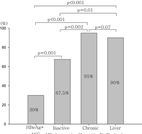
2. Prevalence of the precore/BCP mutations in relation to clinical outcomes Overall, the A1762T/G1764A mutation in the BCP was found in 113 (70.6%) of the 160 patients. In HBeAg-positive asymptomatic carrier, 12 (30%) of 40patients, including 1 patient with mixed type, had A1762T/G1764A mutations in the BCP. In inactive HBsAg carriers, the A1762T/G1764A mutation was found in 27 (67.5%) of 40 patients, including 5 patients with mixed type. In patients with chronic hepatitis B, A1762T/G1764A mutations were found in 38 (95.0%) of 40 patients, including 3 patients with mixed type. In liver cirrhosis, A1762T/G1764A mutations were found in 36 of 40(90%), including 4 patients with mixed type. In HBeAg-positive asymptomatic carriers, A1762T/G1764A mutations in the
BCP were less frequent than in other disease spectrums (p < 0.001). In addition, the prevalence of A1762T/G1764A mutations in the BCP in patients with chronic hepatitis B and liver cirrhosis was higher than in inactive HBsAg carriers. However, there were no significant differences between the patients with chronic hepatitis B and liver cirrhosis patients (p=0.675)(Table 2)(Figure 1).
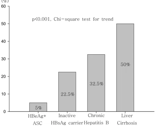
Overall, the prevalence of precore stop codon mutation (G1896A) was 44 (27.5%) of the 160 patients. All patients had T at nucleotide 1858, which makes a base-pair with the 1896 nucleotide in stem loops structure of the pregenomic RNA of HBV. In HBeAg-positive asymptomatic carriers, precore stop codon mutation (G1896A) was found in 2 (5%) of 40 patients. In inactive HBsAg carrier, G1896A mutation was found in 9(22.5%) of 40, including 5 mixed types. In patients with chronic hepatitis B, precore stop mutation was found in 13 (32.5%) of 40, including 8 mixed types. In patients with liver cirrhosis, G1896A mutation was found in 20 (50%), including 8 patients with mixed types. Even though, HBeAg-positive asymptomatic carriers have fewer frequencies of G1896A mutation than other clinical outcomes, (P<0.05). (Table 2), there were no significant differences among the patients with chronic hepatitis B, inactive HBsAg carrier, and liver cirrhosis (Table 2)(Figure 2).
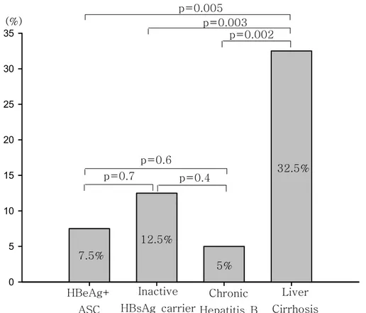
Other mutations were found in the precore/core promoter regions. Of these mutations, T to C or A at nucleotide 1753 (T1753C/A) located at the first TATA-like box was found in 23 (14.4%) of the patients. In addition, 1754 mutation was found in 8 (5.1%) of the patients. The prevalence of these mutations is presented in Table 4. Interestingly, T1753C/A mutation in liver cirrhosis was occurred more frequently than that in other clinical outcomes (P<0.05)(Figure 3).
Another precore mutation, G to A, at nucleotide1899 was detected in 10 (6.25 %) of 160 patients. Eight (80%) of these 10 patients with this mutation were concomitant with G1896A mutation and most of this double mutations
occurred in patients with liver cirrhosis (Table 4).
3. Prevalence of G1896A mutation in the precore and A1762T/G1764A mutations in the BCP in relation to HBeAg status
In relation to HBeAg status, the A1762T/G1764A mutation in the BCP was found in 64.4% (including mixed type) and 78.1% (including mixed type) in HBeAg-positive and HBeAg-negative patients, respectively (p=0.058). When the patients with G1986A mutations in the precore were excluded because they affect expression of the HBeAg status, A1762T/G1764A mutations in the BCP were found in 65.1% (including mixed type) and 70.4% (including mixed type) in HBeAg-positive and HBeAg-negative patients, respectively (p=0.52)(Table 5). However, G1896A mutations in the precore were more frequently found in HBeAg-negative patients compared with HBeAg-positive patients (39.7% vs. 17.2%; p=0.002) (Table 3).
IV. DISCUSSION
In this study, we examined the prevalence of mutations in the precore/BCP region of HBV in 160 consecutive patients infected by hepatitis B virus in Korea according to different clinical manifestations. Thereby, we were to analyze the role of the precore/BCP mutations in the progression of chronic liver diseases.
In our study, we found that Korean patients infected with hepatitis B virus mostly had genotype C regardless of various clinical outcomes. Similar results were found in other Korean studies, too (Lee et al., 2001; Cho et al., 2001). This result may relate to the anthropologic characteristics of the monoracial Korean peoples.
Recently, many investigators were interested in the association between the precore/BCP mutations and severity of liver disease. The BCP has been known that it plays a central role in regulating pregenomic RNA and precore RNA transcription. (Yuh et al., 1992; Chen et al., 1995). It was also reported that the occurrences of the mutations in the precore/BCP region depend on HBV genotypes. The A1762T/G1764A mutation in the BCP were more frequent in genotype C than that in genotype B. On the contrary, G1896A mutation in the precore occurred less in genotype C than in genotype B (Orito et al., 2001; Lindh et al., 1999). In addition, G1896A mutation in the precore was to be restricted in the genotype, which had T at nucleotide 1858 (Chan et al., 1999; Lindh et al., 1997). Several studies reported that these double mutations enhanced the viral replication and suppressed precore RNA expression. (Lindh et al., 1999; Shindo et al., 1999; Moriyama et al., 1996). However, other studies reported that A1762T/G1764A mutations in the BCP region occurred very frequently regardless of HBeAg status and during the progressions of liver disease, but these were not associated with liver disease and viral replication. (Chun et al., 2000; Yoo et al., 2003),
In this study, the prevalence of T1762A/G1764A mutation in the BCP was 70.6%. In Taiwan, 20% of patients infected by hepatitis B virus with genotype C had the core promoter mutation (T1762A/G1764A) (Kao et al., 2003), and in Hong Kong, 87.9% of patients with HBV genotype C had the core promoter mutations (Yuen et al., 2004). Despite of having same genotype, there are different occurrences of the core promoter mutations. However, the exact reason is unknown. It may be associated with the ethnics and geographic differences. We also found that the occurrences of the core promoter mutations were frequent in HBeAg-positive asymptomatic carriers (30%), it may be associated with the transmission of HBV infection, which was the major route of HBV infection in Korea. (Kang et al., 2004)
The A1762T/G1764A mutations have fewer occurrences in HBeAg-positive asymptomatic carriers than other clinical manifestations. However, A1762T/G1764A mutations occurred more frequently in patients with chronic hepatitis B and liver cirrhosis than that in inactive HBsAg carriers, but there were no significant differences between the patients with chronic hepatitis B patients and liver cirrhosis. This finding suggests that A1762T/G1764A mutations occur after immune systems recognize the HBV and attack the HBV. Interestingly, we found that the double mutations (A1762T/G1764A) were comparable between inactive HBsAg carriers and chronic hepatitis B patients. However, there were no significant differences between chronic hepatitis B and liver cirrhosis patients. This finding suggests that these double mutations (T1762A/G1764A) might contribute to the persistence of hepatitis B virus in host, but these mutations by themselves would not be associated with the progression from chronic hepatitis to liver cirrhosis.
It also suggests that during the progression from chronic hepatitis to liver cirrhosis, there was no further accumulation of these mutations. This was quite contrast to the previous results that these mutations were more frequent in liver cirrhosis compared with chronic hepatitis B.
Another common mutation at nucleotide 1896, which makes a premature stop codon and diminishes the production of HBeAg, has the same controversy as core promoter mutations (A1762T/G1764A) (Ikeda et al., 1998; Ballard et al., 2000; Yoo et al., 2003). In our study, there were significant differences between HBeAg positive asymptomatic carriers and other three clinical outcomes in the presences of precore stop codon mutation. Although, there were significant differences between inactive HBsAg carriers and liver cirrhosis patient (22.5% vs. 50.0%, p=0,011), we didn't find statistical differences between chronic hepatitis B patients and inactive HBsAg carriers or liver cirrhosis patients (22.5% vs. 32.5%, p=0.317 or 32.5% vs. 50.0%, p=0.112). But with the progression of the liver disease, the precore mutation was gradually accumulated and this mutation was more frequent in HBeAg negative patients than in HBeAg positive patients (39.7% vs. 17.2%, P=0.002). We found that the presences of the mutations in precore/BCP region were lower in HBeAg positive healthy carriers than that in other clinical manifestations. It may be that HBeAg positive asymptomatic carriers were staying in immune tolerance phase of natural course of hepatitis B virus infection and got less attacks than others from host immune system. But, with the increasing of age, years of infection and immune activity, the hepatitis B virus was attacked by host immune system and made some mutations in its genome to escape from the stress. Through the immune clearance phase of natural course of HBV infection, HBV infected liver disease would clinically convert to inactive HBsAg carrier or chronic hepatitis B.
For understanding, if there exist other mutations to correlate with the severity of liver disease, we also examined the additional mutations in precore/BCP region. Some additional mutations in precore/BCP region were found in our study, such as T to C or A at nucleotide 1753, T to C at nucleotide 1754, G to A at nucleotide 1899 and so on. Of these, T1753A/C mutation and G1899A mutation was found in up to 5% to 32.5% and 2.5% to 20 %, respectively. Previously, it had been reported that approximately 50%
of G1896A mutation was likely to companied G1899A. (Sarin et al., 2002) In our study, we found 20% of G1896A mutations with G1899A mutation, and most of this mutation was found in liver cirrhosis patients. T to C or A at nucleotide 1753 substitution had been supposed that it would be associated with severity of liver disease. (Kidd-Ljunggrenet al, 1997; Takahashi et al., 1998) Our result suggested that mutation at nucleotide 1753 was associated with the progression to liver cirrhosis, because, in our data, the high frequencies of this mutation in liver cirrhosis patients were comparable with either inactive HBsAg carriers or chronic hepatitis B patients. To the contrary, there were no significant differences between inactive HBsAg patients and chronic hepatitis B patients. This mutation will be another genetic risk factor to progress the liver disease, even develop to the hepatocellular carcinoma. (Takahashi et al., 1998)
In HBeAg status, the basal core promoter mutations (T1762A/G1764A) and precore mutation (G1896A) were frequently occurred in both of the groups, but only precore mutation had significant differences in HBeAg status (17.2% vs. 39.7%, p=0.002). And in many HBeAg negative patients also could detect HBV DNA. It suggested that hepatitis B virus express precore stop codon mutant gene to diminish the production of HBeAg, which was the major target of immune system, to escape from the host immune attacks.
From this study, we speculate the progression of chronic liver disease associated with HBV infection as follows: during the natural course of chronic HBV infection from immune tolerant phase to immune clearance phase, manifested as chronic hepatitis or inactive HBsAg carrier, A176T/G1764A mutations in the BCP occurred regardless of clinical manifestation. However, the prevalence of A176T/G1764A mutations did not increase in patients with liver cirrhosis compared with chronic hepatitis. In contrast, G1986A mutation in the precore occurred progressively in due order from the HBeAg-positive asymptomatic carrier, inactive HBsAg carriers, chronic hepatitis B and liver cirrhosis (5% vs. 22.5% vs. 32.5% vs. 50 %). In addition, accompanying
T1753C/A mutation, which did not affect the course of other clinical manifestations, might enhance the progression from chronic hepatitis B to liver cirrhosis.
Primer Sequences Outer primer
Sense 5'-TCACCATATTCTTGGGAACAAGA-3'(2815-2837) Antisense 5'-CGAAC ACTGAACAAATGGC-3' (685-704)
Inner primer Mix 1
Sense 5'-GGCTCCAGTTCCGGAACAGT-3' (67-86) Antisense
Type A specific 5'-CTCGCGGAGATTGACGAGATGT-3'(113-134) Type B specific 5'-CAGGTTGGTGAGTGACTGGAGA-3'(324-345) Type C specific 5'-GGTCCTAGGAATCCTGATGTTG-3'(165-186)
Mix 2 Sense
Type D specific 5'-GCCAACAAGGTAGGAGCT-3' (2979-2996)
Type E specific 5'-CACCAGAAATCCAGATTGGGACCA-3'(2955-2978) Type F specific 5'-GCTACGGTGGGTTACCA-3' (3031-3051)
Antisense 5'-GGAGGCGGATCTGCTGGCAA-3'(3078-3097) Table 1. Primer sequences for HBV genotyping used in this study
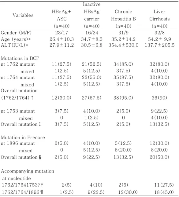
Variables HBeAg+ ASC (n=40) Inactive HBsAg carrier (n=40) Chronic Hepatitis B (n=40) Liver Cirrhosis (n=40) Gender (M/F) 23/17 16/24 31/9 32/8 Age (years)* 26.4±10.3 34.7±8.5 35.2±14.2 54.2± 9.9 ALT(IU/L)* 27.9±11.2 30.5±6.8 354.4±530.0 137.7±205.5 Mutations in BCP nt 1762 mutant mixed 11(27.5) 21(52.5) 34(85.0) 32(80.0) 1(2.5) 5(12.5) 3(7.5) 4(10.0) nt 1764 mutant mixed 11(27.5) 22(55.0) 35(87.5) 32(80.0) 1(2.5) 5(12.5) 3(7.5) 4(10.0) Overall mutation (1762/1764)↑ 12(30.0) 27(67.5) 38(95.0) 36(90) nt 1753 mutant mixed 3(7.5) 4(10.0) 2(5.0) 9(22.5) 0 1(2.5) 0 4(10.0) Overall mutation↕ 3(7.5) 5(12.5) 2(5.0) 13(32.5) Mutation in Precore nt 1896 mutant mixed 2(5.0) 4(10.0) 5(12.5) 12(30.0) 0 5(12.5) 8(20.0) 8(20.0) Overall mutation§ 2(5.0) 9(22.5) 13(32.5) 20(50.0) Accompanying mutation at nucleotide 1762/17641753?‡ 2(5) 4(10) 2(5) 11(27.5) 1762/1764/1896¶ 1(2.5) 9(22.5) 12(30.0) 18(45.0)
Table 2. Characteristics of patients and prevalence(%) of precore/core promoter mutations in relation to clinical outcomes
↑p=0001, HBeAg+ASC vs. inactive HBsAg carrier;
p<0001, HBeAg+ASC vs. Chronic hepatitis B; p<0001, HBeAg+ASC vs. liver cirrhosis;
p=0002, inactive HBsAg carrier vs. Chronic hepatitis B;
p=0014 ,inactive HBsAg carrier vs. liver cirrhosis; p=0675 ,Chronic hepatitis B vs. liver cirrhosis; ↕p=0712, HBeAg+ASC vs. inactive HBsAg carrier; p=0.64, HBeAg+ASC vs. Chronic hepatitis B; p=0.005, HBeAg+ASC vs. liver cirrhosis;
p=0.432, inactive HBsAg carrier vs. Chronic hepatitis B; p=0032 ,inactive HBsAg carrier vs. liver cirrhosis; p=0002 ,Chronic hepatitis B vs. liver cirrhosis; §p=0023, HBeAg+ASC vs. inactive HBsAg carrier; p=0.002, HBeAg+ASC vs. Chronic hepatitis B; p<0001, HBeAg+ASC vs. liver cirrhosis;
p=0.317, inactive HBsAg carrier vs. Chronic hepatitis B; p=0011 ,inactive HBsAg carrier vs. liver cirrhosis; p=0112,Chronic hepatitis B vs. liver cirrhosis; ‡p=0675, HBeAg+ASC vs. inactive HBsAg carrier; p=1.0, HBeAg+ASC vs. Chronic hepatitis B; p=0006, HBeAg+ASC vs. liver cirrhosis;
p=0.675, inactive HBsAg carrier vs. Chronic hepatitis B; p=0045, inactive HBsAg carrier vs. liver cirrhosis; p=0006,Chronic hepatitis B vs. liver cirrhosis; ¶p=0007, HBeAg+ASC vs. inactive HBsAg carrier; p=0.001, HBeAg+ASC vs. Chronic hepatitis B; p<0001, HBeAg+ASC vs. liver cirrhosis;
p=0.446, inactive HBsAg carrier vs. Chronic hepatitis B; p=0033, inactive HBsAg carrier vs. liver cirrhosis; p=0166,Chronic hepatitis B vs. liver cirrhosis; * Expressed as mean±SD.
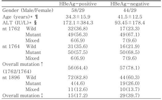
HBeAg-positive HBeAg-negative Gender (Male/Female) 58/29 44/29 Age (years)* ¶ 34.3±15.9 41.5±12.5 ALT (IU/L)* § 172.1±384.3 93.45±178.4 nt 1762 Wild 32(36.8) 17(23.3) Mutant 49(56.3) 49(67.1) Mixed 6(6.9) 7(9.6) nt 1764 Wild 31(35.6) 16(21.9) Mutant 50(57.5) 50(68.5) Mixed 6(6.9) 7(9.6) Overall mutation↑ (1762/1764) 56(64.4) 57(78.1) nt 1896 Wild 72(82.8) 44(60.3) Mutant 4(4.6) 19(26.0) Mixed 11(12.6) 10(13.7) Overall mutation↕ 15(17.2) 29(39.7)
Table 3. Characteristics of patients and prevalence(%) of precore/core promoter mutations in relation to HBeAg status
¶P=0.002, HBeAg-positive vs. HBeAg-negative §P=0.112, HBeAg-positive vs. HBeAg-negative ↑P=0.058, HBeAg-positive vs. HBeAg-negative ↕P=0.002, HBeAg-positive vs. HBeAg-negative * Expressed as mean±SD.
HBeAg+ ASC (n=40) Inactive HBsAg carrier (n=40) Chronic Hepatitis B (n=40) Liver cirrhosis (n=40) nt 1754 Wild 37(92.5) 40(100) 39(97.5) 36(90) Mutant 3(5.0) 0 1(2.5) 4(7.5) nt 1899 Wild 39(97.5) 39(97.5) 40(100) 32(80) Mutant 1(2.5) 1(2.5) 0 8(20) Table 4. Prevalence(%) of other mutations in relation to clinical outcomes
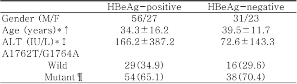
HBeAg-positive HBeAg-negative Gender (M/F 56/27 31/23 Age (years)*↑ 34.3±16.2 39.5±11.7 ALT (IU/L)*↕ 166.2±387.2 72.6±143.3 A1762T/G1764A Wild 29(34.9) 16(29.6) Mutant¶ 54(65.1) 38(70.4)
Table 5. Prevalence(%) of A1762T/G1764A mutations without G1896A mutation in precore in relation to HBeAg status
↑P=0.04, HBeAg-positive vs. HBeAg-negative ↕P=0.09, HBeAg-positive vs. HBeAg-negative ¶P=0.52, HBeAg-positive vs. HBeAg-negative * Expressed as mean SD.
0 20 40 60 80 100
Figure 1. Comparison of the frequency of A1762T/G1764A mutations in relation to clinical outcomes
p=0.001 p=0.07 p=0.002 p=0.01 p<0.001 p<0.001 HBeAg+ ASC Inactive HBsAg carrier Liver Cirrhosis Chronic Hepatitis B (%) 67.5% 30% 95% 90%
0 10 20 30 40 50 60
Figure 2. Comparison of the frequency of the G1896A mutation in relation to clinical outcomes Inactive HBsAg carrier Liver Cirrhosis HBeAg+ ASC Chronic Hepatitis B p<0.001, Chi-square test for trend (%)
5%
22.5%
32.5%
0 5 10 15 20 25 30 35
Figure 3. Comparison of frequency of the T1753C/A mutation in relation to clinical outcomes p=0.7 p=0.002 p=0.6 p=0.4 p=0.005 p=0.003 HBeAg+ ASC Chronic Hepatitis B Inactive HBsAg carrier Liver Cirrhosis 5% 7.5% 12.5% 32.5% (%)
V. REFERENCES
Akarca US, Greene S, Lok AS. 1994. Detection of precore hepatitis B virus mutants in asymptomatic HBsAg-positive family members. Hepatology 19: 1366-1370.
Arauz-Ruiz P, Norder H, Robertson BH, Magnius LO. 2002. Genotype H: a new Amerindian genotype of hepatitis B virus revealed in Central America. J Gen Virol 83: 2059-2073.
Ballard AL, Boxall EH. 2000. Epidemiology of precore mutants of hepatitis B in the United Kingdom. J Med Virol 62: 463-470.
Brunetto MR, Stemler M, Schodel F, Will H, Ottobrelli A, Rizzetto M, Verme G. 1989. Identification of HBV variants which cannot produce pre core derived HBeAg and may be responsible for severe hepatitis. Ital J Gastroenterol 21: 151-154.
Brunetto MR, Stemler M, Bonino F, Schodel F, Oliveri F, Rizzetto M, Verme G, Will H. 1990. A new hepatitis B virus strain in patients with severe anti-HBe positive chronic hepatitis B. J Hepatol 10: 258-261.
Brunetto MR, Giarin MM, Oliveri F, Chiaberge E, Baldi M, Alfarano A, Serra A, Saracco G, Verme G, Will H. 1991. Wild-type and e antigen-minus hepatitis B viruses and course of chronic hepatitis. Proc Natl Acad Sci USA 88: 4186-4190.
Bruix J, Castells A, Bosch J, Feu F, Fuster J, Garcia-Pagan JC, Visa J, Bru C, Rodes J. 1996. Surgical resection of hepatocellular carcinoma in cirrhotic patients: prognostic value of preoperative portal pressure.
Gastroenterology 111: 1018-1022.
Buckwold VE, Xu Z, Chen M, Yen TS, Ou JH. 1996. Effects of a naturally occurring mutation in the hepatitis B virus basal core promoter on precore gene expression and viral replication. J Virol 70: 5845-5851.
Buckwold VE, Xu Z, Yen TS, Ou JH. 1997. Effects of a frequent double-nucleotide basal core promoter mutation and its putative single-nucleotide precursor mutations on hepatitis B virus gene expression and replication. J Gen Virol 78: 2055-2065.
Carman WF, Jacyna MR, Hadziyannis S, Karayiannis P, McGarvey MJ, Makris A, Thomas HC. 1989. Association mutation preventing formation of hepatitis B e antigen in patients with chronic hepatitis B infection. Lancet 2: 588-591.
Carman WF, Fagan EA, Hadziyannis S, Karayiannis P, Tassopoulos NC, Williams R, Thomas HC. 1991. Association of a precore genomic variant of hepatitis B virus with fulminant hepatitis. Hepatology 14: 219-222.
Chan HL, Hussain M, Lok AS.1999. Different hepatitis B virus genotypes are associated with different mutations in the core promoter and precore regions during hepatitis B e antigen seroconversion. Hepatology 29: 976-984.
Chang C, Enders G, Sprengel R, Peters N, Varmus HE, Ganem D. 1987. Expression of the precore region of an avian hepatitis B virus is not required for viral replication. J Virol 61: 3322-3325.
Chen HS, Kew MC, Hornbuckle WE, Tennant BC, Cote PJ, Gerin JL, Purcell RH, Miller RH. 1992. The precore gene of the woodchuck hepatitis virus genome is not essential for viral replication in the natural host. J Virol
66: 5682-5684.
Chen IH, Huang CJ, Ting LP. 1995. Overlapping initiator and TATA box functions in the basal core promoter of hepatitis B virus. J Virol 69: 3647-3657.
Chien RN, Yeh CT, Tsai SL, Chu CM, Liaw YF. 2003. Determinants for sustained HBeAg response to lamivudine therapy. Hepatology 38: 1267-1273.
Cho IH, Song JY, Kim DK, Lim HS, Sheen SS, Kim WS, Lee KM, Hahm KB, Kim JH, Cho SW. 2001. Prevalence of HBV genotypes in Korean patients with chronic hepatitis B. Korean J Hepatol 7: 381- 386.
Chu CJ, Hussain M, Lok AS. 2002. Hepatitis B virus genotype B is associated with earlier HBeAg seroconversion compared with hepatitis B virus genotype C. Gastroenterology 122: 1756-1762.
Chun YK, Kim JY, Woo HJ, Oh SM, Kang I, Ha J, Kim SS. 2000. No significant correlation exists between core promoter mutations, viral replication, and liver damage in chronic hepatitis B infection. Hepatology 32: 1154-1162.
Delius H, Gough NM, Cameron CH, Murray K. 1983. Structure of the Hepatitis B virus genome. J Virol 47: 337-343.
Ding X, Mizokami M, Yao G, Xu B, Orito E, Ueda R, Nakanishi M. 2001. Hepatitis B virus genotype distribution among chronic hepatitis B virus carriers in Shanghai, China. Intervirology 44: 43-47.
Arase Y, Chayama K, Murashima N, Kumada H. 1998. Relationship of hepatocellular carcinogenesis with precore mutant virus and serum hepatitis B virus DNA concentration. A longitudinal analysis of patients with cirrhosis. Hepatol Res 10: 142-155.
Kang HS, Song BC, Cui XJ, Kim SY, and Kim SK. 2004. Serologic markers of hepatitis B virus in pregnant women in Jeju Island. Korean J Hepatol 10:191-196.
Kao JH, Wu NH, Chen PJ, Lai MY, Chen DS. 2000. Hepatitis B genotypes and the response to interferon therapy. J Hepatol 33: 998-1002.
Kao JH, Chen PJ, Lai MY, Chen DS. 2000. Hepatitis B genotypes correlate with clinical outcomes in patients with chronic hepatitis B. Gastroenterology 118: 554-559.
Kao JH, Chen PJ, Lai MY, Chen DS. 2003. Basal core promoter mutations of hepatitis B virus increase the risk of hepatocellular carcinoma in hepatitis B carriers. Gastroenterology 124: 327-334.
Kidd-Ljunggren K, Oberg M, Kidd AH. 1997. Hepatitis B virus X gene 1751 to 1764 mutations: implications for HBeAg status and disease. J Gen Virol 78: 1469-1478.
Laskus T, Persing DH, Nowicki MJ, Mosley JW, Rakela J. 1993. Nucleotide sequence analysis of the precore region in patients with fulminant hepatitis B in the United States. Gastroenterology 105: 1173-1178.
Laskus T, Rakela J, Nowicki MJ, Persing DH. 1995. Hepatitis B virus core promoter sequence analysis in fulminant and chronic hepatitis B.
Gastroenterology 109: 1618-1623.
Lavanchy D. 2004. Hepatitis B virus epidemiology, disease burden, treatment, and current and emerging prevention and control measures. J Viral Hepat 11: 97-107.
Lee SH, Han SH, Cho SC, Roh BJ, Sohn JH, Kim DA, Lee DH, Kee CS. 2001. Distribution of HBV genotypes in patients with chronic HBV infection in Korea. Korean J Hepatol 7: 373-380.
Li JS, Tong SP, Wen YM, Vitvitski L, Zhang Q, Trepo C. 1993. Hepatitis B virus genotype A rarely circulates as an HBe-minus mutant: possible contribution of a single nucleotide in the precore region. J Virol 67: 5402-5410
Liang TJ, Hasegawa K, Rimon N, Wands JR, Ben-Porath E. 1991. A hepatitis B virus mutant associated with an epidemic of fulminant hepatitis. N Engl J Med 324: 1705-1709.
Lindh M, Andersson AS, and Gusdal A. 1997. Genotypes, nt1858 variants, and geographic origin of hepatitis B virus large-scale analysis using a new genotyping method. J Infect Dis 175:1285-1293.
Lindh M, Hannoun C, Dhillon AP, Norkrans G, Horal P. 1999. Core promoter mutations and genotypes in relation to viral replication and liver damage in East Asian hepatitis B virus carriers. J Infect Dis 179: 775-782.
Liu CJ, Kao JH, Lai MY, Chen PJ, Chen DS. 2004. Precore/core promoter mutations and genotypes of hepatitis B virus in chronic hepatitis B patients with fulminant or subfulminant hepatitis. J Med Virol 72: 545-550.
Lo WY, Ting LP. 1994. Repression of enhancer II activity by a negative regulatory element in the hepatitis B virus genome. J Virol 68: 1758-1764.
Locarnini S, McMillan J, Bartholomeusz A. 2003. The hepatitis B virus and common mutants. Semin Liver Dis 23: 5-20.
Lok AS, Akarca U, Greene S. 1994. Mutations in the pre-core region of hepatitis B virus serve to enhance the stability of the secondary structure of the pre-genome encapsidation signal. Proc Natl Acad Sci USA 91: 4077-4081.
Moriyama K, Okamoto H, Tsuda F, Mayumi M. 1996. Reduced precore transcription and enhanced core-pregenome transcription of hepatitis B virus DNA after replacement of the precore-core promoter with sequences associated with e antigen-seronegative persistent infections. Virology 226: 269-280.
Naito H, Hayashi S, Abe K. 2001. Rapid and specific genotyping system for hepatitis B virus corresponding to six major genotypes by PCR using type-specific primers. J Clin Microbiol 39: 362-364.
Norder H, Courouce AM, Magnius LO. 1994. Complete genomes, phylogenetic relatedness, and structural proteins of six strains of the hepatitis B virus, four of which represents two new genotypes. Virology 198: 489-503.
Okamoto H, Tsuda F, Sakugawa H, Sastrosoewignjo RI, Imai M, Miyakawa Y, Mayumi M. 1988. Typing hepatitis B virus by homology in nucleotide sequence: comparison of surface antigen subtypes. J Gen Virol 69: 2575-2583.
Okamoto H, Yotsumoto S, Akahane Y, Yamanaka T, Miyazaki Y, Sugai Y, Tsuda F, Tanaka T, Miyakawa Y, Mayumi M. 1990. Hepatitis B viruses with precore region defects prevail in persistently infected hosts along with seroconversion to the antibody against e antigen. J Virol 64: 1298-1303.
Okamoto H, Tsuda F, Akahane Y, Sugai Y, Yoshiba M, Moriyama K, Tanaka T, Miyakawa Y, Mayumi M. 1994. Hepatitis B virus with mutations in the core promoter for an e antigen-negative phenotype in carriers with antibody to e antigen. J Virol 68: 8102-8110.
Omata M, Ehata T, Yokosuka O, Hosoda K, Ohto M. 1991. Mutations in the precore region of hepatitis B virus DNA in patients with fulminant and severe hepatitis. N Engl J Med 324: 1699-1704.
Orito E, Ichida T, Sakugawa H, Sata M, Horiike N, Hino K, Okita K, Okanoue T, Iino S, Tanaka E, Suzuki K, Watanabe H, Hige S, Mizokami M. 2001. Geographic distribution of hepatitis B virus (HBV) genotype in patients with chronic HBV infection in Japan. Hepatology 34: 590-594.
Orito E, Mizokami M, Sakugawa H, Michitaka K, Ishikawa K, Ichida T, Okanoue T, Yotsuyanagi H, Iino S. 2001. A case-control study for clinical and molecular biological differences between hepatitis B viruses of genotypes B and C. Hepatology 33: 218-223.
Sarin S, Satapathy S, Chauhan R. 2002. Hepatitis B e-antigen negative chronic hepatitis B. J Gastroenterol Hepatol 17: S311-S321.
Sato S, Suzuki K, Akahane Y, Akamatsu K, Akiyama K, Yunomura K, Tsuda F, Tanaka T, Okamoto H, Miyakawa Y. 1995. Hepatitis B virus strains with mutations in the core promoter in patients with fulminant hepatitis. Ann Intern
Med 122: 241-248.
Shindo M, Hamada K, Koya S, Sokawa Y, Okuno T. 1999. The clinical significance of core Promoter and precore mutations during the natural course and interferon therapy in patients with chronic hepatitis B. Am J Gastroenterol 94: 2237-2245.
Sterneck M, Gunther S, Santantonio T, Fischer L, Broelsch CE, Greten H, Will H. 1996. Hepatitis B virus genomes of patients with fulminant hepatitis do not share a specific mutation. Hepatology 24: 300-306.
Stuyver L, De Gendt S, Van Geyt C, Zoulim F, Fried M, Schinazi RF, Rossau R. 2000. A new genotype of hepatitis B virus: complete genome and phylogenetic relatedness. J Gen Virol 81: 67-74.
Sugauchi F, Chutaputti A, Orito E, Kato H, Suzuki S, Ueda R, Mizokami M. 2002. Hepatitis B viral genotypes and clinical manifestation among hepatitis B carriers in Thailand. J Gastroenterol Hepatol 17: 671-676.
Summers J, Mason WS. 1982. Replication of the genome of a hepatitis B-like virus by reverse transcription of an RNA intermediate. Cell 29: 403-415.
Sumi H, Yokosuka O, Seki N, Arai M, Imazeki F, Kurihara T, Kanda T, Fukai K, Kato M, Saisho H. 2003. Influence of hepatitis B virus genotypes on the progression of chronic type B liver disease. Hepatology 37: 19-26.
Takahashi K, Akahane Y, Hino K, Ohta Y, Mishiro S .1998. Hepatitis B virus genomic sequence in the circulation of hepatocellular carcinoma patients: comparative analysis of 40 full-length isolates. Arch Virol 143: 2313-2326.
Thakur V, Guptan RC, Kazim SN, Malhotra V, Sarin SK. 2002. Profile, spectrum and significance of HBV genotypes in chronic liver disease patients in the Indian subcontinent. J Gastroentrol Hepatol 17: 165-170.
Tong SP, Li JS, Vitvitski L, Kay A, Treepo C. 1993. Evidence for a base-paired region of hepatitis B virus pregenome encapsidation signal which influences the patterns of precore mutations abolishing HBe protein expression. J Virol 67: 5651-5655.
Tur-Kaspa R, Klein A, Aharonson S. 1992. Hepatitis B virus precore mutants are identical in carriers from various ethnic origins and are associated with a range of live disease severity. Hepatology 16: 1338-1342.
Wai CT, Chu CJ, Hussain M, Lok AS. 2002. HBV genotype B is associated with better response to interferon therapy in HBeAg (+) chronic hepatitis than genotype C. Hepatology 36: 1425-1430.
Yoo BC, Park JW, Kim HJ, Lee DH, Cha YJ, Park SM. 2003. Precore and core promoter mutations of hepatitis B virus and hepatitis B e antigen-negative chronic hepatitis B in Korea. J Hepatol 38: 98-103.
Yuen MF, Sablon E, Tanaka Y, Kato T, Mizokami M, Doutreloigne J, Yuan HJ, Wong DK, Sum SM, Lai CL. 2004. Epidemiological study of hepatitis B virus genotypes, core promoter and precore mutations of chronic hepatitis B infection in Hong Kong. J Hepatol 41: 119-125.
Yuh CH, Chang YL, Ting LP. 1992. Transcriptional regulation of precore and pregonomic RNAs of hepatitis B virus. J Virol 66: 4073-4084.
V. ABSTRACT IN KOREAN
HBV DNA의 basal core promoter와 precore에서 일어나는 변이 (A1762T/G1764A/G1896A) 및 HBV의 유전자형은 B형 간염 바이러스에 감염 된 만성 간질환의 진행과 밀접한 관계를 가지고 있다고 보고 되였지만 이들 간의 연관성 에 대해서는 아직 까지 많은 논의를 하고 있다. 본 연구에서는 HBV에 감염된 만성 간 질환 환자를 연구대상으로 하여 임상경과에 따라 HBV DNA의 core promoter와 precore에서 일어나는 변이의 축적과 만성 B형 간질환의 진행과의 연관성에 대하여 연구하였다.
본 연구에서는 160명의 만성 B형 간질환 환자를 연구대상으로 하였으며 임상적 진단에 근거하여 네 개의 그룹 즉 활동성보균자(HBeAg+, anti-HBe- HBV DNA+ by hybridization, 그리고 혈청 ALT 정상, n=40), 비활동성 보균자(n=40), 만성간 염환자(n=40), 간경변증환자(n=40)로 분류하였다. HBV DNA의 core promoter와 precore부위의 변이는 직접염기서열방법을 이용하여 측정하였고 HBV DNA의 유전 자형은 각 유전자형에 특이적인 primer를 이용하여 측정하였다. 모든 환자에서 유전 자형이 C인 HBV가 발견 되였으며 그 중 한명에서 유전자형 B와 C의 혼합형인 HBV 가 발견 되였다. Basal core promoter부위에서 A1762T/G1764A변이는 활동성 보 균자에서는 30%, 비활동성 보균자에서는 65.7%, 만성 B형 간염환자에서는 95%, 그 리고 간경변증환자에서는 90%가 발견 되였다. Precore부위에서 G1896A변이는 활 동성 보균자에서는 5%, 비활동성 보균자에서는 22.5%, 만성 B형 간염환자에서는 32.% 그리고 간경변증 환자에서는 50%가 발견 되였다. Basal core promoter부위에 서의 T1753C/A변이와 precore부위에서의 G1899A변이는 간경변증 환자에서의 발 생빈도가 다른 세 그룹에 비하여 현저히 높다(p<0.05).
한국에서의 HBV 유전자형은 대부분 C형이며, 유전자형이 C인 HBV에 감염된 만 성 B형 간질환에서는 임상경과에 관계없이 대부분 환자에서 basal core promoter와 precore부위에서 변이가 발견되며 이들 변이의 축적은 HBV감염과 연관되는 만성간 질환의 진행과정에서 중요한 역할을 할 것이다.
색인단어: B형 간염 바이러스, basal core promoter 변이, precore변이, 유전자형, 만 성 B형 간염