R E S E A R C H
Open Access
First detection of ranavirus in a wild
population of Dybowski
’s brown frog (Rana
dybowskii) in South Korea
Jaejin Park
1, Alejandro Grajal-Puche
2, Nam-Ho Roh
3, Il-Kook Park
4, Nam-Yong Ra
5and Daesik Park
4*Abstract
Background: Ranavirus is an emerging infectious disease which has been linked to mass mortality events in
various amphibian species. In this study, we document the first mass mortality event of an adult population of
Dybowski
’s brown frogs (Rana dybowskii), in 2017, within a mountain valley in South Korea.
Results: We confirmed the presence of ranavirus from all collected frogs (n = 22) via PCR and obtained the 500 bp
major capsid protein (MCP) sequence from 13 individuals. The identified MCP sequence highly resembled Frog
virus 3 (FV3) and was the same haplotype of a previously identified viral sequence collected from Huanren brown
frog (R. huanrenensis) tadpoles in South Korea. Human habitat alteration, by recent erosion control works, may be
partially responsible for this mass mortality event.
Conclusion: We document the first mass mortality event in a wild Korean population of R. dybowskii. We also
suggest, to determine if ranavirus infection is a threat to amphibians, government officials and researchers should
develop continuous, country-wide, ranavirus monitoring programs of Korean amphibian populations.
Keywords: Ranavirus, Rana dybowskii, Mass mortality, Wild population, Major capsid protein
Background
Emerging infectious diseases are one of the key factors
causing rapid global biodiversity declines in this century
(Fey et al.
2015
). Amphibians are particularly vulnerable
to infectious diseases due to their permeable skin and
metamorphic life cycle (Daszak et al.
1999
). Fungal
in-fections by Batrachochytrium dendrobatidis (Bd) and B.
salamandriborans, causing chytridiomycosis, have been
implicated as a primary cause of rapid amphibian
popu-lation declines (Daszak et al.
1999
; Scheele et al.
2019
).
In addition, ranavirus, a double-stranded DNA virus, has
also been identified as a major emerging infectious
dis-ease and is associated with global amphibian declines
(Green et al.
2002
; Carey et al.
2003
). In Northeast Asia,
across China, Japan, and Korea, ranavirus infections have
caused mortality in 17, native and invasive, amphibian
species (4 urodelan and 13 anuran species) as well as
amphibians in the pet trade (Zhang et al.
1996
; Weng
et al.
2002
; Kim et al.
2009
; Une et al.
2009a
; Kolby et al.
2014
; Une et al.
2014
; Duffus et al.
2015
; Kwon et al.
2017
; Park et al.
2017
). Out of the 21 reported ranavirus
infection cases, 18 have been linked to mass mortality
events (Table
1
). Amphibian ranavirus susceptibility and
mortality are often correlated with low environmental
quality, such as habitat destruction and pollution (Carey
et al.
1999
; Gray et al.
2009
; Warne et al.
2011
).
Add-itionally, distinct ranavirus strains may have varying
virulence and infection capabilities (Miller et al.
2011
).
Thus, it is imperative to maintain and update infection
cases, as well as develop country-level screening
proto-cols, to successfully conserve amphibians at national and
global scales (Gray et al.
2009
; Miller et al.
2015
;
García-Díaz et al.
2017
).
© The Author(s). 2021 Open Access This article is licensed under a Creative Commons Attribution 4.0 International License, which permits use, sharing, adaptation, distribution and reproduction in any medium or format, as long as you give appropriate credit to the original author(s) and the source, provide a link to the Creative Commons licence, and indicate if changes were made. The images or other third party material in this article are included in the article's Creative Commons licence, unless indicated otherwise in a credit line to the material. If material is not included in the article's Creative Commons licence and your intended use is not permitted by statutory regulation or exceeds the permitted use, you will need to obtain permission directly from the copyright holder. To view a copy of this licence, visithttp://creativecommons.org/licenses/by/4.0/.
* Correspondence:[email protected]
4Division of Science Education, Kangwon National University, Chuncheon,
Kangwon 24341, Republic of Korea
Within Korea, studies on amphibian infectious
dis-eases have focused on B. dendrobatidis, the fungal
agent causing chytridiomycosis (Yang et al.
2009
;
O’hanlon et al.
2018
). In contrast, studies of
rana-virus infections are limited to a handful of reported
mortality events between four anuran species
includ-ing Gold-spotted pond frog (Pelophylax chosenicus)
tadpoles (Kim et al.
2009
), Huanren brown frog (R.
huanrenensis) tadpoles (Kwon et al.
2017
), adult
Bor-eal digging frogs (Kaloula borBor-ealis), and Japanese
tree frog (Hyla japonica) tadpoles (Park et al.
2017
).
To date, all reported ranavirus strains detected in
Korean amphibians share the major capsid protein
(MCP) DNA sequence, similar to frog virus 3 (FV3),
originally identified from Lithobates pipiens (formerly
R. pipiens; Granoff et al.
1965
) and Atelognathus
patagonicus
samples (Fox et al.
2006
). To understand
the characteristics of ranavirus infections and spread
in Korea and across Northeast Asia, it is necessary
to determine which viral strains are involved in such
mortalities.
In this study, we described a mass mortality event,
which occurred in 2017, in a wild population of
Dybow-ski’s brown frogs (R. dybowskii) in South Korea. All
sam-pled R. dybowskii were PCR-positive for ranavirus.
Additionally, we determined the strain of ranavirus
col-lected from R. dybowskii samples. This is the first known
case of a ranavirus-associated mortality event of adult R.
dybowskii
in South Korea.
Materials and methods
On March 16, 2017, we found 22 dead adult Dybowski’s
brown frogs (R. dybowskii) during a field survey at the
upper region of a stream in Moksang-dong, Gyeyang-gu,
Incheon, South Korea (37° 33′ 34.05″ N, 126° 42′
12.79″ E). We collected 22 less decayed dead frogs in
in-dividual bags, transported the specimens to the
labora-tory, and preserved them at
−20 °C until future use. In
2014, the stream where the frogs were collected, was
heavily modified for erosion control purposes. While
collecting dead frog specimens in 2017, we documented
that the collection site stream was heavily modified and
was lined with stones and concrete along the banks and
bottom, and was planted with trees on either side (Fig.
1
).
Some live adult R. dybowskii individuals were observed
within the stream where we collected the dead individuals;
however, we were unable to collect any live R. dybowskii
due to permitting. At this site, we did not observe any
dis-tinctive external symptoms or erratic behaviors, such as
loss of buoyancy that were described by previous study
(Miller et al.
2015
), from individual live frogs.
Ranavirus detection
Prior to analysis, samples were slowly defrosted in 10 °C
water. Once thawed, we examined any external physical
abnormalities under a dissecting microscope (Sunny
Op-tical Technology, China). Liver tissues, which have often
been used for ranavirus detection (St-Amour and
Les-barrères
2007
), were collected from each frog individual.
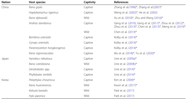
Table 1 List of amphibian ranavirus infection or mortality cases reported in Northeast Asia. Asterisks represent mass mortality event
Nation Host species Captivity References
China Rana grylio Captive Zhang et al.(1996)*, Zhang et al.(2001)* Hoplobatrachus tigerinus Captive Weng et al. (2002)*, He et al. (2002) Rana dybowskii Wild Xu et al. (2010)*, Zhu and Wang (2016)*
Andrias davidianus Captive Geng et al. (2010), Geng et al. (2011)*, Zhou et al. (2012)*, Zhou et al. (2013)*, Chen et al. (2013)*, Meng et al. (2014)* Wild Chen et al. (2013)*
Bombina orientalis Captive Kolby et al. (2014)* Cynops orientalis Captive Kolby et al. (2014)* Paramesotriton hongkongensis Captive Kolby et al. (2014)*
Rana nigromaculata Captive Mu et al. (2018)*, Yu et al. (2020)* Japan Hynobius nebulosus Captive Une et al. (2009a)*
Rana catesbeiana Wild Une et al. (2009b)* Dendrobates spp. Captive Une et al. (2014)* Phyllobates terribilis Captive Une et al. (2014)* Korea Pelophylax chosenicus Captive Kim et al. (2009)* Rana huanrenensis Wild Kwon et al. (2017)* Kaloula borealis Wild Park et al. (2017) Hyla japonica Wild Park et al. (2017)
We extracted whole genomic DNA from 3 to 5 mg of
liver tissue using the Qiagen DNeasy® Blood & Tissue
Kit (Qiagen, Hilden, Germany). For ranavirus strain
identification, the partial sequence of major capsid
pro-tein gene (MCP) was amplified using the specific primer
pairs (MCP4 and MCP5; Mao et al.
1997
). We ran
poly-merase chain reactions (PCR) following Mao et al.
(
1997
) with a negative control using nuclease-free water
and confirmed PCR products on 1% agarose gel by
elec-trophoresis (Mao et al.
1997
; Kwon et al.
2017
). PCR
was run on each sample at least twice to minimize viral
false-positive detection.
Finally, PCR products were purified using an
Accu-Prep® PCR Purification Kit (Bioneer, Daejeon, Korea)
and sequenced using the same primer set (Macrogen,
Seoul, Korea). Sequences were edited and assembled
using Geneious 9.1.8 (Biomatters Ltd., Auckland, New
Zealand), and aligned using ClustalW (Thompson et al.
2003
) for sequence comparison. For genetic relationship
with other iridoviruses, we performed a custom nested
BLAST using Geneious 9.1.8 and Bayesian Inference (BI)
analysis with 18 ranavirus MCP genes, obtained from
GenBank. The TIMef model was selected as the best
Akaike information criterion (AIC) scored model after
testing 56 nucleotide substitution models in
MOTELT-EST v3.7 (Posada and Crandall
1998
). We analyzed the
phylogenetic relationships among the iridoviruses by
ap-plying both maximum likelihood (ML) and Bayesian
in-ference (BI) methods in PAUP v4.0 (Swofford
2001
) and
MrBayes v3.2.47 (Ronquist et al.
2012
), respectively. For
ML analysis and phylogenetic branching, we applied
Bootstrap/Jackknife method, with 1000 bootstraps, and
used the tree-bisection-reconnection (TBR) method. The
BI analysis with Markov Chain Monte Carlo (MCMC)
method was executed using the MrBayes (v3.2.47)
soft-ware. With four random starting trees, we ran 1,000,000
generations, while sampling every 100 tree generations
and discarding the first 5% of the sampled generations
as burn-ins. Therefore, 500 of the 10,000 trees sampled
were discarded.
Results
We found abdominal inflammation and erythema on the
legs of seven collected frog specimens (Fig.
1
). All 22
collected frogs were confirmed infected with ranavirus
by PCR. Out of 22 PCR products, we obtained 13 partial
MCP DNA sequences (> 500 bp) due to low sample
quality. The obtained MCP DNA sequences (505 bp)
were identical and had 100% sequence similarity to the
haplotype (accession number KY264205) collected from
a ranavirus-infected Huanren frog (R. huanrenensis;
Kwon et al.
2017
). In addition, the MCP sequence
showed
99.8%
similarity
with
ranavirus
KRV-1
(HM133594) from South Korea, Rana catesbeiana virus
(AB474588) from Japan, and 99.6% similarity for FV3
(FJ459783) a soft-shelled turtle iridovirus (DQ335253;
Table
2
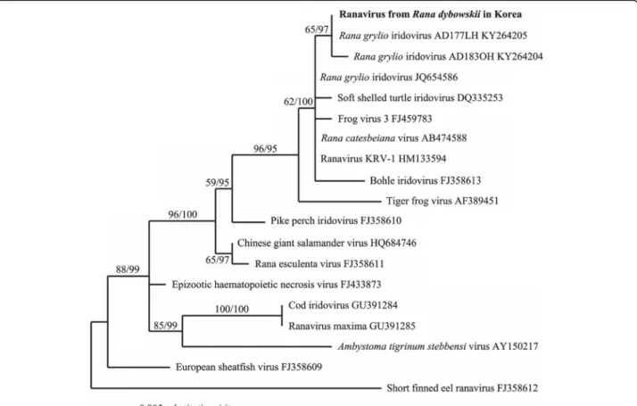
). The Bayesian inference (BI) phylogenetic
ana-lysis revealed that our sequenced ranavirus grouped with
a FV3-like virus including Rana grylio virus (RGV), FV3,
and Bohle iridovirus. The nested BLAST analysis was
consistent with our phylogenetic results.
Discussion
Diagnosing the physical symptoms of ranavirus infection
was often difficult due to the decomposition status of
the collected frog samples. Nevertheless, we found
ab-dominal inflammation and erythema on the legs of seven
frog specimens. Considering that erythema and skin
ul-cerations on the legs and ventrum, in amphibians, are
known external characteristics of ranavirus infection
(Gray et al.
2009
; Park et al.
2017
), we suspected our
specimens might be infected with ranavirus.
Results of our molecular analyses corroborated our
suspicions, as all frog specimens were confirmed infected
with ranavirus. These results suggest that FV3-like
rana-virus infections may be correlated with mass mortality
events in populations of adult R. dybowskii. This fact is
Fig. 1 The mountain valley (a) where the mass mortality of Rana dybowskii’s adults (b) occurred, associated with ranavirus-infection at Gyeyang-gu, Incheon, South Korea. Yellow arrowheads indicate dead frogs and in the insert, white arrowheads indicate red skins, a possible symptom of ranavirus infection
nothing new, as recent amphibian mass mortality events
have been correlated with ranavirus infections across
several studies (Weng et al.
2002
; Une et al.
2009a
;
Kwon et al.
2017
; Yu et al.
2020
). Within the pet trade,
ranavirus has been detected in a large number of
am-phibians, including cases in Hong Kong and Japan
(Kolby et al.
2014
; Une et al.
2014
). Ranavirus strains,
detected in the pet trade, were similar to the common
midwife toad virus (CMTV) and FV3-like viruses, like
the virus detected here. FV3-like viruses have been
doc-umented worldwide including Northeast Asia (Kim et al.
2009
; Xu et al.
2010
), Europe (de Matos et al.
2011
),
North and South America (Granoff et al.
1965
; Fox et al.
2006
), and Australia (Hengstberger et al.
1993
). To date,
at least 11 mass mortality events have been documented
in Northeast Asia and were caused by FV3-like viruses
(Zhang et al.
1996
; Zhou et al.
2012
). To this regard,
dis-tribution patterns of specific ranavirus strains across
Northeast Asia are still under investigation (Duffus et al.
2015
). FV3-like viruses have also been found in other
taxa such as turtles (Chen et al.
1999
) and fish (Ahne
et al.
1989
), highlighting the importance of ranavirus
screening across taxa.
Although the majority of ranavirus-associated
amphib-ian mortalities have occurred in captivity (Table
1
; Meng
et al.
2014
; Mu et al.
2018
), there have also been
con-firmed cases in wild populations. Wild
ranavirus-associated mortality events have occurred across Asia,
including the Heilongjiang, Jiangxi, and Henan provinces
in China (Xu et al.
2010
; Chen et al.
2013
; Zhu and
Wang
2016
), the western part of Japan (Une et al.
2009b
), and in Gangwon-do, Gyeongsangnam-do, and
Daejeon in South Korea (Kwon et al.
2017
; Park et al.
2017
). Various environmental factors are known to
in-crease ranavirus susceptibility and virulence in
amphib-ians and facilitate mortality (Brunner et al.
2015
). In this
study, two environmental factors may have contributed
to the mortality of adult R. dybowskii. First, the discovery
site was heavily altered with concrete for erosion
protec-tion 3 years prior, causing water stagnaprotec-tion. Stagnant
water during early spring drought periods may contain
high concentrations of various ions and pollutants (Kang
et al.
2016
), possibly increasing stress hormones and
making R. dybowskii individuals more susceptible to
in-fection (Gahl and Calhoun
2010
; Leduc
2014
). In a
pre-vious study, dead adult boreal digging frogs were
discovered in concrete walled, low circulation waterways
(Park et al.
2017
), similar to the environment observed in
this study. Future studies should determine if surface
alter-ations may influence amphibian-ranavirus susceptibility.
Second, amphibian mortality due to ranavirus has
often been correlated with elevated stress hormone
levels. Distinct life-history stages, including
metamor-phosis and reproduction, may be periods where frogs
have elevated stress, thus increasing their susceptibility
to infection (Green et al.
2002
; Duffus et al.
2008
; Gray
et al.
2009
). For example, ranavirus-linked mortality
events occurred during the metamorphosis stage of P.
chosenicus
and during the breeding season of K. borealis
(Kim et al.
2009
; Park et al.
2017
). Rana dybowskii is an
explosive breeding species that communally spawns
(Yoo and Jang
2012
), possibly resulting in elevated stress
hormones (Norris and Jones
2012
). Thus, highlighting a
need to understand how amphibian life history patterns
influence viral susceptibility and virulence.
Conclusion
Here, we document the first mass mortality event of R.
dybowskii
in the wild. All collected individuals were PCR
positive for ranavirus, possibly indicating that these
indi-viduals died due to viral infection. Elevated stress levels
by erosion control works and/or from natural
life-history stages may have contributed to ranavirus
infec-tion and mortality. To understand if ranavirus infecinfec-tion
is a threat to Korean amphibians there are three
conser-vation strategies, which should be implemented. First,
there is a need for continuous, country-wide, monitoring
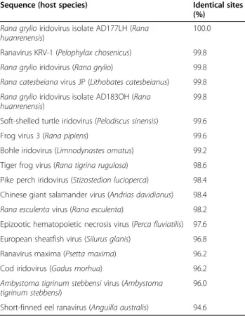
Table 2 Results of custom BLAST using partial major capsid
protein (MCP) DNA sequence (500 bp) of the ranavirus from
Rana dybowskii in this study. The accession numbers of the
sequences used for custom BLAST analysis are shown in Fig.
2
Sequence (host species) Identical sites (%)
Rana grylio iridovirus isolate AD177LH (Rana huanrenensis)
100.0 Ranavirus KRV-1 (Pelophylax chosenicus) 99.8 Rana grylio iridovirus (Rana grylio) 99.8 Rana catesbeiana virus JP (Lithobates catesbeianus) 99.8 Rana grylio iridovirus isolate AD183OH (Rana
huanrenensis)
99.8 Soft-shelled turtle iridovirus (Pelodiscus sinensis) 99.6 Frog virus 3 (Rana pipiens) 99.6 Bohle iridovirus (Limnodynastes ornatus) 99.2 Tiger frog virus (Rana tigrina rugulosa) 98.6 Pike perch iridovirus (Stizostedion lucioperca) 98.4 Chinese giant salamander virus (Andrias davidianus) 98.4 Rana esculenta virus (Rana esculenta) 98.2 Epizootic hematopoietic necrosis virus (Perca fluviatilis) 97.6 European sheatfish virus (Silurus glanis) 96.8 Ranavirus maxima (Psetta maxima) 96.2 Cod iridovirus (Gadus morhua) 96.2 Ambystoma tigrinum stebbensi virus (Ambystoma
tigrinum stebbensi)
96.0 Short-finned eel ranavirus (Anguilla australis) 94.6
of amphibian populations. Second, ranavirus screening
should be conducted across various taxa and not
rele-gated to just amphibians. Third, government officials or
researchers must identify which environmental factors
may increase amphibian susceptibility to ranavirus. By
implementing these three strategies, government officials
and researchers may be able to successfully protect
am-phibians from ranavirus infections in Korea and perhaps
globally.
Supplementary Information
The online version contains supplementary material available athttps://doi. org/10.1186/s41610-020-00179-2.
Additional file 1 PCR detection of the MCP sequence of ranavirus from the liver tissues of dead Rana dybowskii’s adults. The numbers on the bands represent individual frogs. P, positive MCP sequence control of ranavirus from Rana huarenensis tadpoles and N, negative control, which used nuclease-free water instead of extracted DNA in PCR process.
Abbreviations
AIC:Akaike information criterion; Bd: Batrachochytrium dendrobatidis; BI: Bayesian inference; CMTV: Common midwife toad virus; FV3: Frog virus 3; MCP: Major capsid protein; MCMC: Markov Chain Monte Carlo; ML: Maximum
likelihood; PCR: Polymerase chain reaction; RGV: Rana grylio virus; TBR: Tree-bisection-reconnection
Acknowledgements Not applicable
Authors’ contributions
JP analyzed experimental data and wrote the manuscript draft. IK performed the molecular experiment. NY performed the field survey. NH and AGP reviewed/edited the manuscript. DP designed the study and reviewed/ edited the manuscript draft. The authors read and approved the final manuscript.
Funding
This research was supported by the Basic Science Research Program through the National Research Foundation of Korea (NRF) funded by the Ministry of Education (No. 2020R1A6A3A1306094911).
Availability of data and materials
The datasets generated during and/or analyzed during the current study are available from the corresponding author on reasonable request.
Ethics approval and consent to participate
This study was approved by the Institutional Animal Care and Use Committee in Kangwon National University (KW-200618-3).
Consent for publication Not applicable.
Fig. 2 Bayesian inference (BI) tree based on the 19 partial major capsid protein (MCP) DNA sequences (500 bp) of the ranavirus obtained from dead Rana dybowskii and from GenBank. We used short-finned eel ranavirus as the outgroup. The accession number of iridoviruses, which was obtained from GenBank, indicated after each species name. On each branch, analyzed values (ML bootstrap value/Bayesian posterior probabilities) are denoted
Competing interests
The authors declare that they have no competing interests.
Author details
1Department of Regional Innovation, Kangwon National University,
Chuncheon, Kangwon 24341, Republic of Korea.2Department of Biological
Sciences, Northern Arizona University, Flagstaff, AZ 86011, USA.3Natural
Environmental Restoration Institute Co. Ltd., Daejeon, Republic of Korea.
4Division of Science Education, Kangwon National University, Chuncheon,
Kangwon 24341, Republic of Korea.5Rana Eco-Consultant, Namyangju,
Gyeonggi 12141, Republic of Korea.
Received: 14 September 2020 Accepted: 25 December 2020
References
Ahne W, Schlotfeldt HJ, Thomsen I. Fish viruses: isolation of an icosahedral cytoplasmic deoxyribovirus from sheatfish (Silurus glanis). J Vet Med B. 1989; 36(1-10):333–6.
Brunner JL, Storfer A, Gray MJ, Hoverman JT. Ranavirus ecology and evolution: from epidemiology to extinction. In: Gray MJ, Chinchar VG, editors. Ranaviruses. Cham: Springer; 2015. p. 71–104.
Carey C, Bradford DF, Brunner JL, Collins JP, Davidson EW, Longcore JE, et al. Biotic factors in amphibian population declines. In: Linder G, Krest SK, Sparling DW, editors. Amphibian decline: an integrated analysis of multiple stressor effects. Pensacola: SETAC Press; 2003. p. 153–208.
Carey C, Cohen N, Rollins-Smith L. Amphibian declines: an immunological perspective. Dev Comp Immunol. 1999;23(6):459–72.
Chen Z, Gui J, Gao X, Pei C, Hong Y, Zhang Q. Genome architecture changes and major gene variations of Andrias davidianus ranavirus (ADRV). Vet Res. 2013; 44(1):1–13.
Chen ZX, Zheng JC, Jiang YL. A new iridovirus isolated from soft-shelled turtle. Virus Res. 1999;63(1-2):147–51.
Daszak P, Berger L, Cunningham AA, Hyatt AD, Green DE, Speare R. Emerging infectious diseases and amphibian population declines. Emerg Infect Dis. 1999;5(6):735.
de Matos APA, da Silva Trabucho MFA, Papp T, Matos BADCA, Correia ACL, Marschang RE. New viruses from Lacerta monticola (Serra da Estrela, Portugal): further evidence for a new group of nucleo-cytoplasmic large deoxyriboviruses. Microsc Microanal. 2011;17(1):101–8.
Duffus ALJ, Pauli BD, Wozney K, Brunetti CR, Berrill M. Frog virus 3-like infections in aquatic amphibian communities. J Wildlife Dis. 2008;44(1):109–20. Duffus ALJ, Waltzek TB, Stöhr AC, Allender MC, Gotesman M, Whittington RJ,
et al. Distribution and host range of ranaviruses. In: Gray MJ, Chinchar VG, editors. Ranaviruses. Cham: Springer; 2015. p. 9–57.
Fey SB, Siepielski AM, Nusslé S, Cervantes-Yoshida K, Hwan JL, Huber ER, et al. Recent shifts in the occurrence, cause, and magnitude of animal mass mortality events. P Natl Acad Sci USA. 2015;112(4):1083–8.
Fox SF, Greer AL, Torres-Cervantes R, Collins JP. First case of ranavirus-associated morbidity and mortality in natural populations of the South American frog Atelognathus patagonicus. Dis Aquat Organ. 2006;72(1):87–92.
Gahl MK, Calhoun AJK. The role of multiple stressors in ranavirus-caused amphibian mortalities in Acadia National Park wetlands. Can J Zool. 2010; 88(1):108–21.
García-Díaz P, Ross JV, Woolnough AP, Cassey P. Managing the risk of wildlife disease introduction: pathway-level biosecurity for preventing the introduction of alien ranaviruses. J Appl Ecol. 2017;54(1):234–41.
Geng Y, Wang K, Li C, Wang J, Liao Y, Huang J. Zhou Z. PCR detection and electron microscopic observation of bred Chinese giant salamander infected with ranavirus associated with mass mortality. Vet. Sci China. 2010;40(8):817–21. Geng Y, Wang KY, Zhou ZY, Li CW, Wang J, He M, et al. First report of a ranavirus
associated with morbidity and mortality in farmed Chinese giant salamanders (Andrias davidianus). J Comp Pathol. 2011;145(1):95–102. Granoff A, Came PE, Rafferty KA Jr. The isolation and properties of viruses from
Rana pipiens: their possible relationship to the renal adenocarcinoma of the leopard frog. Ann NY Acad Sci. 1965;126(1):237–55.
Gray MJ, Miller DL, Hoverman JT. Ecology and pathology of amphibian ranaviruses. Dis Aquat Organ. 2009;87(3):243–66.
Green DE, Converse KA, Schrader AK. Epizootiology of sixty-four amphibian morbidity and mortality events in the USA, 1996-2001. Ann NY Acad Sci. 2002;969(1):323–39.
He JG, Lü L, Deng M, He HH, Weng SP, Wang XH, et al. Sequence analysis of the complete genome of an iridovirus isolated from the tiger frog. Virology. 2002;292(2):185–97.
Hengstberger SG, Hyatt AD, Speare R, Coupar BEH. Comparison of epizootic haematopoietic necrosis and Bohle iridoviruses, recently isolated Australian iridoviruses. Dis Aquat Organ. 1993;15(2):93–107.
Kang MJ, Kim KD, Oh KS, Park JW, Park JH. Analysis of forest environmental factors on torrent erosion control work area in Gyeongsangnam-do: focus on erosion control dam and stream conservation. J Agric & Life Sci. 2016;50(5): 111–20 (in Korean).
Kim S, Sim MY, Eom AH, Park D, Ra NY. PCR detection of ranavirus in gold-spotted pond frogs (Rana plancyi chosenica) from Korea. Korean J Environ Biol. 2009;27(1):110–3.
Kolby JE, Smith KM, Berger L, Karesh WB, Preston A, Pessier AP, Skerratt LF. First evidence of amphibian chytrid fungus (Batrachochytrium dendrobatidis) and ranavirus in Hong Kong amphibian trade. PloS one. 2014;9(3):e90750. Kwon S, Park J, Choi WJ, Koo KS, Lee JG, Park D. First case of ranavirus-associated
mass mortality in a natural population of the Huanren frog (Rana huanrenensis) tadpoles in South Korea. Anim Cells Syst. 2017;21(5):358–64. Leduc J. Life-history trade-offs in Northern leopard frog (Lithobates [Rana] pipiens)
tadpoles: interactions of trace metals, temperature, and ranavirus. PhD Thesis, Laurentian University of Sudbury; 2014.
Mao J, Hedrick RP, Chinchar VG. Molecular characterization, sequence analysis, and taxonomic position of newly isolated fish iridoviruses. Virology. 1997;229(1):212–20. Meng Y, Ma J, Jiang N, Zeng LB, Xiao HB. Pathological and microbiological
findings from mortality of the Chinese giant salamander (Andrias davidianus). Arch Virol. 2014;159(6):1403–12.
Miller D, Gray M, Storfer A. Ecopathology of ranaviruses infecting amphibians. Viruses. 2011;3(11):2351–73.
Miller D, Pessier A, Hick P, Whittington R. 2015. Comparative pathology of ranaviruses and diagnostic techniques. In: Gray MJ, Chinchar VG. Ranaviruses. Cham: Springer; 2015. p. 171-208.
Mu WH, Geng Y, Yu ZH, Wang KY, Huang XL, Ou YP, et al. FV3-like ranavirus infection outbreak in black-spotted pond frogs (Rana nigromaculata) in China. Microb Pathogenesis. 2018;123:111–4.
Norris DO, Jones RE. Hormones and reproduction in fishes, amphibians, and reptiles. Berlin: Springer Science & Business Media; 2012.
O’hanlon SJ, Rieux A, Farrer RA, Rosa GM, Waldman B, Bataille A, et al. Recent Asian origin of chytrid fungi causing global amphibian declines. Science. 2018;360(6389):621–7.
Park IK, Koo KS, Moon KY, Lee JG. Park D. PCR detection of ranavirus from dead Kaloula borealis and sick Hyla japonica tadpoles in the wild. Korean. J Herpetol. 2017;8:10–4.
Posada D, Crandall KA. Modeltest: testing the model of DNA substitution. Bioinformatics. 1998;14(9):817–8.
Ronquist F, Teslenko M, Van Der Mark P, Ayres DL, Darling A, Höhna S, et al. MrBayes 3.2: efficient Bayesian phylogenetic inference and model choice across a large model space. Syst Biol. 2012;61(3):539–42.
Scheele BC, Pasmans F, Skerratt LF, Berger L, Martel A, Beukema W, et al. Amphibian fungal panzootic causes catastrophic and ongoing loss of biodiversity. Science. 2019;363(6434):1459–63.
St-Amour V, Lesbarrères D. Genetic evidence of Ranavirus in toe clips: an alternative to lethal sampling methods. Conserv Genet. 2007;8(5):1247. Swofford DL. Paup*: Phylogenetic analysis using parsimony (and other methods).
Sunderland: Sinauer Associates; 2001. Ver.4.0.b5.
Thompson JD, Gibson TJ, Higgins DG. Multiple sequence alignment using ClustalW and ClustalX. Curr Protoc Bioinformatics. 2003;(1):2–3.
Une Y, Kudo T, Tamukai KI, Murakami M. Epidemic ranaviral disease in imported captive frogs (Dendrobates and Phyllobates spp.), Japan, 2012: a first report. JMM Case Rep. 2014;1(3):e001198.
Une Y, Nakajima K, Taharaguchi S, Ogihara K, Murakami M. Ranavirus infection outbreak in the salamander (Hynobius nebulosus) in Japan. J Comp Pathol. 2009a;4(141):310. Une Y, Sakuma A, Matsueda H, Nakai K, Murakami M. Ranavirus outbreak in North
American bullfrogs (Rana catesbeiana), Japan, 2008. Emerg Infect Dis. 2009b; 15(7):1146.
Warne RW, Crespi EJ, Brunner JL. Escape from the pond: stress and
developmental responses to ranavirus infection in wood frog tadpoles. Funct Ecol. 2011;25(1):139–46.
Weng SP, He JG, Wang XH, Lü L, Deng M, Chan SM. Outbreaks of an iridovirus disease in cultured tiger frog, Rana tigrina rugulosa. in southern China. J Fish Dis. 2002;25(7):423–7.
Xu K, Zhu DZ, Wei Y, Schloegel LM, Chen XF, Wang XL. Broad distribution of ranavirus in free-ranging Rana dybowskii in Heilongjiang, China. EcoHealth. 2010;7(1):18–23.
Yang H, Baek H, Speare R, Webb R, Park S, Kim T, et al. First detection of the amphibian chytrid fungus Batrachochytrium dendrobatidis in free-ranging populations of amphibians on mainland Asia: survey in South Korea. Dis Aquat Organ. 2009;86(1):9–13.
Yoo E, Jang Y. Abiotic effects on calling phenology of three frog species in Korea. Anim Cells Syst. 2012;16(3):260–7.
Yu Z, Mou W, Geng Y, Wang K, Chen D, Huang X, et al. Characterization and genomic analysis of a ranavirus associated with cultured black-spotted pond frogs (Rana nigromaculata) tadpoles mortalities in China. Transbound Emerg Dis. 2020.https://doi.org/10.1111/tbed.13534.
Zhang QY, Li ZQ, Jiang YL, Liang SC, Gui JF. Preliminary studies on virus isolation and cell infection from disease frog Rana grylio. Acta Hydrobiol Sin. 1996; 20(4):390–2 (in Chinese).
Zhang QY, Xiao F, Li ZQ, Gui JF, Mao J, Chinchar VG. Characterization of an iridovirus from the cultured pig frog Rana grylio with lethal syndrome. Dis Aquat Organ. 2001;48(1):27–36.
Zhou ZY, Geng Y, Liu XX, Ren SY, Zhou Y, Wang KY, Huang XL, et al. Characterization of a ranavirus isolated from the Chinese giant salamander (Andrias davidianus, Blanchard, 1871) in China. Aquaculture. 2013;384:66–73. Zhou ZY, Geng Y, Ren SY, Wang KY, Huang XL, Chen DF, et al. Ranavirus (family Iridoviridae) detection by polymerase chain reaction (PCR) in Chinese giant salamander (Andrias davidianus, Blanchard, 1871), China. Afr J Biotechnol. 2012;11(85):15130–4.
Zhu YQ, Wang XL. Genetic diversity of ranaviruses in amphibians in China: 10 new isolates and their implications. Pak J Zool. 2016;48:107–14.
Publisher
’s Note
Springer Nature remains neutral with regard to jurisdictional claims in published maps and institutional affiliations.