원발성 대장-결장암 환자의 치료 전 PET/CT 스캔에서
FDG 섭취 정도와 병리학적 및 면역조직화학적
지표들과의 비교
가톨릭대학교 의과대학 방사선과학교실1, 병원병리학교실2
나세정
1․정용안
1․맹이소
2․김기준
1․손경명
1․김성훈
1․손형선
1․정수교
1Comparison between FDG Uptake and Pathologic or Immunohistochemical
Parametersin Pre-operative PET/CT Scan of Patient with Primary Colorectal
Cancer
Sae-Jung Na, M.D.
1, Yong-An Chung, M.D.
1, Lee-So Maeng, M.D.
2, Ki-Jun Kim, M.D.
1,
Kyung-Myung Sohn, M.D.
1, Sung-Hoon Kim, M.D.
1, Hyung-Sun Sohn, M.D.
1, and Soo-Kyo Chung, M.D.
1Departmentsof 1Radiology and 2Hospital Pathology, College of Medicine, The Catholic University of Korea, Seoul, Korea
Purpose: To evaluate the relationship between F-18 FDG uptake of tumor in PET/CT scan and pathological or immunohistochemial parameters of colorectal cancer. Materials and Methods: 147 colorectal cancer patients who underwent both pre-operative F-18 FDG PET/CT scan and surgery were included. In cases with perceptible FDG uptake in primary tumor, the maximum standardized uptake value (SUVmax) was calculated. The pathologic results such as site, size, depth of invasion (T stage), growth pattern, differentiation of primary tumor, lymph node metastasis and Dukes-Astler & Coller stage and immunohistochemical markers such as expression of EGFR, MLH1, MSH2 and Ki-67 index were reviewed. Results: 146 out of 147 PET/CT scans with colorectal cancer showed perceptible focal FDG uptake. SUVmax showed mild positive linear correlation with size of primary tumor (r=0.277, p=0.001) and Ki-67 index (r=0.226, p=0.019). No significant difference in F-18 FDG uptake was found according to site, depth of invasion (T stage), growth pattern, differentiation of primary tumor, presence of lymph node metastasis, Dukes-Astler & Coller stage and expression of EGFR. Conclusion: The degree of F-18 FDG uptake in colorectal cancer was associated with the size and the degree of Ki-67 index of primary tumor. It could be thought that FDG uptake of primary tumor has a correlation with macroscopic and microscopic tumor growth. (Nucl Med Mol Imaging 2009;43(6):557-564)
Key Words: Colorectal cancer, F-18 FDG, PET/CT
∙Received: 2009. 7. 24. ∙Revised: 2009. 9. 18. ∙Accepted: 2009. 12. 8.
∙Address for reprints: Ki-Jun Kim, M.D., Ph.D., Department of Radiology, Incheon Saint Mary’s Hospital, The Catholic University of Korea, 665 Bupyeong-dong, Bupyeong-gu, Incheon 403-720, Korea Tel: 82-32-510-5849, Fax: 82-32-529-0964 E-mail: [email protected] ※이 논문은 2009년도 정부(교육과학기술부)의 재원으로 한국연구 재단-신기술융합형 성장동력사업의 지원을 받아 수행된 연구임 (No. 2009-0082087).
서 론
대장-결장암(Colorectal cancer, 이하 CRC)은 서양에서
암 관련 사망률의 3번째 순위에 있으며, 2005년에 약 오만
명 이상이 사망한 것으로 보고되었다.
1)우리 나라에서도
2005년 기준으로 인구 10만 명당 36.2명의 새로운 환자가 발
생하여 발생 빈도가 빠르게 증가하고 있는 암 가운데 하나
이며 발생 빈도와 암 사망률 모두 4번째로 높은 암이다.
2)F-18 fluorodeoxyglucose (F-18 FDG)을 이용한 양전자
방출단층촬영술(positron emission tomography, 이하
PET)은 암의 진단, 병기 평가, 그리고 치료 후 반응평가
등에 있어 세계적으로 널리 사용되고 있으며 최근 몇 년
동안 CRC 환자에서 PET 이용은 증가되고 있는 추세이다.
CRC 환자의 치료를 위한 계획과 성공을 위해서는 정확
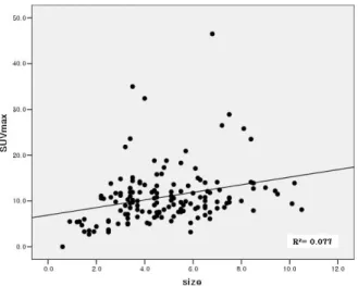
Figure 1. This graph present SUVmax of primary tumor showed mild positive linear correlation with size of primary tumor.
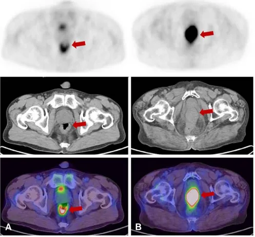
Figure 2. A 56-year-old female underwent pre-operative PET/CT (A). In the axial PET/CT images, focal FDG uptake is noted in the rectum (SUVmax 3.2). 2.5 cm sized rectal cancer (T3) with lymph node metastasis was confirmed by operation. A 74-year-old female underwent pre-operative PET/CT (B). In the axial PET/CT images, focal FDG uptake is noted in the rectum (SUVmax 23.5). 8.4 cm sized rectal cancer (T3) with lymph node metastasis was confirmed by operation.
적 목적으로도 유용할 뿐만 아니라 재발의 발견에도 도움
이 되어 치료 방침 결정에 중요한 역할을 하는 것으로 잘
알려져 있다.
4-8)Gambhir 등이 여러 연구들을 분석하여 보
고한 결과를 보면, CRC의 진단 및 병기 설정을 하는데 있
어 민감도는 85%, 특이도는 71%이며, 재발을 진단하는데
있어서는 민감도는 94%, 특이도는 87%였다.
9)또한 어떤
연구들에서는 FDG 섭취 정도에 따라 예후나 치료 반응을
예측할 수 있다고 보고하기도 하였다.
10,11)이에 저자들은 현재 일반적으로 예후와 관련성이 있다
고 알려져 있거나 관련성이 의심이 되는 병리학적, 면역조
직화학적 지표들과 원발성 CRC의 F-18 FDG 섭취 정도와
어떤 연관이 있는지를 후향적으로 분석해 보았다.
대상 및 방법
1. 환자군
2008년 1월부터 2008년 12월까지 대장 내시경 또는 다른
영상 검사에서 CRC가 진단되거나 의심되어 초기 병기 설정
을 위해 F-18 FDG PET/CT를 시행한 사람들 중에서 다른
치료 없이 수술을 받은 환자들을 대상으로 하였다. 수술 검체
조직 검사상 종양 세포가 남아 있지 않은 4명, 악성 림프종
으로 진단된 2명, 심한 폐쇄성 종양으로 우회 수술만 한 1
명은 환자군에서 제외시켰다. 총 147명(여자 67명, 남자
A
B
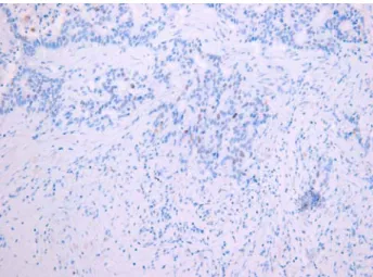
Figure 3. Ki-67showsreactivityless than.
Pathologic parameters Number SUVmax
(meanSD) p value Location Ascending colon Transverse colon Descending colon Sigmoid colon Rectosigmoid Rectum 26 5 4 41 6 65 12.15.9 6.03.4 9.31.7 10.47.9 10.03.0 10.75.5 0.182
Tumor invasion Tis, T1, T2
T3 and T4
27 120
9.46.1
11.36.2 0.111
Tumor growth pattern
Expanding Expanding and infiltrative
Infiltrative 21 118 8 9.35.4 11.36.3 10.66.2 0.312 LN metastasis Negative Positive 54 90 11.37.4 11.05.3 0.727 Differentiation Well Moderate Poor 4 122 12 7.45.1 10.96.2 11.65.1 0.496
Dukes-Astler & Coller stage
A B C D 4 51 77 12 9.64.4 11.47.5 11.25.6 9.53.1 0.780 Table 1. The SUVmax of Primary Tumor According to Pathological Parameters in Colorectal Cancer
80명)이었고, 평균 나이는 62±11세(범위 31-93세)이었다.
대상군의 조직아형은 138명은 선암종이었으며, 9명은 점액
성 선암종이었다. 선암종 138명 중 4명은 mucin component,
한 명은signet ring cell component가 포함되어 있었다.
2. 영상 획득 및 분석
모든 환자들은 F-18 FDG PET/CT 스캔(Biograph Duo,
Biograph Truepoint; Siemens Medical Solutions,
Knoxville, TN, USA)을 시행하였고, 검사 시행 전 최소 6
시간 이상 금식을 하고, 검사 전 혈당치는 130 mg/dL 이
하였다. 환자들은 F-18 FDG 370~550 MBq을 정맥 주사
후 1시간 동안 침대에서 안정을 취하였다. PET/CT 스캔
은 환자 두개골 기저부에서 근위 대퇴부까지 감쇠보정용
CT를 먼저 얻었고, 조영제 는 사용하지 않았다. PET은 베
드 당 2.5분씩 영상을 얻었다. 얻어진 영상 자료들은
OSEM (168x168 matrix, 3.5 mm slice thickness, subset:
8, Iteration: 4, Gaussian-Filter: 5.0 FWHM (mm),
Diameter: 70 cm) 방식을 통해 재구성 하였다. 원발 부위
의 F-18 FDG 섭취 정도를 반정량적으로 평가하기 위하여
주변 대장-직장 조직과 구별되는 인지 가능한 원발 부위에
관심영역 (region of interest, ROI)를 그리고, 최대 표준화
섭취계수(maximum standardized uptake value, 이하
SUVmax)를 구하였다. PET/CT를 시행하고 수술까지의
시간은 평균 3.93.1일(범위 0-20일)이었다.
3. 병리학적 및 면역조직화학적 지표
수술 후 원발 종양의 위치, 크기, 침습 정도(T 병기), 종
양 성장 패턴, 림프절 전이 여부, Dukes-Astler & Coller 병
기, 분화도를 기술한 환자의 병리 기록지를 검토하였다. 총
147명 중 림프절박리가 시행되지 않은 3명은 림프절 전이
여부와 Dukes-Astler & Coller 병기 자료를 결손치로 처리
하였다. 분화도는 점액성 선암종인 9명에서는 확인이 되지
않아 역시 결손치로 처리하였다. Epidermal Growth Factor
receptor (EGFR), MutL homolog 1 (MLH1), mutS
homolog 2 (MSH2) 그리고 Ki-67 와 같은 면역조직화학적
지표들을 일부의 환자들에서 확인하여 함께 분석하였다.
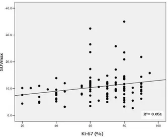
Figure 4. This graph present SUVmax of primary tumor showed mild positive linear correlation with Ki-67 index.
각 환자의 파라핀 포매조직을 5m두께로 연속 절편을 만
들어 pH7.4 Tris 완충액 수세를 한 후, 5분간 3% 과산화수
소에 작용시켜 내인성 peroxidase의 작용을 차단시켰다. 그
후 다시 Tris 완충액으로 수세 및 정상 염소 혈청으로 5분
간의 작용을 통해 비특이적 단백질의 결합을 차단하였다.
그리고 각각의 조직에 Ki-67 (1:50, Dako, USA), MLH-1
(1:25, BD Bioscience, USA), MHS-2 (1:50, BD
Bioscience, USA) 단백에 대한 일차항체를 40C에서 20분
간 반응시켰다. 다시 pH7.6인 Tris완충액으로 수세한 다음,
biotin이 부착된 이차항체를 40C에서 10분간 반응한 후
Tris 완충액으로 수세하였고, streptoavidine-peroxidase 와
10분간 반응시켰다.. Tris 완충액 세척 후 3-amino-9-ethyl
carbazole로 발색한 후 10% Mayer’s hematoxlyin으로 대
조염색하여 수세하였다. EGFR pharmDx kit
(Dakocyto-mation, carpinteria, CA)로 염색하였는데 간략하게 정리하
면 조직은 함수 한 후 proteinase K 처리하고 peroxidase
처리 후 일차항체를 넣어 배양하였다. 다음 과정은 위와 동
일하다. 염색 결과 판독은 2명의 병리 전문의에 의하여 과
거의 조직병리학적, 임상적 정보 없이 시행되었다. Ki-67의
경우 1000개의 세포 격자를 이용하여 양성으로 나온 종양
세포의 비율을 퍼센트로 정하여, 5~49%는 저증식, 50%이
상을 고증식으로 분류하였다. EGFR은 종양세포들중 1%
이상 세포막 발현을 보인 경우를 양성으로 판정하였고
MLH-1와 MSH-2는 종양세포의 핵에서 전혀 발현되지 않
은 것을 음성으로 판정하였다.
4. 통계 분석
각 환자들의 병리학적 및 면역조직화학적 지표들에 따
라 CRC의 F-18 FDG 섭취 정도가 차이가 있는지를 알아
보기 위하여 Kruskal-wallis test, Mann-whitney U test,
이변량 상관분석(bivariate correlation)을 이용하여 분석하
였다. 통계분석용 프로그램(SPSS version 13.0, SPSS Inc.,
Chicago, IL)을 이용하였고, 통계적 유의성은 p값 0.05를
기준으로 하였다.
결 과
총 147명의 환자 중 8명에서 수술 후 대장-결장 내 원발
병소가 두 군데에서 발견되었다. 이 환자들에서 발견된 두
개의 대장-결장 종양 중 크기가 큰 종양의 조직 소견을 기
준으로 하여 분석하였다. 수술 후 두 개의 원발 병소가 확
인된 환자 8명 중 7명의 PET/CT에서 각각의 병소와 일치
하는 인지 가능한 국소 FDG 섭취가 두 군데에서 모두 보
였고, 크기가 작은 종양들의 SUVmax는 평균 8.1±3.4(범위
3.1~13.9)로 측정되었다. 나머지 한 명의 PET/CT에서는
큰 병소에서만 FDG 섭취를 보이고 나머지 작은 병소에서
는 인지 가능한 FDG 섭취를 보이지 않았다.
총 147명 중 한 환자의 PET/CT 에서 인지 가능한
FDG 섭취를 보이지 않았고, 나머지 146명의 환자에서는
SUVmax가 평균 11.0±6.1 (범위 2.7~46.5)으로 관찰되었
다. 대장-결장 종양의 SUVmax는 종양의 크기와 약한 양
적 선형 관계를 보였다(r=0.277, p=0.001)(Fig1, 2). 그 외
종양의 위치, 침습 정도, 종양 성장 패턴, 림프절 전이 여
부, Dukes-Astler & Coller 병기, 분화도에 따라 살펴본 원
발 종양의 SUVmax는 모두 통계적으로 유의한 차이를 보
이지 않았다(Table 1).
면역화학적 지표들 중 Ki-67 표지율은(Fig 3) 147명
중 108명에서 분석 가능하였고, 그 결과 표지율이 낮은
그룹(<50%, n=23)과 높은 그룹(≥50%, n=85)간에서
SUVmax가 유의한 차이를 보였다(7.6±2.8 vs. 11.6±5.8,
p=0.002)(Fig. 4). 또한 약한 양적 선형 관계가 있음을 알
수 있었다(r=0.226, p 0.019). EGFR 발현 여부는 147명 중
120명에서 검사하였고 양성을 보인 환자가 71명, 음성을 보
인 환자가 49명이었다. T3 이상의 조직 침습을 보인 경우와
림프절 전이를 보인 환자는 EGFR 양성 그룹에서 61명
(85.9%), 47명(66.2%)이었고, 음성 그룹에서 36명(73.5%),
32명(65.3%)이었다. EFGR 발현 여부에 따른 두 그룹 간
의 FDG 섭취는 유의한 차이를 보이지 않았다(p=0.534).
MLH1과 MSH2의 발현 여부는 119명에서 확인이 가능하
였고 발현된 그룹과 발현되지 않은 그룹 사이에서 FDG 섭
취의 유의한 차이를 보이지 않았다(Table 2).
Immunohistochemical parameters Number SUVmax (meanSD) p value EGFRa) Negative Positive 49 71 10.35.0 10.95.7 0.808 MLH1b) Negative Positive 8 111 14.05.2 10.55.4 0.077 MSH2c) NegativePositive 1154 10.43.810.85.5 0.881 Ki-67 < 50%> 50% 2385 11.65.87.62.8 0.000 a)
Epidermal Growth Factor receptor, b)MutL homolog 1, c)mutS homolog 2
Table 2. The SUVmax of Primary Tumor According to Expression of Immunohistochemical Parameters in the Colorectal Cancer Figure 5. A 53-year-old male underwent pre-operative PET/CT (A). In the axial PET/CT images, focal FDG uptake is noted in the rectum (SUVmax 5.2). 4.1 cm sized rectal cancer (T3) without lymph node metastasis was confirmed by operation, and Ki-67 index was 40 % in immunohistochemistry. A 77-year-old female underwent pre-operative PET/CT (B). In the axial PET/CT images, focal FDG uptake is noted in the rectum (SUVmax 13.5). 4.0 cm sized rectal cancer (T1) without lymph node metastasis was confirmed by operation, and Ki-67 index was 80 % in immunohistochemistry.
고 찰
재발이 의심되거나 간 전이가 의심되는 CRC 환자의
F-18 FDG PET 연구는 매우 많고 그 유용성은 널리 알려
져 있다. 또한 최근에는 CRC 환자의 새로운 진단을 위한
수술 전 연구도 그 유용성에 대해 보고 되고 있다.
Abdel-Nabi 등은 CT와 F-18 FDG PET을 CRC 환자에서
비교하였고, 원발 종양에 대한 PET의 민감도가 100%, 특
이도가 43%, CT는 민감도와 특이도가 각각 37%, 83%로
보고하였다.
12)또한 Mukai 등은 CRC의 원발 병소 진단 민
A
B
감도를 96%로 보고하였다.
13)이번 저자들의 연구에서는
총 147명 중 1명만이 원발 병소를 확인 할 수 없었고 나머
지 146명에서는 모두 CRC 원발 병소를 확인하여 민감도
99%로 F-18 FDG PET의 유용성을 확인할 수 있었다.
이번 저자들의 주 연구 목적은 CRC를 대상으로, 원발
종양의 F-18 FDG 섭취에 영향을 미치는 인자들을 알아보
고자 하였다. 총 147명 중 유일하게 PET/CT 스캔에서 인
지 가능한 FDG 섭취를 보이지 않았던 한 명은 원발 종양
의 크기가 0.6cm로 대상 환자 중 종양의 크기가 가장 작은
환자였다. 나머지 146명에서 종양의 크기와 FDG 섭취의
관계를 보았을 때 CRC의 크기가 증가할수록 FDG 섭취 정
도도 의미 있게 증가하였다(r=0.277, p=0.001). Jinyu 등
14)은 CRC 환자에서 종양의 크기와 침습 정도가 FDG 섭취
와 연관성이 있다고 보고하였다. 이는 종양의 크기가 클수
록 종양 내의 세포 수가 증가하게 되고, 따라서 FDG 섭취
도 비례하여 증가한다고 생각할 수 있다. 종양 크기와
FDG 섭취의 관련성을 언급하기 위해서는 부분 용적 효과
(partial volume effect)를 고려하지 않을 수 없지만 이 연
구에 포함된 종양의 크기가 평균 4.90±2.1cm(범위 0.6~
10.5cm)이었고 147명 중 137명(93%)에서 종양의 크기가
1cm 이상으로 크다는 점을 생각해 보았을 때 부분 용적 효
과의 영향은 큰 고려의 대상이 아닐 듯 하다. 하지만 보다
정확한 결과를 얻기 위해서는 부분 용적 효과의 교정을 시
행한 연구가 추후에 필요할 것으로 생각된다. 그 외 이번
연구에서 함께 분석한 종양의 위치, 침습 정도(T 병기), 성
장 패턴, 분화도, 림프절 전이 여부, Dukes-Astler & Coller
병기와 같은 다른 인자들은 원발 종양의 FDG 섭취와 유의
한 연관성이 없는 것으로 나타났다.
Ki-67 표지율은 세포 증식과 관련된 인자로, G0의 휴지
기를 제외한 증식하고 있는 세포의 핵에서 발현된다. 따라
서 조직에서의 성장 분획을 측정하는 지표로 사용할 수 있
고, 이에 대한 여러 연구에서 세포 증식 지표로 Ki-67 표지
율이 종양의 진행과 관련이 있고, CRC 예후 예측 가능 인
자로 사용될 수 있다고 보고하였다.
15-19)이 연구에서는
Ki-67 표지율이 낮은 그룹(50% 미만)과 높은 그룹(50%
이상) 사이에 유의한 FDG의 섭취의 차이(p=002)를 보였
을 뿐 아니라 약한 양적 선형 관계를 보여 증식하는 세포
의 정도에 따라 FDG 섭취가 증가함을 알 수 있다.
EGFR은 세포 표면에 있는 수용체로 세포의 분열과 생
존에 관련된 신호 전달 체계에 관여하는데 CRC의 환자에
서 발현률이 25%에서 77%정도 증가되어 있다고 알려져
있다.
20-22)일부의 연구에서 EGFR의 발현은 좀 더 침습적
인 병과 관련이 있어, 전이의 위험도, 종양의 침습 정도, 림
프절 전이의 가능성이 증가한다고 보고 하였지만, 많은 연
구에서 불량한 예후 인자들과 EGFR의 관련이 없다고 보
고하고 있다.
21-25)이 연구의 결과 역시 FDG 섭취가 EGFR
발현 여부와 관련성이 떨어지는 것으로 나타났다.
산발성 CRC 환자의 15%에서 반복서열 불안정성
(microsatellite instability, MSI)가 관찰되고, 이는 MLH1,
MSH2와 같은 DNA 틀린 짝 수복(mismatch repair) 단백
질의 결함으로 인해 발생한다고 알려져 있다.
26)많은 연구
결과에서 MSI는 좋은 예후와 관련이 있는 것으로 보고되
었다.
27-29)이 연구에서 MLH1, MSH2 음성인 그룹과 양성
인 그룹 간에 유의한 FDG 섭취의 차이를 보이지 않았다.
이는 PET에서 FDG 섭취 정도로 평가할 수 있는 세포의
대사 활성도는 원발 종양의 MLH1, MSH2의 결손 여부와
직접적인 관련을 없음을 시사한다고 볼 수 있겠다. 또한
MLH1, MSH2가 MSI와 관련이 있긴 하지만 MSI 여부와
정도를 직접적으로 나타내는 것이 아니므로 예후와 직접적
인 관련이 있는 MSI 정도와 FDG 섭취를 비교하는 연구가
더 필요할 것으로 생각된다.
이 연구의 제한점은 후향적 연구의 제약으로 연구대상
의 선택에 있어 정확한 포함 기준과 제외 기준을 제시할
수 없어 선택오차에 대한 문제가 있을 수 있다. 그리고 세
포의 FDG 섭취와 직접적인 연관이 있는 포도당 운반체와
(glucose transporter)와 헥소키나제(hexokinase)와 같은
면역조직화학적 지표들에 대한 연구가 함께 이루어 지지
않았다는 점이다. 또한 CRC 환자에서 원발 종양의 FDG
섭취가 실제 환자의 생존율이나 예후와 어떤 상관 관계를
가지는 지를 직접적으로 평가하지 못하였다. 따라서 이들
에 대하여는 추후에 추가적인 연구가 필요할 것으로 생각
된다.
결론적으로 CRC 환자 147명 중 한 명을 제외한 나머지
146명에서 인지 가능한 F-18 FDG 섭취를 보여 높은 진단
능을 나타내었다. 그리고 FDG의 섭취 정도는 종양의 크기,
Ki-67 표지율과 통계적으로 유의한 연관성이 있었다. 이는
종양 세포의 수가 많을수록 또한 증식하는 세포가 많을수
록 FDG 섭취가 증가한 것으로 생각할 수 있다. 그러나 원
발 종양의 FDG 섭취는 종양의 위치나 일반적으로 예후와
관련이 있는 침습 정도(T 병기), 림프절 전이 여부, 분화도
등과는 뚜렷한 상관 관계를 보이지 않았다.
요 약
목적: CRC 환자의PET/CT 스캔에서 FDG 섭취에 영
향을 주는 임상병리학적 및 면역조직화학적 지표들이 있는
지 알아보고자 하였다. 대상 및 방법: 본원에 내원하여 치
료 전 F-18 FDG PET/CT 스캔을 시행한 147명의 CRC
환자를 대상으로 하였다. PET/CT 영상에서 원발 종양의
SUVmax를 구한 후 종양의 위치, 크기, 침습 정도(T 병
기), 종양 성장 패턴, 림프절 전이 여부, Dukes-Astler &
Coller 병기, 분화도, EGFR, MLH1, MSH2 발현 유무 그리
고 Ki-67 표지율과 같은 임상병리학적 및 면역조직화학적
지표들과의 상관 관계를 분석하였다. 결과: CRC 환자 147
명의 치료 전 PET/CT 스캔 중 146명의 스캔에서 인지 가
능한 FDG 섭취가 확인되었다. FDG 섭취는 종양의 크기와
약한 양적 선형 관계를 보였다(r=0.277, p=0.001). 원발
종양 조직에서 Ki-67 표지율이 50% 이상인 그룹에서의
SUVmax는 50% 미만인 그룹에서의 SUVmax 보다 의미
있게 높았고(7.62.8 vs. 11.65.8, p=0.002), Ki-67 표지율 정
도와 FDG 섭취 간에 약한 양적 선형 관계를 보였다
(r=0.226, p=0.019). 그 외 종양의 위치, 침습 정도(T 병
기), 종양 성장 패턴, 림프절 전이 여부, Dukes-Astler &
Coller 병기, 분화도, EGFR, MLH1, MSH2 발현 유무에 따
른 CRC의 SUVmax는 통계학적으로 유의한 차이가 없었
다. 결론: CRC에서 F-18 FDG의 섭취 정도는 종양의 크
기, Ki-67 표지율과 연관성이 있어 종양의 거시적 그리고
미시적 성장에 따라 FDG 섭취가 증가함을 알 수 있었다.
References
1. Jemal A, Murray T, Ward E, Samuels A, Tiwari RC, Ghafoor A, et al. Cancer statistics, 2005. CA Cancer J Clin 2005;55:10-30. 2. Ministry for Health WaFA. Annual report of cancer incidence
(2005) and survival (1993-2005) in Korea. Seoul. Ministry for Health, Welfare and Family Affairs; 2008.
3. Bipat S, Glas AS, Slors FJ, Zwinderman AH, Bossuyt PM, Stoker J. Rectal cancer: local staging and assessment of lymph node involvement with endoluminal US, CT, and MR imaging--a meta-analysis. Radiology 2004;232:773-83.
4. Fletcher JW, Djulbegovic B, Soares HP, Siegel BA, Lowe VJ, Lyman GH, et al. Recommendations on the use of 18F-FDG PET in oncology. J Nucl Med 2008;49:480-508.
5. Meta J, Seltzer M, Schiepers C, Silverman DH, Ariannejad M, Gambhir SS, et al. Impact of 18F-FDG PET on managing patients with colorectal cancer: the referring physician's perspective. J Nucl Med 2001;42:586-90.
6. Kalff V, Hicks RJ, Ware RE, Hogg A, Binns D, McKenzie AF. The clinical impact of 18F-FDG PET in patients with suspected or confirmed recurrence of colorectal cancer: a prospective study. J Nucl Med 2002;43:492-9.
7. Votrubova J, Belohlavek O, Jaruskova M, Oliverius M, Lohynska R, Trskova K, et al. The role of FDG-PET/CT in the detection of recurrent colorectal cancer. Eur J Nucl Med Mol Imaging 2006;33:779-84.
8. Lonneux M, Reffad AM, Detry R, Kartheuser A, Gigot JF,
Pauwels S. FDG-PET improves the staging and selection of patients with recurrent colorectal cancer. Eur J Nucl Med Mol Imaging 2002;29:915-21.
9. Gambhir SS, Czernin J, Schwimmer J, Silverman DH, Coleman RE, Phelps ME. A tabulated summary of the FDG PET literature. J Nucl Med 2001;42:1S-93S.
10. Dimitrakopoulou-Strauss A, Strauss LG, Burger C, Ruhl A, Irngartinger G, Stremmel W, et al. Prognostic aspects of 18F-FDG PET kinetics in patients with metastatic colorectal carcinoma receiving FOLFOX chemotherapy. J Nucl Med 2004;45:1480-7. 11. Cascini GL, Avallone A, Delrio P, Guida C, Tatangelo F,
Marone P, et al. 18F-FDG PET is an early predictor of pathologic tumor response to preoperative radiochemotherapy in locally advanced rectal cancer. J Nucl Med 2006;47:1241-8.
12. Abdel-Nabi H, Doerr RJ, Lamonica DM, Cronin VR, Galantowicz PJ, Carbone GM, et al. Staging of primary colorectal carcinomas with fluorine-18 fluorodeoxyglucose whole- body PET: correlation with histopathologic and CT findings. Radiology 1998;206:755-60.
13. Mukai M, Sadahiro S, Yasuda S, Ishida H, Tokunaga N, Tajima T, et al. Preoperative evaluation by whole-body 18 F-fluorodeoxy-glucose positron emission tomography in patients with primary colorectal cancer. Oncol Rep 2000;7:85-7.
14. Gu J, Yamamoto H, Fukunaga H, Danno K, Takemasa I, Ikeda M, et al. Correlation of GLUT-1 overexpression, tumor size, and depth of invasion with 18F-2-fluoro-2-deoxy-D-glucose uptake by positron emission tomography in colorectal cancer. Dig Dis Sci 2006;51:2198-205.
15. Jakob C, Liersch T, Meyer W, Becker H,Baretton GB, Aust DE. Predictive value of Ki67 and p53 in locally advanced rectal cancer: correlation with thymidylate synthase and histopatholo-gical tumor regression after neoadjuvant 5-FU-based chemoradio-therapy. World J Gastroenterol 2008;14:1060-6.
16. Valera V, Yokoyama N, Walter B, Okamoto H, Suda T, Hatakeyama K. Clinical significance of Ki-67 proliferation index in disease progression and prognosis of patients with resected colorectal carcinoma. Br J Surg 2005;92:1002-7.
17. Salminen E, Palmu S, Vahlberg T, Roberts PJ, Soderstrom KO. Increased proliferation activity measured by immunoreactive Ki67 is associated with survival improvement in rectal/recto sigmoid cancer. World J Gastroenterol 2005;11:3245-9.
18. Oshima CT, Iriya K, Forones NM. Ki-67 as a prognostic marker in colorectal cancer but not in gastric cancer. Neoplasma 2005;52:420-4.
19. Rosati G, Chiacchio R, Reggiardo G, De Sanctis D, Manzione L. Thymidylate synthase expression, p53, bcl-2, Ki-67 and p27 in colorectal cancer: relationships with tumor recurrence and survival. Tumour Biol 2004;25:258-63.
20. Spano JP, Lagorce C, Atlan D, Milano G, Domont J, Benamouzig R, et al. Impact of EGFR expression on colorectal cancer patient prognosis and survival. Ann Oncol 2005;16:102-8. 21. Goldstein NS, Armin M. Epidermal growth factor receptor
immunohistochemical reactivity in patients with American Joint Committee on Cancer Stage IV colon adenocarcinoma: implications for a standardized scoring system. Cancer 2001;92: 1331-46.
22. Messa C, Russo F, Caruso MG, Di Leo A. EGF, TGF-alpha, and EGF-R in human colorectal adenocarcinoma. Acta Oncol 1998; 37:285-9.
23. Radinsky R, Risin S, Fan D, Dong Z, Bielenberg D, Bucana CD, et al. Level and function of epidermal growth factor receptor
predict the metastatic potential of human colon carcinoma cells. Clin Cancer Res 1995;1:19-31.
24. Baselga J. Targeting the epidermal growth factor receptor with tyrosine kinase inhibitors: small molecules, big hopes. J Clin Oncol 2002;20:2217-9.
25. Waksal HW. Role of an anti-epidermal growth factor receptor in treating cancer. Cancer Metastasis Rev 1999;18:427-36.
26. Grady WM, Carethers JM. Genomic and epigenetic instability in colorectal cancer pathogenesis. Gastroenterology 2008;135:1079- 99.
27. Gryfe R, Kim H, Hsieh ET, Aronson MD, Holowaty EJ, Bull SB, et al. Tumor microsatellite instability and clinical outcome in young patients with colorectal cancer. N Engl J Med 2000; 342:69-77.
28. Sankila R, Aaltonen LA, Jarvinen HJ, Mecklin JP. Better survival rates in patients with MLH1-associated hereditary colorectal cancer. Gastroenterology 1996;110:682-7.
29. Thibodeau SN, Bren G, Schaid D. Microsatellite instability in cancer of the proximal colon. Science 1993;260:816-9.