www.kjvr.org 1 / 11 2021 The Korean Society of Veterinary Science.
This is an open-access article distributed under the terms of the Creative Commons Attribution Non-Commercial license (http://creativecommons. org/licenses/by-nc/4.0/), which permits unrestrict-ed non-commercial use, distribution, and repro-duction in any medium, provided the original work is properly cited.
*Corresponding author: Kyoung Won Seo
Department of Veterinary Internal Medicine, College of Veterinary Medicine, Chungnam National University, 99 Daehak-ro, Yuseong-gu, Daejeon 34134, Korea
Tel: +82-42-821-6756 Fax: +82-42-821-8903 E-mail: [email protected]
Current affiliation: Department of Veterinary Internal Medicine, College of Veterinary Medicine, Seoul National University, 1 Gwanak-ro, Gwanak-gu, Seoul 08826, Korea Tel: +82-2-880-1180
E-mail: [email protected] ORCID:
https://orcid.org/0000-0002-1561-3278 Conflict of interest:
The authors declare no conflict of interest. Received: December 18, 2020
Revised: February 09, 2021 Accepted: February 25, 2021
Original Article
Introduction
Toceranib phosphate, a small molecule tyrosine kinase inhibitor, is commonly used in veterinary oncology. Receptor tyrosine kinases (RTKs) are good candi-dates for targeted molecular treatment as they play important roles in cell survival and proliferation and are dysregulated in a diverse range of malignancies through overexpression, activating mutations, and autocrine activation loops [1]. Toceran-ib inhToceran-ibits both normal and mutated RTKs by competitive inhToceran-ibition of adenos-ine triphosphate binding, which is need to phosphorylation and downstream sig-naling. The targets of toceranib are split-kinase family elements such as the FMS-like tyrosine kinase-3, KIT, vascular endothelial growth factor receptor 2, and platelet-derived growth factor receptor β, in a similar manner to sunitinib malate, another small-molecule inhibitor of RTKs [2,3]. Toceranib has not only antitu-mor but also antiangiogenic effects, and therefore has the potential to treat vari-ous tumor types [1,4-6].
Toceranib was approved by the Food and Drug Administration for the treat-pISSN 2466-1384 · eISSN 2466-1392
Korean J Vet Res 2021;61(1):e10
https://doi.org/10.14405/kjvr.2021.61.e10
phosphate (Palladia) for treatment of
different tumor types in 31 dogs
Seo-In Choi
1, Ye-Lim Nam
2, Jin-Kyoung Kim
2, Hyung-Jin Park
3,
Kun-Ho Song
1, Kyoung Won Seo
1,*1Department of Veterinary Internal Medicine, College of Veterinary Medicine, Chungnam National
University, Daejeon 34134, Korea
2Haemaru Referral Animal Hospital, Seongnam 13590, Korea
3Sungsim Animal Medical Center, Daejeon 34187, Korea
The purpose of this retrospective study was to provide additional data on the use of toceranib in a wide variety of tumor types in small breed dogs, especially < 8 kg (ex-cept 5 dogs). This was a retrospective study of 31 dogs with malignant tumors treated with a 2.5 mg/kg median dose of toceranib (Palladia; Zoetis, USA) on a Monday– Wednesday–Friday schedule. Clinical benefit was observed in 13 of 15 dogs (86.7%, 3 with complete response, 4 with partial response, 6 with stable disease) with gross dis-ease. Distant metastasis, response to treatment, and treatment setting were signifi-cantly associated with survival time. Negative prognostic factors were multiple che-motherapy and distant metastasis (affecting progression-free survival [PFS]), surgery, regional enlarged lymph nodes, underlying disease, and toxicity (affecting median sur-vival time [MST]). Positive prognostic factors were epithelial and round cell tumor (af-fecting PFS), epithelial tumor, microscopic disease, no evidence of disease response, and stable disease (MST). In conclusion, a clinical benefit from toceranib treatment was noted in most of the dogs with gross disease in our study. This study suggested that the toceranib is probably selective treatment to various tumor types in small breed dogs.
ment of canine mast cell tumors (MCTs) in 2009. Its safety and activity were first assessed in a Phase 1 clinical trial involving a total of 57 dogs with a variety of tumors [5]. In this study, 31 dogs had an objective response (54.4%; 6 with complete re-sponse [CR], 10 with partial rere-sponse [PR], and 15 with stable disease [SD]), demonstrating the potential biological activity of toceranib. The highest response rate was observed in 22 dogs with MCTs (59.1%, 13/22), including 11 dogs with KIT muta-tions (90.9%, 10/11) and sarcomas, carcinomas, melanomas, and myeloma also responded to the treatment [5]. Various re-ports have described the evaluation of toceranib [7,8]. Apart from MCTs [6,9], off-label uses have been described in a vari-ety of tumors, including those of epithelial origin (apocrine gland anal sac adenocarcinoma [AGASA] [10,11], squamous cell carcinoma [SCC] [12], hepatocellular carcinoma [13], nasal carcinoma [11], thyroid carcinoma [14], and mammary gland tumor [15]), mesenchymal origin (osteosarcoma [OSA] [11,16,17], gastrointestinal stromal tumor [GIST] [18], and melanoma [5]), and round cell origin (lymphoma [19]). In one study that reported toceranib’s use in the treatment of solid tu-mors (AGASA, OSAs, thyroid carcinoma, and head and neck carcinoma), a clinical benefit was observed in 63/85 (74.1%) dogs [11].
Studies evaluated prognostic factors for the application of to-ceranib to general tumors were not enough and no cases were previously applied to majority of small breed dogs. Therefore, the purpose of the following retrospective study was to provide additional data on the use of toceranib in a wide variety of tu-mor types in small breed dogs, especially < 8 kg (except 5 dogs). This study describes the responses of toceranib treat-ment to various tumors, which has been reported for the first time in republic of Korea.
Materials and Methods
Case selection and treatment procedures
The client-owned dogs in this study were treated at the Vet-erinary Medical Teaching Hospital College of Chungnam Na-tional University or local referral veterinary clinics in Korea be-tween January 2016 and September 2020. Medical charts with the owner’s consent to the use of patient information prior to medical care were used in this study. Authors declare no IA-CUC or other approval was needed for our study. We identified dogs that received diagnoses of malignant tumors by reviewing the medical records, from which we also recorded signalment (age, sex, and breed), physical examination, complete blood count (CBC), serum biochemistry profile, urinalysis,
diagnos-tic imaging, histopathological assessment, toceranib dose and schedule (interval and duration), concomitant medications, and follow-up information including adverse events (AEs) and response to treatment. Lymph node (LN) assessment was per-formed in all patients through ultrasonography (US) and dis-tant metastasis was evaluated by computed tomography (CT). In addition, the basal conditions of all patients were recorded before the treatment began. Dogs were excluded from the study (n = 7; 6 epithelial origin and 1 mesenchymal origin) if they were treated with toceranib for less than 28 days because it is difficult to assess the effects in short courses of treatment, as has been previously reported in solid tumors (except round cell populations) [1,16].
Re-evaluations, including physical examination, CBC, serum biochemistry, and urinalysis, were performed within a month of starting toceranib (usually within two weeks). Tumors were restaged based on direct measurement of gross mass or imag-ing (radiography, US, or CT) and this continued up to every 3 months periodically until toceranib ceased.
Toceranib (Palladia; Zoetis, Florham Park, NJ, USA) was ad-ministrated at a dose of 2.4 to 2.9 mg/kg which causes sufficient target inhibition and the treatment schedule was on a Monday– Wednesday–Friday basis in most dogs. In some cases, the dose and schedule were adjusted depending on patient’s condition [20]. In the case of symptoms of toxicity (lethargy, weight loss, inappetence, diarrhea, vomiting, fever, syncope), toceranib was discontinued until side effects improved, and then re-adminis-tration began with a dose reduced by 0.5 mg/kg.
Most of dogs (20/31) with surgical removal of tumor burden or biopsy were confirmed by histopathological examination. The surgical margin, mitotic count, and vascular invasion of tumor were evaluated for each malignant tumor according to the histopathology report. Some dogs (11/31) were tentatively diagnosed by cytology based on fine needle aspiration in most-ly MCTs (6 of 11). Toceranib was administrated first by the choice of owner who was burdened with surgery or intravenous injection of antitumor drug or by the presence of distant me-tastasis or to prevent recurrence after surgery. Vinblastine and prednisolone with or without lomustine were used as the first dugs for MCT treatment and replaced or combined with tocer-anib due to side effects, no treatment response or recurrence of tumor. Also, electrochemotherapy was applied to gross tumor (relapsed or not) with toceranib. At the time of treatment, 10 dogs had other existing underlying diseases.
Response to therapy
diag-nostic images (radiographs, US, or CT) and direct measure-ment of the tumor diameter if possible. The responses of gross (macroscopic) tumors in dogs without surgical intervention or with recurrence of resected tumor were defined as CR (com-plete regression of the target tumor, no new lesions), PR (partial regression of the target tumor, ≥30% decrease in the longest diameter of the target tumor, no new lesions), progressive dis-ease (PD, > 20% incrdis-ease in the longest diameter of the target tumor, progression of nontarget lesions and new lesions), or SD (SD, decrease of the target tumor of less than 30% or increase of the target tumors of less than 20%, no progression of nontar-get lesions and no new lesions for at least 10 weeks) [21]. The responses of tumors in dogs with microscopic disease after sur-gical intervention were defined as no evidence of disease (NED, at initiation of treatment, no relapse) or PD (metastasis or re-currence of tumor that has been surgically removed or presence of new lesions).
Progression-free survival (PFS) was defined as the time from the initiation of toceranib to PD or death from any cause. The median survival time (MST) was defined as the time from ini-tiation of toceranib to death from any cause. If the dog was alive at the time of writing this manuscript, the survival time was calculated using the last date recorded in the medical notes. PFS and MST were assessed in the total population (31 dogs) by reviewing the medical records. Clinical benefit was deter-mined by response to treatment and was defined as CR or PR of any duration, or SD for at least 10 weeks in dogs with gross disease.
Assessment of AEs
All dogs receiving toceranib were evaluated by physical ex-amination, CBC, serum biochemistry, and urinalysis before treatment and then at intervals that gradually increased from 2 weeks to 1 to 3 months if AEs were not found. All AEs were classified by the attending clinician according to the Veterinary Co-operative Oncology Group’s common terminology criteria for AEs (VCOG-CTCAE v1.1) [22]. Multiple selection was al-lowed for each dog. For gastrointestinal AEs, supportive care included antiemetics, antidiarrheal agents, and gastric pro-tectants. When neutropenia occurred, prophylactic antibiotics were prescribed until the next evaluation.
Statistical analysis
The Kaplan–Meier method was used to examine PFS and MST according to the studied characteristics of the dogs. Medi-an PFS could not be estimated as tumors recurred in nine dogs (29.0%). Therefore, mean PFS was used. Cox regression
analy-sis was performed to determine factors affecting the recurrence of tumor or death after initiation of toceranib treatment. Vari-ables were selected by a backward elimination method. All sta-tistical analyses were performed using IBM SPSS Statistics for Windows, version 25.0 (IBM Corp., USA). A p-value of < 0.05 was considered statistically significant.
Results
Study population
Thirty-one dogs with tumors were treated with toceranib ei-ther alone or in combination with metronomic chemoei-therapy, surgery, or both for the study period. The population included 21 female dogs (18 spayed) and 10 male dogs (8 castrated). Fourteen different breeds were presented (Table 1). The medi-an age was 13 years (rmedi-ange, 6 to 18 years) medi-and the medimedi-an body weight was 5 kg (range 2.3 to 42 kg). The types of tumor char-acterized in the study population consisted of epithelial tumors (n = 12), mesenchymal tumors (n = 10), and round cell tu-mors (n = 9). The majority of the dogs (51.6%) were treated with toceranib in the absence of evidence of mass after surgical resection of a tumor. The remaining dogs (48.4%) were treated in the presence of primary tumor, metastasis, recurrent tumor, or a combination thereof. Metastasis located in distant organs was present in only 3 dogs (9.7%) at initiation of toceranib, but 14 dogs (45.2%) had enlarged LNs around the target tumor. Detailed patient characteristics are presented in Table 2.
Overview table with following information for each dog en-rolled in the study was described in the supplement.
Table 1. Summary of breeds included in this study (n = 31)
Breed Number of dogs Percentage (%)
Maltese 8 25.8 Shih Tzu 5 16.1 Mixed 5 16.1 Yorkshire Terrier 2 6.5 Miniature Pinscher 2 6.5 Miniature Schnauzer 1 3.2 Golden Retriever 1 3.2 Labrador Retriever 1 3.2 Welsh Corgi 1 3.2 Bichon Frise 1 3.2 Jindo 1 3.2 Beagle 1 3.2 Poodle 1 3.2 Pug 1 3.2
Treatments
The median dose of toceranib used in treated dogs was 2.5 mg/kg (range, 2.2 to 3 mg/kg). Most dogs (87.1%, 27/31) were administered toceranib three days per week (Monday–Wednes-day–Friday), but one dog was administered toceranib every second day and three dogs two days per week (Monday–Thurs-day).
Nineteen dogs (61.3%) underwent surgery before initiation of treatment. In four of these dogs, regional enlarged LNs re-moved during surgery were confirmed to have metastases by histopathology. An additional ten dogs had regional enlarged LNs suspected to have metastases after CT or US. Three dogs (9.7%) had distant metastasis of lungs or abdominal
carcino-matosis. Histopathologic details such as margin and mitotic count were well characterized (Table 3). There was no vascular invasion of tumor in any of the dogs examined.
Of the 31 dogs in the study population, 21 were treated solely with toceranib and 10 were treated with other chemotherapies during toceranib treatment. Other chemotherapy agents cluded electrochemotherapy with bleomycin (0.3 mg/kg), in-tratumor injection (n = 3; SCC, intraperitoneal liposarcoma, MCT), vinblastine (n = 3; MCTs), carboplatin (n = 1; SCC), prednisolone (n = 5; four MCT, one MGT), and non-steroidal anti-inflammatory drugs (n = 2; SCC, MGT).
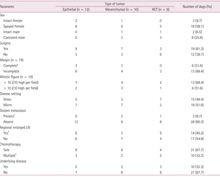
Table 2. Patient characteristics and additional tumor information (n = 31)
Parameter Type of tumor Number of dogs (%)
Epithelial (n = 12) Mesenchymal (n = 10) RCT (n = 9) Sex Intact female 2 1 0 3 (9.7) Spayed female 8 5 5 18 (58.1) Intact male 0 1 1 2 (6.5) Castrated male 2 3 3 8 (25.8) Surgery Yes 9 7 3 19 (61.3) No 3 3 6 12 (38.7) Margin (n = 19) Complete* 3 3 0 6 (31.6) Incomplete 6 4 3 13 (68.4) Mitotic figure (n = 19)
< 10 (/10 high per field) 7 4 2 13 (68.4)
≥ 10 (/10 high per field) 2 3 1 6 (31.6)
Disease setting Gross 5 3 7 15 (48.4) Micro 7 7 2 16 (51.6) Distant metastasis Present† 0 2 1 3 (9.7) Absent 12 8 8 28 (90.3) Regional enlarged LN Yes‡ 6 3 5 14 (45.2) No 6 7 4 17 (54.8) Chemotherapy Sole 9 8 4 21 (67.7) Multiple§ 3 2 5 10 (32.3) Underlying disease Yes 5 2 3 10 (32.3) No 7 8 6 21 (67.7)
RCT, round cell tumor; LN, lymph node.
*Complete margin was defined by the histopathology report.
†Metastasis includes regional lymph nodes or lungs, confirmed by histopathology and computed tomography.
‡Enlarged lymph nodes (mesenteric, superficial etc.) suspected of metastasis were detected around the tumor.
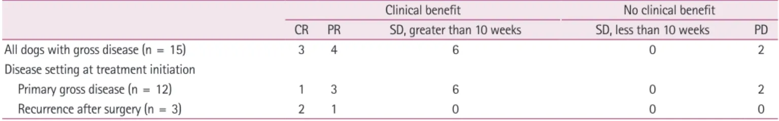
Treatment outcome
Clinical benefit was observed in 13 of 15 dogs (86.7%; 3 with CR, 4 with PR, 6 with SD) with gross disease. The remaining two dogs (13.3%) had PD (Table 3). Of the 13 dogs for which toceranib treatment showed benefit, 11 presented prolonged clinical benefit (median 35 days; range, 7 to 490 days) from ces-sation of toceranib to death or last date reported. 2 dogs in these groups were expired during toceranib treatment; 1 had PR, which then turned to PD, and 1 with SD died due to dete-rioration from toceranib-related side effects. The 16 dogs (51.6%) with microscopic disease were defined as having NED at the initiation of toceranib treatment. Seven dogs with NED had PD during toceranib treatment or after discontinuation of toceranib.
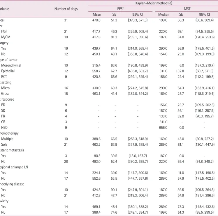
In the 31 treated dogs, the toceranib-related mean PFS and median MST were 471 and 199 days, respectively, based on the Kaplan–Meier product of survival time (Fig. 1). Distant metas-tasis was significantly associated with PFS (p < 0.05). Response to treatment and treatment setting (macro- or micro-scopic) were significantly associated with MST (p < 0.001 and p < 0.05,
respectively). The median MSTs for dogs with CR, PR, SD, and PD were 311, 133, 187, and 156 days, respectively. The mean PFS and median MST were 440 and 290 days, respectively, for dogs that underwent surgery and 450 and 154 days, respective-ly, for dogs that did not undergo surgery. The mean PFS and median MST were 315 and 199 days, respectively, for dogs with mesenchymal tumors, 588 and 311 days, respectively, for dogs with epithelial tumors, and 421 and 156 days, respectively, for dogs with round cell tumors. The mean PFS and median MST were 410 and 290 days, respectively, for dogs with microscopic disease, compared with 463 and 169 days, respectively, for dogs with gross disease. The mean PFS and median MST were 389 and 169 days, respectively, for dogs that received multiple che-motherapeutic drugs, compared with 463 and 289 days, respec-tively, for dogs that received toceranib alone. The mean PFS and median MST were 90 and 187 days, respectively, for dogs with distant metastasis, compared with 493 and 220 days, re-spectively, for dogs with no distant metastasis. The mean PFS and median MST were 224 and 169 days, respectively, for dogs with regional enlarged LNs, compared with 553 and 289 days,
Table 3. Clinical benefits of toceranib administration in 15 dogs with gross disease
Clinical benefit No clinical benefit
CR PR SD, greater than 10 weeks SD, less than 10 weeks PD
All dogs with gross disease (n = 15) 3 4 6 0 2
Disease setting at treatment initiation
Primary gross disease (n = 12) 1 3 6 0 2
Recurrence after surgery (n = 3) 2 1 0 0 0
CR, complete response; PR, partial response; SD, stable disease; PD, progressive disease.
Fig. 1. (A) Kaplan–Meier progression free survival curve based on the time of toceranib initiation (mean 471 days, range 370-571); (B)
Kaplan–Meier median survival time curve based on the time of toceranib treatment initiation (median 199 days, range 89-309). All censored dogs are marked with a cross.
A
1.0 0.8 0.6 0.4 0.2 0.0 Overall Censored 0 200 400 600pregression free survival
Time (days)
B
1.0 0.8 0.6 0.4 0.2 0.0 Overall Censored 0 200 400 600 survival probability Time (days)respectively, for dogs which did not have regional enlarged LNs. The mean PFS and median MST were 425 and 187 days, respectively, for dogs with underlying disease, compared with 413 and 289 days, respectively, for dogs without underlying dis-ease. The mean PFS and median MST were 469 and 289 days, respectively, for dogs with symptoms of toxicity to toceranib, compared with 388 and 199 days, respectively, for dogs without toxicity to toceranib (Table 4).
Prognostic factors
Multiple factors were assessed for their influence on progno-sis using a multivariable Cox proportional hazard model for PFS (Table 5) and MST (Table 6). Multiple chemotherapy and distant metastasis (in PFS) and surgery, regional enlarged LN, underlying disease, and toxicity (in MST) negatively affected prognosis. Conversely, epithelial and round cell tumors and
Table 4. Progression-free survival (PFS) and median survival time (MST) based on the Kaplan–Meier method
Variable Number of dogs
Kaplan–Meier method (d) PFS* MST Mean SE 95% CI Median SE 95% CI Total 31 470.8 51.3 (370.3, 571.3) 199.0 56.3 (88.6, 309.4) Sex F/SF 21 417.7 46.3 (326.9, 508.4) 220.0 69.1 (84.5, 355.5) M/CM 10 417.8 91.2 (239.1, 596.6) 187.0 34.0 (120.4, 253.6) Surgery Yes 19 439.7 64.1 (314.0, 565.4) 290.0 56.9 (178.5, 401.5) No 12 450.1 49.1 (353.8, 546.4) 154.0 23.0 (109.0, 199.0) Type of tumor Mesenchymal 10 315.4 63.6 (190.8, 439.9) 199.0 6.0 (187.3, 210.7) Epithelial 12 558.7 62.7 (435.8, 681.7) 311.0 132.8 (50.7, 571.3) RCT 9 420.8 65.6 (292.1, 549.4) 156.0 22.4 (112.2, 199.8) Tx setting Micro 16 410.0 69.3 (274.2, 545.8) 290.0 64.3 (163.9, 416.1) Gross 15 463.1 41.4 (382.0, 544.2) 169.0 25.7 (118.6, 219.4) Tx response PD 9 - - - 156.0 23.7 (109.5, 202.5) SD 6 - - - 187.0 36.1 (116.1, 257.9) PR 4 - - - 133.0 32.0 (70.3, 195.7) CR 3 - - - 311.0 - NED 9 - - - 656.0 0.0 -Chemotherapy Multiple 10 388.6 66.5 (258.3, 518.9) 169.0 45.0 (80.8, 257.2) Sole 21 463.2 63.9 (337.9, 588.4) 289.0 81.1 (130.1, 447.9) Distant metastasis Yes 3 90.3 39.5 (13.0, 167.7) 187.0 0.0 No 28 493.0 52.4 (390.2, 595.7) 220.0 65.4 (91.8, 348.2) Regional enlarged LN Yes 14 224.1 39.0 (147.7, 300.6) 169.0 11.0 (147.5, 190.5) No 17 552.6 53.5 (447.7, 657.6) 289.0 57.9 (175.5, 402.5) Underlying disease Yes 10 424.5 90.1 (247.9, 601.1) 187.0 39.5 (109.5, 264.5) No 21 412.8 47.7 (319.3, 506.4) 289.0 54.9 (181.4, 396.6) Toxicity Yes 14 469.1 45.4 (380.1, 558.2) 289.0 73.3 (145.4, 432.6) No 17 388.4 74.6 (242.1, 534.7) 199.0 51.3 (98.5, 299.5)
SE, standard error; CI, confidence interval; F, female; SF, spayed female; M, male; CM, castrated male; RCT, round cell tumor; Tx, treatment; PD, progressive disease; SD, stable disease; PR, partial response; CR, complete response; NED, no evidence of disease; LN, lymph node.
duration of toceranib (in PFS) and epithelial tumors, micro-scopic disease, NED, SD, and duration of toceranib (in MST) positively affected prognosis.
In dogs that underwent surgery, none of the examined fac-tors were significantly associated with MST (Table 7).
Adverse events
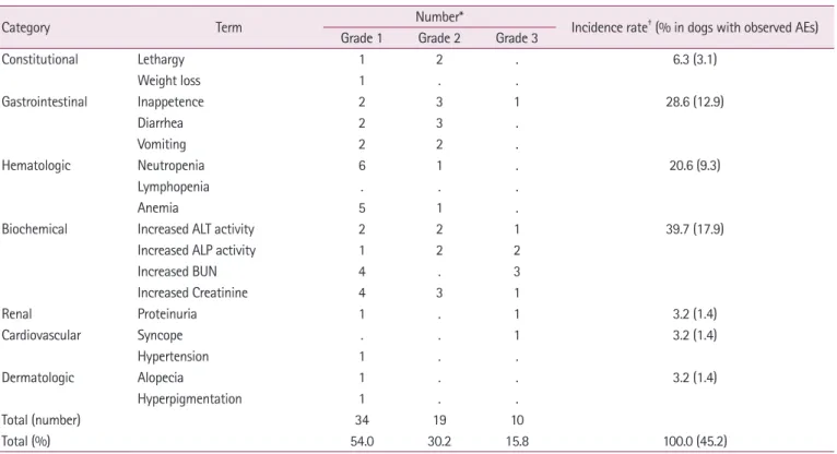
AEs occurred in 14/31 (45.2%) of the dogs treated with toc-eranib. The categories, grades (1 to 3), and incidence rate per category of these AEs are described in Table 8. There were no grade 4 or 5 AEs observed in any dogs. The majority of the AEs was grade 1 (54.0%) and categorized as biochemical (39.7%) or gastrointestinal (28.6%) AEs. Most of the gastrointestinal AEs (8/14) were classified as low grade (grades 1 to 2) and were managed quickly by supportive therapy with discontinuation of toceranib. In 6/14 (42.9%) dogs, toceranib administration was discontinued due to AEs (3 biochemical: increased blood urea nitrogen and creatinine; 2 hematologic: 1 neutropenia and 1 anemia; 1 gastrointestinal).
Discussion
This retrospective study describes the responses of toceranib treatment to various tumors, which has been reported for the first time in republic of Korea. The biological effect of toceranib on various types of tumors was evaluated in mostly small breed dogs. Most of the treated dogs with gross disease (48.4% of the total cohort) presented with clinical benefit (86.7%). This treat-ment response is consistent with previous studies [10,11,18]. Most previous reports tested the effect of toceranib on one type of tumor while our study supports the use of toceranib in vari-ous types of tumor. There have been few studies of the
relation-ship between tumor type and prognosis, but our study shows that epithelial tumors have a low recurrence rate and a high survival rate. In the previous studies assessing toceranib, clini-cal benefits have mainly been shown in epithelial tumors (‘-car-cinoma’) and MCTs [1,6,10,11,13,14,18]. Conversely, few stud-ies have reported effects on mesenchymal tumors such as GIST, OSA, injection site sarcoma, and histiocytic sarcoma, and these studies were limited by small populations [11,18,23-25]. Muta-tions in c-KIT, an important factor which is the target of
Table 5. Prognostic factors based on Cox proportional hazard model for progression-free survival (PFS)
Variable† PFS HR 95% CI p-value Surgery 69.06 (0.86, 5,560.44) 0.06 Type of tumor Epithelial 0.02 (0.00, 0.68) 0.03* RCT 0.01 (0.00, 0.61) 0.03* Chemotherapy 42.66 (1.83, 991.88) 0.02* Distant metastasis 663.52 (2.87, 153,515.22) 0.02* Regional enlarged LN 4.88 (0.48, 49.67) 0.18 Duration of administration 0.97 (0.95, 1.00) 0.02* Toxicity 0.11 (0.01, 2.03) 0.14
HR, hazard ratio; CI, confidence interval; RCT, round cell tumor; LN, lymph node.
†Reference category: surgery (no = 0), type of tumor (mesenchymal = 0), chemotherapy (sole = 0), distant metastasis (no = 0), regional enlarged LN (no
= 0), toxicity (no = 0). *p < 0.05.
Table 6. Prognostic factors based on Cox proportional hazard model
for median survival time (MST)
Variable† MST HR 95% CI p-value Surgery 91.62 (1.17, 7,149.10) 0.04* Type of tumor Epithelial 0.02 (0.00, 0.62) 0.03* RCT 0.30 (0.02, 3.50) 0.33 Tx setting 0.00 (0.00, 0.05) < 0.01** Tx response SD 0.00 (0.00, 0.05) < 0.01** PR 0.09 (0.00, 3.18) 0.19 CR 0.00 (0.00, 1.91) 0.07 NED 0.02 (0.00, 0.41) 0.01* Regional enlarged LN 6.86 (1.05, 44.87) 0.04* Duration of administration 0.97 (0.95, 1.00) 0.03* Underlying disease 17.27 (2.00, 149.06) < 0.01** Toxicity 22.07 (1.85, 263.21) 0.01*
HR, hazard ratio; CI, confidence interval; RCT, round cell tumor; Tx, treatment; SD, stable disease; PR, partial response; CR, complete response; NED, no evidence of disease; LN, lymph node; PD, progressive disease. †
Reference category: surgery (no = 0), type of tumor (mesenchymal = 0), Tx setting (gross = 0), Tx response (PD = 0), Regional enlarged LN (no = 0), underlying disease (no = 0), toxicity (no = 0).
ceranib. A previous report showed that PR and SD were posi-tive factors of prognosis [9]. Since our study involved 31 dogs and was limited to three centers, a direct comparison with pre-vious retrospective studies may not be appropriate. However, the following points may explain why the effect of PR on prog-nosis found in our study differed from that in other studies. In our study, a total of four dogs (one with SCC, and three with MCT, stage 3) that experienced PR all died of each tumor, and their median MST was 133 days. Of these, the first responded to secondary toceranib treatment after several trials with other chemotherapies, but had recurrence two weeks before death and the patient’s condition was deteriorated quickly. The sec-ond dog also experienced recurrence two weeks before death from a distant metastasis (carcinomatosis). The third dog ini-tially showed PR, but the tumor continued to increase, and leading to PD. The fourth was already in a state of relapse after surgery when toceranib was initiated. Therefore, the dogs that experienced PR all showed rapid relapse and deterioration of patient’s condition even though a clinical benefit from the treatment was recorded. Overall, the fact that our study had conditions differing from the earlier report needs to be fully considered and interpreted.
small-molecule tyrosine kinase inhibitors in veterinary medi-cine, have been found in MCTs (grades 1 to 3) in dogs, and in GIST and mammary gland tumor (grades 2 to 3) in dogs and humans [15,26-28]. In our study, dogs with epithelial tumors with sufficient clinical evidence had the highest representation (12 of 31 dogs, 38.7%) and showed clinical benefit from tocer-anib treatment, including two dogs with CR.
Using a Cox regression analysis, we identified that NED or SD response to treatment had a significantly positive effect on prognosis (in MST). Unexpectedly, CR and PR did not have a significant effect on prognosis, despite the clinical benefit of
to-Table 8. Adverse events (AEs) in dogs treated with toceranib (45.2% of the total study cohort)
Category Term Number* Incidence rate† (% in dogs with observed AEs)
Grade 1 Grade 2 Grade 3
Constitutional Lethargy 1 2 . 6.3 (3.1) Weight loss 1 . . Gastrointestinal Inappetence 2 3 1 28.6 (12.9) Diarrhea 2 3 . Vomiting 2 2 . Hematologic Neutropenia 6 1 . 20.6 (9.3) Lymphopenia . . . Anemia 5 1 .
Biochemical Increased ALT activity 2 2 1 39.7 (17.9)
Increased ALP activity 1 2 2
Increased BUN 4 . 3 Increased Creatinine 4 3 1 Renal Proteinuria 1 . 1 3.2 (1.4) Cardiovascular Syncope . . 1 3.2 (1.4) Hypertension 1 . . Dermatologic Alopecia 1 . . 3.2 (1.4) Hyperpigmentation 1 . . Total (number) 34 19 10 Total (%) 54.0 30.2 15.8 100.0 (45.2)
ALT, alanine aminotransferase; ALP, alkaline phosphatase; BUN, blood urea nitrogen. *Number includes overlap selection.
†Incidence rate was defined as percentage of occurrence on the category among all adverse events and after that percentage in the brackets indicates
occurrence on dogs in which AEs were observed.
Table 7. Prognostic factors based on Cox proportional hazard model
for median survival time (MST) in the group that underwent surgery
Variable* MST HR 95% CI p-value Mitotic count 1.02 (0.98, 1.07) 0.33 Margin 0.20 (0.02, 2.26) 0.19 Distant metastasis 0.00 (0.00, -) 0.99 Regional enlarged LN 2.01 (0.35, 11.46) 0.43
HR, hazard ratio; CI, confidence interval; LN, lymph node.
*Reference category: margin count (incomplete = 0), distant metastasis (no = 0), regional enlarged LN (no = 0).
munohistochemistry [31].
In conclusion, a clinical benefit of toceranib treatment was noted in the majority of the 15 dogs with gross disease in our study (86.7%). In the 31 treated dogs, the toceranib-related mean PFS was 471 days and the median MST was 199 days. Complex chemotherapy and distant metastasis (PFS), surgery, regional enlarged LN, underlying disease, and toxicity (MST) were negative prognostic factors. Our results provide additional information about the clinical efficacy of toceranib and suggest that the toceranib is probably selective treatment to various tu-mor types in small breed dogs. Further study is required to evaluate its clinical activity in a large number of small breed dogs.
Acknowledgements
We are thanks to the Cooperative Research Program of Center for Companion Animal Research (Project no. PJ01404502): Ru-ral Development Administration, Republic of Korea for the sup-port.
ORCID
Seo-In Choi, https://orcid.org/0000-0003-0328-8874 Ye-Lim Nam, https://orcid.org/0000-0001-5658-1893 Jin-Kyoung Kim, https://orcid.org/0000-0002-3618-2884 Hyung-Jin Park, https://orcid.org/0000-0002-4383-1037 Kun-Ho Song, https://orcid.org/0000-0001-8478-2035 Kyoung Won Seo, https://orcid.org/0000-0002-1561-3278
References
1. Pryer NK, Lee LB, Zadovaskaya R, Yu X, Sukbuntherng J, Cherrington JM, London CA. Proof of target for SU11654: inhibition of KIT phosphorylation in canine mast cell tumors. Clin Cancer Res 2003;9:5729-5734.
2. Chow LQ, Eckhardt SG. Sunitinib: from rational design to clinical efficacy. J Clin Oncol 2007;25:884-896.
3. Papaetis GS, Syrigos KN. Sunitinib: a multitargeted receptor tyrosine kinase inhibitor in the era of molecular cancer thera-pies. BioDrugs 2009;23:377-389.
4. Vail DM, Thamm DH, Liptak JM. Withrow and MacEwen’s Small Animal Clinical Oncology. 6th ed. pp. 197-198, Elsevier, St. Louis, 2020.
5. London CA, Hannah AL, Zadovoskaya R, Chien MB, Kol-lias-Baker C, Rosenberg M, Downing S, Post G, Boucher J, Shenoy N, Mendel DB, McMahon G, Cherrington JM. Phase In our study, dogs that underwent surgery had a worse
prog-nosis than those that did not, and dogs with microscopic dis-ease had a better prognosis than those without. In a previous report, the effect of surgery on prognosis was not statistically significant [10]. In our study, of the dogs that underwent sur-gery, 16/19 initiated toceranib treatment in a microscopic dis-ease setting and recurrence occurred in three dogs after sur-gery, resulting in gross disease (3/19).
Considering the result that the microscopic condition had a significant positive effect on the prognosis, these dogs relapsed after surgery had poor MST, even if clinical benefit (two with CR and one with PR) was observed. So, surgery was considered a negative factor of prognosis in our study.
In dogs with regional enlarged LN and distant metastasis, the prognosis (MST) was poor. Previous studies have shown results consistent with this finding [10,17,18,29]. Conversely, in one report related to grade 2 MCTs, the survival time between dogs with and without regional LN metastasis was no significant dif-ference. However, prolonged survival time was observed in dogs in which metastatic LNs were removed [30]. Our study identified regional enlarged LN was removed in only 4 of 14 dogs.
The median dose of toceranib used in treated dogs was 2.5 mg/kg (range, 2.2 to 3 mg/kg). According to existing reports, low doses of 2.5 to 2.9 mg/kg every other day are well tolerated without grade 3 or 4 AEs, compared with dosing at 3.25 mg/kg every other day [20]. In our study, 10 dogs had existing under-lying diseases (three with heart failure, one with chronic kidney disease, four with both heart and kidney disease, and two with chronic pancreatitis). Depending on these concomitant diseas-es, main side effects were occurred and therefore biochemistry AEs was the most common (39.7%), unlikely to another re-ports. Gastrointestinal AEs were the second most common type of AEs, and these were effectively managed in grades 1 to 2 through drug holidays, supportive medicine, and dose adjust-ments to avoid drug withdrawal. The dogs that died from se-vere side effects developed worsening chronic kidney disease, severe pancreatitis, or anemia and did not recover.
There were several limitations of our study. Not only was the overall population size small, but the numbers in the subgroup analyses were also small. These findings should be further con-firmed in a large number of small breed dogs. As this study had a retrospective design, it was impossible to match baseline characteristics such as sex, biochemistry, and underlying dis-eases. Additionally, toceranib affecting the tumor microenvi-ronment (TME) in various tumor types may be assessed by molecular biology in other ways, such as Western blots or
im-16. Laver T, London CA, Vail DM, Biller BJ, Coy J, Thamm DH. Prospective evaluation of toceranib phosphate in metastatic canine osteosarcoma. Vet Comp Oncol 2018;16:E23-E29. 17. Kim C, Matsuyama A, Mutsaers AJ, Woods JP. Retrospective
evaluation of toceranib (Palladia) treatment for canine metastat-ic appendmetastat-icular osteosarcoma. Can Vet J 2017;58:1059-1064. 18. Berger EP, Johannes CM, Jergens AE, Allenspach K, Powers
BE, Du Y, Mochel JP, Fox LE, Musser ML. Retrospective eval-uation of toceranib phosphate (Palladia®) use in the treatment of gastrointestinal stromal tumors of dogs. J Vet Intern Med 2018;32:2045-2053.
19. Yamazaki H, Miura N, Lai YC, Takahashi M, Goto-Koshino Y, Yasuyuki M, Nakaichi M, Tsujimoto H, Setoguchi A, Endo Y. Effects of toceranib phosphate (Palladia) monotherapy on mul-tidrug resistant lymphoma in dogs. J Vet Med Sci 2017;79:1225-1229.
20. Bernabe LF, Portela R, Nguyen S, Kisseberth WC, Pennell M, Yancey MF, London CA. Evaluation of the adverse event pro-file and pharmacodynamics of toceranib phosphate adminis-tered to dogs with solid tumors at doses below the maximum tolerated dose. BMC Vet Res 2013;9:190.
21. Nguyen SM, Thamm DH, Vail DM, London CA. Response evaluation criteria for solid tumours in dogs (v1.0): a Veteri-nary Cooperative Oncology Group (VCOG) consensus docu-ment. Vet Comp Oncol 2015;13:176-183.
22. Veterinary Cooperative Oncology Group-Common Termi-nology Criteria for Adverse Events (VCOG-CTCAE) follow-ing chemotherapy or biological antineoplastic therapy in dogs and cats v1.1. Vet Comp Oncol 2016;14:417-446.
23. Elliott JW, Swinbourne F, Parry A, Baines L. Successful treatment of a metastatic, gastrointestinal stromal tumour in a dog with to-ceranib phosphate (Palladia). J Small Anim Pract 2017;58:416-418.
24. Hong H, Lim S, Shin HR, Choi H, Lee H, Song KH, Seo KW. Metronomic chemotherapy with toceranib phosphate for a disseminated histiocytic sarcoma in a Miniature Schnauzer dog. J Vet Clin 2017;34:441-444.
25. Jacobs TM, Poehlmann CE, Kiupel M. Injection-site sarcoma in a dog: clinical and pathological findings. Case Rep Vet Med 2017;2017:6952634.
26. Frost D, Lasota J, Miettinen M. Gastrointestinal stromal tu-mors and leiomyomas in the dog: a histopathologic, immuno-histochemical, and molecular genetic study of 50 cases. Vet Pathol 2003;40:42-54.
27. Patruno R, Marech I, Zizzo N, Ammendola M, Nardulli P, Gadaleta C, Introna M, Capriuolo G, Rubini RA, Ribatti D, Gadaleta CD, Ranieri G. c-Kit expression, angiogenesis, and I dose-escalating study of SU11654, a small molecule receptor
tyrosine kinase inhibitor, in dogs with spontaneous malignan-cies. Clin Cancer Res 2003;9:2755-2768.
6. London CA, Malpas PB, Wood-Follis SL, Boucher JF, Rusk AW, Rosenberg MP, Henry CJ, Mitchener KL, Klein MK, Hintermeister JG, Bergman PJ, Couto GC, Mauldin GN, Mi-chels GM. Multi-center, placebo-controlled, double-blind, randomized study of oral toceranib phosphate (SU11654), a receptor tyrosine kinase inhibitor, for the treatment of dogs with recurrent (either local or distant) mast cell tumor follow-ing surgical excision. Clin Cancer Res 2009;15:3856-3865. 7. Shin HR, Kim JS, Kim SM, Song KH, Seo KW. Metronomic
chemotherapy with toceranib phosphate for treatment of a chemodectoma in a dog. J Vet Clin 2017;34:43-46.
8. Musser ML, Taikowski KL, Johannes CM, Bergman PJ. Retro-spective evaluation of toceranib phosphate (Palladia®) use in the treatment of inoperable, metastatic, or recurrent canine pheochromocytomas: 5 dogs (2014-2017). BMC Vet Res 2018;14:272.
9. Koshino A, Mauldin GE, Dickinson RM, Mauldin GN. A clinical oncology case report. Can Vet J 2011;52:899-902. 10. Heaton CM, Fernandes AFA, Jark PC, Pan X. Evaluation of
toceranib for treatment of apocrine gland anal sac adenocar-cinoma in dogs. J Vet Intern Med 2020;34:873-881.
11. London C, Mathie T, Stingle N, Clifford C, Haney S, Klein MK, Beaver L, Vickery K, Vail DM, Hershey B, Ettinger S, Vaughan A, Alvarez F, Hillman L, Kiselow M, Thamm D, Higginbotham ML, Gauthier M, Krick E, Phillips B, Ladue T, Jones P, Bryan J, Gill V, Novasad A, Fulton L, Carreras J, Mc-Neill C, Henry C, Gillings S. Preliminary evidence for biologic activity of toceranib phosphate (Palladia(®)) in solid tumours. Vet Comp Oncol 2012;10:194-205.
12. de Vos J, Ramos Vega S, Noorman E, de Vos P. Primary frontal sinus squamous cell carcinoma in three dogs treated with piroxicam combined with carboplatin or toceranib. Vet Comp Oncol 2012;10:206-213.
13. Heishima K, Iwasaki R, Kawabe M, Murakami M, Sakai H, Maruo K, Mori T. Short-term administration of single-agent toceranib in six cases of inoperable massive canine hepatocel-lular carcinoma. J Am Anim Hosp Assoc 2019;55:35-41. 14. Sheppard-Olivares S, Bello NM, Wood E, Szivek A, Biller B,
Hocker S, Wouda RM. Toceranib phosphate in the treatment of canine thyroid carcinoma: 42 cases (2009-2018). Vet Comp Oncol 2020;18:519-527.
15. Koltai Z, Szabó B, Jakus J, Vajdovich P. Tyrosine kinase expres-sion analyses in canine mammary gland tumours: a pilot study. Acta Vet Hung 2018;66:294-308.
nosis in malignant mammary tumors of dogs. J Vet Intern Med 2015;29:1360-1367.
30. Baginski H, Davis G, Bastian RP. The prognostic value of lymph node metastasis with grade 2 MCTs in dogs: 55 cases (2001-2010). J Am Anim Hosp Assoc 2014;50:89-95.
31. Yamazaki H, Tanaka T, Mie K, Nishida H, Miura N, Akiyoshi H. Assessment of postoperative adjuvant treatment using toc-eranib phosphate against adenocarcinoma in dogs. J Vet In-tern Med 2020;34:1272-1281.
grading in canine mast cell tumour: a unique model to study c- Kit driven human malignancies. Biomed Res Int 2014;2014: 730246.
28. Kubo K, Matsuyama S, Katayama K, Tsutsumi C, Yonezawa K, Shimada T, Kotani T, Sakuma S, Ohashi F, Takamori Y. Fre-quent expression of the c-kit proto-oncogene in canine malig-nant mammary tumor. J Vet Med Sci 1998;60:1335-1340. 29. de Araújo MR, Campos LC, Ferreira E, Cassali GD.