저작자표시-비영리-변경금지 2.0 대한민국 이용자는 아래의 조건을 따르는 경우에 한하여 자유롭게 l 이 저작물을 복제, 배포, 전송, 전시, 공연 및 방송할 수 있습니다. 다음과 같은 조건을 따라야 합니다: l 귀하는, 이 저작물의 재이용이나 배포의 경우, 이 저작물에 적용된 이용허락조건 을 명확하게 나타내어야 합니다. l 저작권자로부터 별도의 허가를 받으면 이러한 조건들은 적용되지 않습니다. 저작권법에 따른 이용자의 권리는 위의 내용에 의하여 영향을 받지 않습니다. 이것은 이용허락규약(Legal Code)을 이해하기 쉽게 요약한 것입니다. Disclaimer 저작자표시. 귀하는 원저작자를 표시하여야 합니다. 비영리. 귀하는 이 저작물을 영리 목적으로 이용할 수 없습니다. 변경금지. 귀하는 이 저작물을 개작, 변형 또는 가공할 수 없습니다.
의학 석사학위 논문
Traumatic arterial dissection of the
lower extremity: computed
tomography findings and endovascular
management
아 주 대 학 교 대 학 원
의 학 과
Traumatic arterial dissection of the
lower extremity: computed
tomography findings and endovascular
management
지도교수 원 제 환
이 논문을 의학 석사학위 논문으로 제출함.
2018 년 8 월
아 주 대 학 교 대 학 원
의 학 과
서 배 선
서배선의 의학 석사학위 논문을 인준함.
심사위원장 원 제 환 (인)
심 사 위 원 선 주 성 (인)
심 사 위 원 이 제 희 (인)
아 주 대 학 교 대 학 원
2018년 7월 6일
i
- ABSTRACT -
Traumatic arterial dissection of the lower extremity: computed
tomography findings and endovascular management
Introduction: To report imaging findings and endovascular management in traumatic
arterial dissection of lower extremity.
Material and Methods: Nine patients who underwent endovascular treatment for
traumatic arterial dissection of the lower extremity from January 2010 to July 2016 were retrospectively reviewed. Pre-procedural multiphasic CT angiography or single-phase abdomen CT was performed. Balloon angioplasty was primarily performed while stenting was reserved for cases of ineffective initial balloon angioplasty. Computed tomography (CT) findings, treatment results, and patient outcomes were assessed. Technical success was defined as recanalization of injured vessels in completion angiography, and clinical success as an improvement of ischemic signs.
Results: On contrast enhanced CT, segmental occlusion accompanied by the ‘crescent
sign’ was seen in seven patients while dissection flaps were clearly visible in two. Progressive contrast filling in the segment with ‘crescent sign’ was noted on delayed phase of enhancement. Types of endovascular procedures included balloon angioplasty alone (n=5), bare-metal stent insertion (n=2), and stent-graft insertion (n=2). The technical success rate was 100% and the clinical success rate was 88.9%. All treated vessels were patent on follow-up CT angiogram. Pseudoaneurysm was seen in one patient during follow-up.
Conclusion: Characteristic features on CT angiography may aid diagnosis of traumatic
arterial dissection, in which case balloon angioplasty can be attempted initially and stenting can be reserved as a bailout strategy.
ii
Keywords: trauma, dissection, lower extremity, CT, endovascular
iii
TABLE OF CONTENTS
ABSTRACT ... i
TABLE OF CONTENTS ... iii
LIST OF FIGURES ... iv
LIST OF TABLES ... v
INTRODUCTION ... 1
MATERIAL AND METHODS ... 1
A. Patient selection ... 1
B. CT techniques and image interpretation ... 2
C. Endovascular techniques ... 2
D. Outcome analysis ... 3
RESULTS ... 4
A. Patient demographics and lesion characteristics ... 4
B. CT findings ... 6 C. Endovascular treatment ... 8 D. Patient outcomes ... 10 DISCUSSION ... 12 CONCLUSION ... 14 REFERENCES ... 15 국문요약 ... 17
iv
LIST OF FIGURES
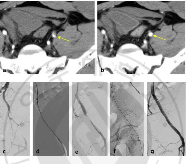
Fig. 1. A 40-year-old male patient was injured following a motorcycle accident resulting in dissection that extended from the left external iliac to the common femoral artery ... 7
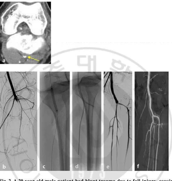
Fig. 2. A 20-year-old male patient with blunt trauma due to fall injury, causing left popliteal artery dissection ... 9
v
LIST OF TABLES
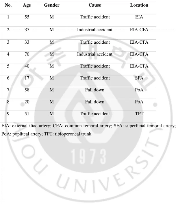
Table 1. Summary of the patient population and lesion characteristics ... 5
- 1 -
I. INTRODUCTION
Trauma has become an important social problem with the development of industry and traffic in modern societies. It accounts for about 10% of deaths worldwide and is increasing steadily, especially in developing countries, according to World Health Organization (WHO) reports. [1] Although vascular injuries, especially arterial dissection following trauma, are not common, they can lead to organ or limb loss and even death without appropriate management.
Surgical management is effective in repairing vascular injuries in the lower extremities. However, it requires dissection of the surrounding post-traumatic tissues and can lead to post-operative complications, such as peripheral nerve damage. [2, 3] In recent studies the role of endovascular treatment has increased due to its many advantages over surgery, such as its minimally invasive nature, shorter procedure time, and smaller amount of blood loss. [4] Several studies reported endovascular treatment of arterial dissection such as involvement of aorta, superior mesenteric artery [5], renal artery [6], and axillary artery [7]. However, its role specifically in the lower extremities has not been reported.
The purpose of this retrospective study was to assess imaging features of traumatic arterial dissections on pre-procedural CT angiography and their outcome after endovascular management.
II. MATERIALS AND METHODS
A. Patient selectionStudy approval was obtained from institutional review board of our hospital and patient consent was waived. We retrospectively reviewed medical records of patients with traumatic vascular injuries from January 2010 to July 2016 who were referred from our level 1 trauma center. We included nine patients who were diagnosed with arterial dissection in the lower extremity and underwent endovascular
- 2 -
treatment.
B. CT techniques and image interpretation
All patients underwent CT angiography before their endovascular procedures. Multiphasic CT angiography of lower extremity was performed for the patients whose major clinical concerns were ischemic signs. It was performed using 128-slice dual source CT (SOMATOM Definition Flash; Siemens, Erlangen, Germany), ranging from xyphoid process to toes. The CT scan parameters included 64 × 0.625mm section collimation, 5mm slice thickness, 120kVp tube voltage, 150mAs tube current, pitch 0.891. After a pre-contrast scan, 150mL pre-contrast media (iohexol, Omnihexol 350, Korea United Pharm Inc., Seoul, Korea) was administrated intravenously at a rate of 3.5mL/sec. Arterial and delayed phase images were obtained automatically using bolus-tracking technique, 3 seconds and 130 seconds each after attenuation of abdominal aorta reached a threshold of 150 HU.
For the patients with suspected accompanying intra-abdominal organ injury, single phase abdomen CT scan was performed (Somatom Definition Edge; Siemens Healthcare, Forchheim, Germany), ranging from xyphoid process to pubic symphysis. The CT scan parameters included 64 × 0.625mm section collimation, 5mm slice thickness, 120kVp tube voltage, 300mAs tube current, pitch 0.891. Contrast-enhanced images were obtained 90 seconds after the contrast media injection, 150mL at a rate of 2.5mL/sec.
Three radiologists retrospectively interpreted the radiologic characteristics of the injured vessels and surrounding structures.
C. Endovascular techniques
All angiographic images were obtained using high -quality C-arm fluoroscopy system (Allura Xper FD 20; Philips Medical Systems, Best, The Netherlands). After performing digital subtraction angiography (DSA), balloon angioplasty was initially performed in all patients. In the presence of residual stenosis of >30%, prolonged balloon inflation was performed. If there was elastic recoil or residual stenosis of more than 30% despite prolonged balloon angioplasty, a bare-metal stent (Complete SE stent,
- 3 -
Medtronic Vascular, Santa Rosa, CA, USA) or stent-graft (Viabahn, W.L. Gore & Associates, Flagstaff, AZ) was deployed. The latter was used in cases of long segmental dissection accompanied by a large amount of thrombus.
D. Outcome analysis
Technical success of endovascular treatment was defined as successful restoration of distal blood flow on completion angiography. Clinical success was defined as an immediate improvement in clinical signs induced by arterial dissection. Physical examination was performed by clinicians to evaluate restoration of blood flow of dorsalis pedis artery or improvement of skin color change. Follow-up CT scans were performed in all patients to evaluate patency of injured vessels and possible development of complications.
- 4 -
III. RESULTS
A. Patient demographics and lesion characteristicsAll were males and their mean age was 42.3 (range, 17 –70) years. All of the injuries were a result of blunt trauma. The major causes of trauma were traffic accidents (n=5, 55.6%), industrial accidents (n=2, 22.2%), and falls (n=2, 22.2%). All patients showed ischemic signs, six of whom initially presented with absent or weak pulsation in the dorsalis pedis artery, while three presented with pallor or cyanosis in the lower extremities. Patient characteristics and anatomical locations of the arterial dissections are presented in Table 1.
- 5 -
Table 1. Summary of the patient population and lesion characteristics
No. Age Gender Cause Location
1 55 M Traffic accident EIA
2 37 M Industrial accident EIA-CFA
3 33 M Traffic accident EIA-CFA
4 70 M Industrial accident EIA-CFA
5 40 M Traffic accident EIA-CFA
6 17 M Traffic accident SFA
7 58 M Fall down PoA
8 20 M Fall down PoA
9 51 M Traffic accident TPT
EIA: external iliac artery; CFA: common femoral artery; SFA: superficial femoral artery; PoA: popliteal artery; TPT: tibioperoneal trunk.
- 6 - B. CT findings
Dissection flaps were visible in two patients, demonstrated as a linear low-density structure inside the enhancing arterial lumen. Seven patients showed segmental occlusion of the injured arteries accompanied by crescent-shaped luminal contrast filling in the proximal aspect of the occluded segments (the ‘crescent sign’). The perivascular fat plane was preserved without contrast extravasation or surrounding hematoma. Four underwent three-phase CT scans which demonstrated the crescent sign during the arterial phase, and progressive contrast filling at the level of stenosis during the delayed phase (Fig. 1).
- 7 -
Fig. 1. A 40-year-old male patient was injured following a motorcycle accident resulting in dissection that extended from the left external iliac to the common femoral artery. Arterial-phase CT image shows severe eccentric stenosis with
crescent-shaped luminal contrast filling (crescent sign) at the proximal level of the occluded segment (arrow) (A). There is no hematoma around the lesion. Delayed-phase axial CT scan at the same level shows delayed contrast filling (arrow) (B). Initial DSA reveals a long segmental occlusion (C). Balloon angioplasty is attempted first (D) but there is a large amount of thrombus remaining along the treated segment (E). A stent-graft is deployed after the balloon angioplasty (F) and successful recanalization is seen on follow-up angiography (G).
a
b
- 8 - C. Endovascular treatment
On initial digital subtraction angiography, eight patients showed segmental occlusion of injured vessel with delayed contrast filling. One other patient showed localized pooling of contrast media into the dissected false lumen.
The blood flow was restored in five patients after balloon angioplasty alone (Fig. 2). Two patients with dissection either in the external iliac artery or superficial femoral artery demonstrated residual stenosis of >30% due to elastic recoil of the thrombosed false lumen, and were consequently treated with bare-metal stents. In two other patients who demonstrated such recoil after angioplasty, stent-grafts were used to treat long-segmental dissections accompanied by a large amount of thrombus (Fig. 1).
- 9 -
Fig. 2. A 20-year-old male patient had blunt trauma due to fall injury, causing left popliteal artery dissection. Contrast-enhanced CT image shows the crescent sign
in the left popliteal artery (arrow) (A). Initial DSA reveals occlusion of the left popliteal artery with delayed contrast pooling in the false lumen (B). Balloon angioplasty is initially performed in the popliteal artery (C), and two balloons are used in “kissing” configuration in the tibioperoneal trunk to treat dissection involving the tibial bifurcation (D). DSA after angioplasty shows restored distal blood flow with minimal residual stenosis in the treated segment (E). The blood flow is well preserved on the CT angiogram which is performed six months later (F).
a
- 10 - D. Patient outcomes
Technical success was achieved in all patients (technical success rate = 100%). Eight patients experienced improvement in ischemic signs after treatment (clinical success rate = 88.9%). Only one patient showed persistent weak pulsation in the dorsalis pedis artery. In this patient, intravenous infusion of dopamine fluid was initiated after admission in order to maintain his blood pressure. Anticoagulation drugs were also administered in this patient due to distally migrated multifocal residual thrombus at below-the-knee arteries. DSA was repeated after 5 days, but no occlusive lesion was found.
Seven patients underwent follow-up CT scans after an average follow-up period of 6 months, on which the treated vessels were demonstrated to be patent. Major complication occurred in only one patient in whom a pseudoaneurysm had developed in the tibioperoneal trunk after balloon angioplasty. The pseudoaneurysm was seen to grow in size during follow -up and was therefore treated with percutaneous injection of thrombin.
- 11 -
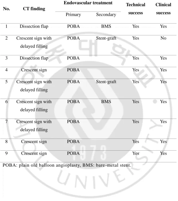
Table 2. Summary of CT findings, endovascular techniques, and clinical outcomes
No. CT finding
Endovascular treatment Technical success
Clinical success
Primary Secondary
1 Dissection flap POBA BMS Yes Yes
2 Crescent sign with delayed filling
POBA Stent-graft Yes No
3 Dissection flap POBA Yes Yes
4 Crescent sign POBA Yes Yes
5 Crescent sign with delayed filling
POBA Stent-graft Yes Yes
6 Crescent sign with delayed filling
POBA BMS Yes Yes
7 Crescent sign with delayed filling
POBA Yes Yes
8 Crescent sign POBA Yes Yes
9 Crescent sign POBA Yes Yes
- 12 -
IV. DISCUSSION
All of our patients were males at a mean age of 42.3 years, which is similar to the population described in previous reports. [8] Such results reflect the typically active and aggressive lifestyle of young adult males. In modern societies, trauma occurs mainly in connection with traffic accidents and, thus, blunt trauma is the most common mechanism. Blunt vascular injury is often found in the setting of pelvic bone trauma which undergoes strong external force during traumatic situations. Rotation of the pelvic bone increases mobility of the iliac and femoral vessels, making them more susceptible to shear stress, causing dissection of the external iliac or common femoral artery.
Careful assessment of CT may provide evaluable information that may help to differentiate arterial dissection from other types of vascular injury. With reference to descriptions on vertebral artery dissection, the pattern of dissection is commonly divided into aneurysmal and steno-occlusive patterns. [9] Most of the patients in our study showed a steno-occlusive pattern, and all of them showed eccentric stenosis with a crescent-shaped deformation of the lumen due to hematoma, “crescent sign”. Rieger et al. [10] previously reported similar findings on CT angiogram and stated that diagnosing dissection was slightly less reliable, especially in occluded segments. Interestingly, four patients in our study, who underwent three-phase dynamic CT exams showed delayed contrast filling in the occluded segment at the level of eccentric stenosis. This appearance may reflect slow contrast filling in the false lumen. This result may indicate the importance of a multi-phase dynamic CT study in distinguishing arterial dissection accompanied by luminal occlusion from other causes of thrombotic occlusion. With regard to the differential diagnosis of arterial transection, preservation of the fat plane surrounding the injured artery along with lack of contrast extravasation helps to exclude the likelihood of transection.
Although past reports describe stenting for the treatment of blunt arterial injuries [11, 12], traumatic vascular injuries are frequent in young adults. Considering the age of such population and their long-term prognosis, it may be preferable to treat them without the use of permanent implant. For this reason, we attempted balloon angioplasty first and
- 13 -
performed prolonged balloon dilatation in case of elastic recoil. Patients who were successfully treated with balloon angioplasty only showed good patency during a mean follow-up period of 5.4 months. Although the exact mechanism is poorly understood, it is presumed that increased flow in the true lumen with stenting or balloon angioplasty may change the dynamics of blood flow, resulting in decreased flow into the false lumen. [13] In addition, such procedures may help to seal the dissection flap onto the vessel wall and maintain this state of restored blood flow. In two of our patients, we used stent-grafts in order to treat extensive dissections extending from the external iliac artery to the common femoral artery. Stent-grafts were chosen over bare-metal stents in consideration of large amounts of thrombus that were retained after initial balloon angioplasty. Furthermore, it was more suitable due to the distal extent of the dissection which ran across the hip joint.
The outcome result from our study is similar to that described for surgical intervention [14-17]. However, reports on surgical outcome include various types of arterial injury and are not confined to arterial dissection. In this regard, a comparison of outcome between endovascular and surgical treatment cannot be made.
Our study has several limitations. First, it is a single-center study with a retrospective design. Secondly, we can neither assess the sensitivity and specificity of CT features nor compare the outcome of endovascular versus surgical treatment owing to the limited number of patients. Lastly, the patients were only available for short-term follow-up after treatment. Longer-term follow-up is needed in future studies in order to support the use of endovascular techniques for the treatment of patients with traumatic arterial dissection.
- 14 -
V. CONCLUSION
In conclusion, CT angiography may aid the diagnosis of traumatic arterial dissection in the lower extremity in the presence of a visible dissection flap or crescent sign. Progressive contrast filling in the injured segment with crescent sign on multiphasic CT angiography may give additional diagnostic information. Balloon angioplasty may be initially considered for the treatment of arterial dissection, whereas stenting may be performed in cases of elastic recoil after balloon angioplasty.
- 15 -
REFERENCES
1. Organization WH. Injuries and violence: the facts. Injuries and violence: the facts. 2010.
2. Rozycki GS, Tremblay LN, Feliciano DV, McClelland WB. Blunt vascular trauma in the extremity: diagnosis, management, and outcome. J Trauma. 2003;55(5):814-24. 3. Ekim H, Tuncer M. Management of traumatic brachial artery injuries: a report on 49 patients. Ann Saudi Med. 2009;29(2):105-9.
4. Reuben BC, Whitten MG, Sarfati M, Kraiss LW. Increasing use of endovascular therapy in acute arterial injuries: analysis of the National Trauma Data Bank. Journal of vascular surgery. 2007;46(6):1222-26.
5. Sirignano P, Setacci F, Galzerano G, Setacci C. Endovascular Treatment of Isolated Dissection of Superior Mesenteric Artery. Acta chirurgica Belgica. 2015;115(4):319-21.
6. Abu-Gazala M, Shussman N, Abu-Gazala S, Elazary R, Bala M, Rozenberg S, et al. Endovascular management of blunt renal artery trauma. The Israel Medical Association journal : IMAJ. 2013;15(5):210-5.
7. Cvjetko I, Staresinic M, Hlevnjak D, Bakota B, Dovzak I. Axillary artery dissection after scapular fracture. Annals of vascular surgery. 2011;25(6):837.e5-7.
8. Kauvar DS, Sarfati MR, Kraiss LW. National trauma databank analysis of mortality and limb loss in isolated lower extremity vascular trauma. Journal of vascular surgery. 2011;53(6):1598-603.
9. Shin JH, Suh DC, Choi CG, Leei HK. Vertebral artery dissection: spectrum of imaging findings with emphasis on angiography and correlation with clinical presentation. Radiographics. 2000;20(6):1687-96.
10. Rieger M, Mallouhi A, Tauscher T, Lutz M, Jaschke WR. Traumatic arterial injuries of the extremities: initial evaluation with MDCT angiography. AJR Am J Roentgenol. 2006;186(3):656-64.
11. Piffaretti G, Tozzi M, Lomazzi C, Rivolta N, Caronno R, Lagana D, et al. Endovascular treatment for traumatic injuries of the peripheral arteries following blunt
- 16 -
trauma. Injury. 2007;38(9):1091-7.
12. Brandt MM, Kazanjian S, Wahl WL. The utility of endovascular stents in the treatment of blunt arterial injuries. J Trauma. 2001;51(5):901-5.
13. Ahn JY, Han IB, Kim TG, Yoon PH, Lee YJ, Lee BH, et al. Endovascular treatment of intracranial vertebral artery dissections with stent placement or stent-assisted coiling. AJNR Am J Neuroradiol. 2006;27(7):1514-20.
14. McCready RA, Logan NM, Daugherty ME, Mattingly SS, Crocker C, Hyde GL. Long-term results with autogenous tissue repair of traumatic extremity vascular injuries. Annals of surgery. 1987;206(6):804-8.
15. Dorweiler B, Neufang A, Schmiedt W, Hessmann MH, Rudig L, Rommens PM, et al. Limb trauma with arterial injury: long-term performance of venous interposition grafts. The Thoracic and cardiovascular surgeon. 2003;51(2):67-72.
16. Klocker J, Bertoldi A, Benda B, Pellegrini L, Gorny O, Fraedrich G. Outcome after interposition of vein grafts for arterial repair of extremity injuries in civilians. Journal of vascular surgery. 2014;59(6):1633-7.
17. Liang NL, Alarcon LH, Jeyabalan G, Avgerinos ED, Makaroun MS, Chaer RA. Contemporary outcomes of civilian lower extremity arterial trauma. Journal of vascular surgery. 2016;64(3):731-6.
- 17 - - 국문요약 -
하지 외상성 동맥 박리에서의 CT 소견 및 혈관 중재시술의 역
할
아주대학교 대학원 의학과 서 배 선 (지도교수: 원 제 환) 서론: 외상으로 인한 하지의 동맥 박리에서의 전산화 단층 촬영 (CT) 소견과 혈관 중재시술의 방법 및 환자 예후에 대해 보고하고자 한다. 연구방법: 2010년 1월부터 2016년 7월까지 외상으로 인한 하지 동맥 박리 환 자 중 혈관 중재시술로 치료를 받은 환자 9명을 후향적으로 분석하였다. 환자 는 시술 전에 다중위상 혈관조영 CT 또는 조영 증강 복부 단층 촬영을 시행하 였다. 박리된 혈관에 대해 모든 환자에서 풍선 혈관성형술을 우선적으로 시행 하였으며, 효과가 없는 경우 스텐트 삽입술을 시행하였다. CT 소견, 치료 결과 및 환자 예후를 분석하였다. 기술적 성공은 시술 후 혈관조영술에서 손상된 혈 관이 성공적으로 재개통 된 경우로 정의하였고, 임상적 성공은 하지의 허혈성 증상이 개선된 경우로 정의하였다. 연구결과: 조영 증강 CT에서 7명의 환자에서 분절형 폐쇄와 동반된 초승달 모 양의 동맥 내경이 관찰되었으며, 2명의 환자에서는 일자형의 박리판이 관찰되 었다. 다중위상 혈관조영 CT을 시행한 4명의 환자에서는 초승달 모양의 동맥- 18 - 내경을 보인 손상 부위가 점진적으로 조영 증강되는 모습을 보였다. 5명의 환 자에서는 풍선 성형술만으로 재개통 되었으며, 2명에서는 금속 스텐트 삽입, 나 머지 2명에서 인조혈관 스텐트 삽입으로 재개통 되었다. 기술적 성공률은 100%, 임상적 성공률은 88.9%로 확인되었다. 치료받은 모든 혈관에서 추적 관찰 시 시행한 CT상 혈관 개통이 유지되었으며, 1명의 환자에서 합병증으로 가성동맥류가 발생하였다. 결론: 분절형 폐쇄와 동반된 초승달 모양의 동맥 내경 또는 일자형의 박리판으 로 관찰되는 특징적인 CT 소견은 하지의 외상성 동맥 박리를 진단하는데 도움 이 될 수 있다. 풍선 혈관성형술이 우선적으로 시행될 수 있고, 효과적이지 않 은 경우 스텐트 삽입술이 도움이 되겠다. 핵심어: 외상, 동맥 박리, 하지, 전산화 단층 촬영, 중재시술