Copyright ⓒ 2012 by the The Korean Society for Biochemistry and Molecular Biology
This is an open-access article distributed under the terms of the Creative Commons Attribution Non-Commercial License (http://creativecommons.org/licenses/ by-nc/3.0) which permits unrestricted non-commercial use, distribution, and reproduction in any medium, provided the original work is properly cited.
Increased p190RhoGEF expression in activated B cells
correlates with the induction of the plasma cell differentiation
Yun Jung Ha
1, Ji Hye Jeong
1, Yuna Park
1and Jong Ran Lee
1,2,3,41
Division of Life and Pharmaceutical Sciences 2
Department of Life Science College of Natural Sciences 3
Center for Cell Signaling and Drug Discovery Research Ewha Womans University
Seoul 120-750, Korea 4
Corresponding author: Tel, 82-2-3277-3762; Fax, 82-2-3277-3760; E-mail, [email protected] http://dx.doi.org/10.3858/emm.2012.44.2.009 Accepted 22 November 2011
Available Online 23 November 2011
Abbreviations: AID, activation-induced deaminase; Bcl-6, B-cell lymphoma protein-6; Blimp-1, B lymphocyte-induced matura-tion protein-1; CA, constitutively active; DN, dominant negative; EV, empty vector; GC, germinal center; HA, hemagglutinin; IB, immunoblot; IRF-4, interferon regulatory factor-4; MFI, mean flu-orescence intensities; Pax5, paired-box-protein 5; PC, plasma cell; PE, phycoerythrin; PerCP, peridinin chlorophyll protein; p190RhoGEF, p190 Rho guanine nucleotide exchange factor; XBP-1, X-box-binding protein-1
Abstract
Previously, we demonstrated that the p190 Rho
gua-nine nucleotide exchange factor (p190RhoGEF) was
induced following CD40 stimulation of B cells. In this
study, we examined whether p190RhoGEF and a
down-stream effector molecule RhoA are required for B cell
differentiation. Expression of p190RhoGEF positively
correlated with the expression of surface markers and
transcriptional regulators that are characteristic of
ma-ture B cells with plasma cell (PC) phenotypes.
Moreover, either the overexpression of p190RhoGEF
or the expression of a constitutively active RhoA drove
cellular differentiation toward PC phenotypes. B cell
maturation was abrogated in cells that overexpressed
p190RhoGEF and a dominant-negative form of RhoA
simultaneously. CD40-mediated maturation events
were also abrogated in cells that overexpressed either
dominant-negative p190RhoGEF or RhoA. Together,
these data provide evidence that p190RhoGEF
signal-ing through RhoA in CD40-activated B cells drives the
induction of the PC differentiation.
Keywords: antigens, CD40; ARHGAP35 protein,
hu-man; B-lymphocytes; CD40; cell differentiation;
p190RhoGEF; plasma cells; rhoA GTP-binding
pro-tein
Introduction
Many of the signaling molecules and transcription factors that regulate B cell fate decisions have been well characterized. Key transcriptional regu-lators, including B lymphocyte-induced maturation protein-1 (Blimp-1), X-box-binding protein-1 (XBP-1), and interferon regulatory factor-4 (IRF-4), are re-quired for plasma cell (PC) differentiation and func-tion (Calame, 2001; Shapiro-Shelef and Calame, 2005). Blimp-1 is a major regulator of PC differ-entiation and generation of Ab-secreting cells (Turner et al., 1994; Shaffer et al., 2002, 2004; Shapiro- Shelef et al., 2003). Blimp-1 suppresses the transcriptional repressor paired- box-protein 5 (Pax5), which is essential for the commitment of lymphoid progenitors to the B cell lineage, and also functions in late B cell development and activation by inducing activation-induced deaminase (AID) expression (Delogu et al., 2006; Klein et al., 2006; Nera et al., 2006). AID, a novel member of the RNA editing cytidine deaminase family, is specifi-cally expressed in germinal center (GC) B cells and is essential for somatic hypermutation and iso-type switching/class switch recombination (Gray et
al., 1994; Muramatsu et al., 2000; Xu et al., 2007).
Blimp-1 suppresses another transcriptional re-pressor, B cell lymphoma protein-6 (Bcl-6), a major regulator of GC B cells (Ye et al., 1997; Delogu et
al., 2006; Nera et al., 2006). Blimp-1 is thought to
act upstream of XBP-1, a transcriptional regulator of the secretory pathway in PC (Reimold et al., 2001). Recently, IRF-4 has also been identified as a second major gene in PC differentiation (Klein et
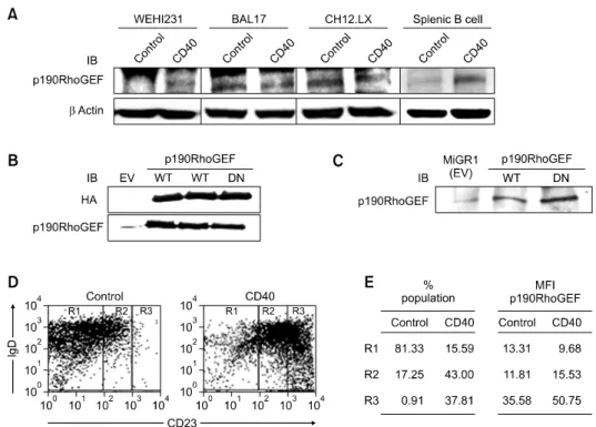
Figure 1. p190RhoGEF protein levels and changes after CD40 stimulation of B cells of various maturational stages. (A) Mouse splenic B cells (1 × 10⁶/ml)
and the B cell lines WEHI 231, BAL17, CH12.LX (5 × 10⁵/ml) were stimulated with isotype control Ig or anti-CD40 at a concentration of 1 μg/ml for 48 h. (B) HEK 293T cells were transfected with plasmids carrying an empty vector (EV), WT, or DN p190RhoGEF (HA). (C) WEHI 231 B cells were stably trans-fected with MIGR1 (EV), WT, or DN p190RhoGEF. Lysates were prepared from these cells, subjected to a SDS-PAGE (6% or 8% gels), transferred to ni-trocellulose, and probed with an Ab specific for p190RhoGEF (A-C) or HA (B). Equal loading of the protein samples was demonstrated by anti-β actin IB in (A). (D, E) Mouse splenic B cells (1 × 10⁶/ml) were stimulated with control Ig or anti-CD40 at a concentration of 1 μg/ml. After 48 h of stimulation, cells were fixed, permeabilized, and stained for endogenous p190RhoGEF, CD23, and IgD. The fluorescence intensities of cells stained with an isotype control Ab or antiserum were all corrected (data not shown). The results from flow cytometry are presented as a dot plot, showing IgD versus CD23 positive cells. For the cell populations in R1, R2, and R3 as indicated in (D), the population of cells (%) and the expression level of p190RhoGEF as mean fluorescence intensity (MFI) in the absence (control) and presence of CD40 stimulation is presented in (E). The results shown are representative of three independent experiments.
CD40 is an important cell surface receptor for Ag-dependent and T helper cell-regulated terminal differentiation of naïve B cells into Ab-secreting plasma and memory cells. The molecular mecha-nisms underlying the developmental coordination of CD40 stimulation with Ig secretion has been well characterized by many groups (Gray et al., 1994; Kawabe et al., 1994; Benson et al., 2007). However, the context in which CD40-CD154 signaling acts to activate transcription factors that regulate B cell fate decisions is not clearly understood.
Previously, we identified the p190 Rho guanine nucleotide exchange factor (p190RhoGEF) as a protein that is strongly induced in response to CD40 stimulation of B cells (Lee et al., 2003). We also demonstrated that the expression of p190RhoGEF is correlated with changes in cellular structure fol-lowing CD40 stimulation in WEHI 231 B cells (Lee
et al., 2003). Furthermore, cells that transiently
overexpressed p190RhoGEF behaved as if they expressed a constitutively active (CA) form of RhoA (Q63L), whereas transient overexpression of
dominant negative (DN) forms of p190RhoGEF (Y1003A) or RhoA (T19N) blocked the CD40-medi-ated activation effects (Lee et al., 2003). These re-sults suggest that there may exist a relationship between p190RhoGEF, the morphological changes in CD40-activated B cells, and additional B cell ma-turation and differentiation events.
In the present study, we examined whether p190RhoGEF expression is correlated with the ex-pression of specific surface markers and transcrip-tional regulators that are characteristic of both ma-ture and differentiated B cells. We also examined whether the activities of p190RhoGEF and RhoA are required for CD40-mediated B cell maturation and differentiation. Our results demonstrate that the expression of p190RhoGEF correlates with the expression of the cell surface markers CD138 (syndecan-1) and CXCR4, as well as the tran-scription factors Blimp-1, XBP-1, and IRF-4. The expression of these molecules was also induced following CD40 stimulation, as well as by over-expressing either wild type (WT) p190RhoGEF or a
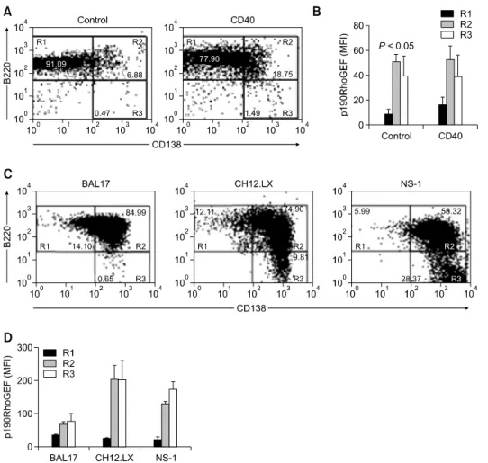
Figure 2. Expression of p190RhoGEF, CD138, and B220 in B cells at various maturational stages. (A, B) Mouse splenic B cells (1 × 10⁶/ml) were
stimu-lated with control Ig or anti-CD40 at a concentration of 1 μg/ml. After 72 h of stimulation, cells were fixed, permeabilized, and stained for endogenous p190RhoGEF, B220, and CD138. The fluorescence intensities of cells stained with an isotype control Ab or antiserum were all corrected (data not shown). The results from flow cytometry are presented as a dot plot, showing the B220 positive cells versus the CD138 positive cells. The data shown are resentative of three independent experiments. For the cell populations indicated in (A), expression of p190RhoGEF is presented as MFI in (B). Data are pre-sented as the mean (n = 4) ± SD. (C, D) The B cell lines, BAL17, CH12.LX, and NS-1 were fixed, permeabilized, and stained for endogenous p190RhoGEF, CD138, and B220. The results were obtained as described above. For the cell populations indicated in (C), expression of p190RhoGEF is presented as MFI in (D). Data are presented as the mean (n = 3) ± SD.
CA form of RhoA (Q63L). Collectively, these data suggest a specific contribution of p190RhoGEF to CD40-mediated differentiation and that p190RhoGEF may be a crucial component of T cell-dependent B cell maturation and differentiation.
Results
Changes in expression of surface markers and
p190RhoGEF by CD40 stimulation
We previously reported that a 48 h stimulation of CD40 leads to increases in p190RhoGEF expression and causes characteristic structural changes in B cells (Lee et al., 2003). Because the cellular struc-ture of B cells changes during cell maturation, we tested whether p190RhoGEF expression
corre-lates with stages in the maturation of B cells follow-ing CD40 stimulation. Analysis of the protein levels of p190RhoGEF in B cells of various maturational stages also demonstrated that there was an in-crease in p190RhoGEF protein levels following CD40 stimulation of WEHI 231 and splenic B cells, but not the mature B cell lines, BAL17 and CH12.LX (Figure 1A). The specificity of the poly-clonal Ab generated against p190RhoGEF was characterized in HEK 293T cells transfected with hemagglutinin (HA)-p190RhoGEF expression plas-mids (Figure 1B) and in WEHI 231 B cells in which p190RhoGEF was overexpressed (Figure 1C). As B cell maturation progresses, expression of CD23 is increased and expression of IgD is decreased. We further analyzed expression of the surface markers IgD and CD23. We used flow
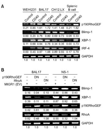
cy-Figure 3. Expression changes of transcriptional regulators after CD40 stimulation and in response to p190RhoGEF and RhoA down-regulation in B cells at various maturational stages. (A) cDNA was prepared from B cells that were either untreated (control) or stimulated for 24 h with an-ti-CD40 (1 μg/mL). (B) cDNA was prepared from BAL17 and NS-1 cells that were stably transfected with MIGR1 (EV), p190RhoGEF (Y1003A) or RhoA (T19N). The PCR products for p190RhoGEF, RhoA, Blimp-1, XBP-1, IRF-4, and GAPDH were separated as described in “Methods.” The data shown are representative of three independent experiments. Values indicate normalization for GAPDH.
tometry to analyze whether CD40 stimulation caused changes in the expression of IgD and CD23. Changes in the expression level of IgD ver-sus CD23 in mouse splenic B cells treated with an-ti-CD40 are shown in Figure 1D. B cell population was defined as three groups (R1: CD23low, R2:
CD23medium, R3: CD23low) based on the level of
CD23 expression. Results in Figure 1D and 1E demonstrate that CD40 stimulation of these B cells results in a change in maturation stage, indicated as a percentage of cell population in R1-R3. p190RhoGEF expression correlates to changes in cellular maturation both in unstimulated control and CD40 stimulation of splenic B cells (Figure 1E).
Expression of p190RhoGEF is correlated with the
expression of CD138 and transcriptional regulators
driving PC differentiation
To determine whether p190RhoGEF expression duced by CD40-stimulated B cell activation is in-volved in PC differentiation, we correlated the ex-pression of p190RhoGEF with the exex-pression of a specific surface molecule, CD138, which has been previously implicated as a PC marker (Calame, 2001; Shapiro-Shelef and Calame, 2005). B cell population was separated by the expression level of B220 and CD138 (R1: B220highCD138low, R2: B220highCD138high, R3: B220lowCD138high). By flow cytometry, we found that following 72 h of CD40 stimulation, the B cell population expressing CD138 on the surface increased from -7.4% to -20% (R2 and R3) in mouse splenic B cells (Figure 2A). Accordingly, the expression of p190RhoGEF was analyzed in the regions (R1, R2, and R3) sepa-rated by the expression level of B220 and CD138. As shown in Figure 2B, p190RhoGEF expression increased in R2 and R3 in mouse splenic B cells over the expression level in R1 both in unstimulated controls and following CD40 stimulation. CD40 stimulation also increased p190RhoGEF expression -2-fold in R1compared to the level of unstimulated controls.
To directly correlate the expression of p190RhoGEF with PC differentiation, we analyzed the expression levels of B220 versus CD138 in the mature B cell lines with PC phenotypes, BAL17 and CH12.LX, and in plasmacytoma NS-1 cells. As shown in Figure 2C, populations demonstrating distinct maturation stages differ slightly when R1-R3 are compared. Further analysis of the ex-pression of p190RhoGEF in these cell populations demonstrated that p190RhoGEF expression is much higher in cells that show more characteristics of PC with higher population in R3 (CH12.LX and NS-1 cells with higher population in R3 vs. BAL17
cells with lower population in R3 (Figure 2D). Because the transcriptional regulators, Blimp-1, XBP-1, and IRF-4 are known to play key roles in PC development (Turner et al., 1994; Reimold et
al., 2001; Shaffer et al., 2002; Shapiro-Shelef et al.,
2003; Shaffer et al., 2004; Klein et al., 2006; Sciammas et al., 2006), we next examined whether p190RhoGEF expression also correlated with the expression of these transcriptional regulators. The expression of Blimp-1, XBP-1, and IRF-4 was com-pared with the CD40-induced changes in these regulators compared to the unstimulated controls. As shown in Figure 3A, CD40 stimulation greatly enhanced the expression of these transcripts in WEHI 231 and splenic B cells. In contrast, the ex-pression of Blimp-1, XBP-1, and IRF-4 is high in BAL17 and CH12.LX cells, independent of CD40 stimulation (Figure 3A).
Similarly, p190RhoGEF expression was con-stitutively high in BAL17 and CH12.LX cells, but was induced in WEHI 231 and splenic B cells after
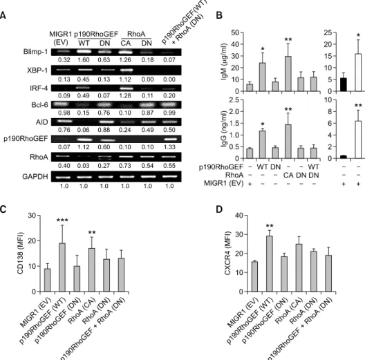
Figure 4. Direct effects of manipulating the activity of p190RhoGEF and RhoA on B cell maturation and downstream transcriptional regulators. WEHI 231 B cells were stably transfected with MIGR1 (EV), WT p190RhoGEF, DN p190RhoGEF (Y1003A), CA RhoA (Q63L), DN RhoA (T19N), or a combination of WT p190RhoGEF and DN RhoA (T19N). (A) cDNA was prepared from these stably transfected cells. The PCR products for p190RhoGEF, RhoA, Blimp-1, XBP-1, IRF-4, Bcl-6, AID, and GAPDH were separated as described in “Methods.” The data shown are representative of three independent experiments. Values indicate normalization for GAPDH. (B) These stably transfected cells were removed from the cultures and then washed, counted, and recultured in fresh medium for 24 h at 1 × 10⁶ cells/ml. Some EV-transfected cells were also incubated with a medium alone (black bars) or with a combination of CD40 and IL4 as positive controls (white bars). Supernatants were harvested and serially diluted, and secreted Ig was measured by anti-IgM and anti-IgG ELISA as described in “Methods.” Values of the optical densities at 490 nm in triplicate supernatant samples were collected in each independent experiment. Data are presented as the mean of three independent experiments (n = 3) ± SD, *P < 0.05, **P < 0.01, compared with cells transfected with EV. (C, D) These stably transfected cells were fixed, stained with anti-CD138 or anti-CXCR4, and analyzed by standard flow cytometry. The fluo-rescence intensities of cells stained with an isotype control Ab or antiserum were all corrected (data not shown).The bar graphs show the expression of CD138 (C) or CXCR4 (D), as indicated by MFI. Data are presented as the mean of three independent experiments (n = 3) ± SEM, **P < 0.01, ***P < 0.001, compared with cells transfected with EV.
CD40 stimulation (Figures 1A and 3A). These re-sults strongly suggest that the expression of p190RhoGEF mediates the induction of key tran-scriptional regulators, Blimp-1, XBP-1, and IRF-4, which regulate B cell maturation and differentiation. Because p190RhoGEF specifically activates RhoA (Gebbink et al., 1997; van Horck et al., 2001), we next examined whether the activities of p190RhoGEF and its downstream effector RhoA are required during PC differentiation. As shown in Figure 3B, the expression levels of the major transcriptional regulators of PC maturation and
differentiation, Blimp-1 and XBP-1 were greatly reduced in BAL17 cells transfected with a DN form of p190RhoGEF (Y1003A) or RhoA (T19N). These results strongly suggest that one function of p190RhoGEF is to maintain the expression of key transcriptional regulators of B cell maturation and differentiation. In contrast, the DN form of either p190RhoGEF or RhoA had no effect on the expression of these transcription factors in NS-1 cells (Figure 3B). These results also suggest that p190RhoGEF functions in maturing B cells, but not in completely differentiated plasmacytoma cells.
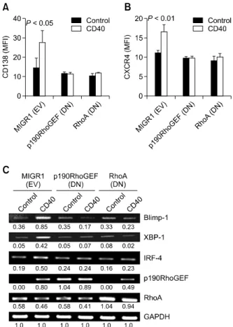
Figure 5. Effects of the overexpressed DN forms of p190RhoGEF or RhoA on the expression of CD138, CXCR4, and transcriptional regu-lators after CD40 stimulation. WEHI 231 B cells were stably transfected with either MIGR1 (EV), a plasmid encoding DN p190RhoGEF (Y1003A), or a plasmid encoding DN RhoA (T19N). (A, B) After CD40 stimulation for 48 h, cells were fixed, stained with anti-CD138 or anti-CXCR4, and measured by flow cytometry. The fluorescence intensities of cells stained with an isotype control Ab or antiserum were all corrected (data not shown). The results show the expression intensities of CD138 (A) or CXCR4 (B). Data are presented as the mean of three independent ex-periments (n = 3) ± SD. (C) cDNA was prepared from these stably transfected cells, after being stimulated for 48 h with anti-CD40 Ab (1 μg/ml). The PCR products for p190RhoGEF, RhoA, Blimp-1, XBP-1, IRF-4, and GAPDH were separated as described in “Methods.” The data shown are representative of three independent experiments. Values in-dicate normalization for GAPDH.
Expression of p190RhoGEF and activation of RhoA
mimic CD40-induced B cell maturation and
differentiation
We next determined whether p190RhoGEF ex-pression induced by CD40-stimulated B cell activa-tion is involved in the PC differentiaactiva-tion process. To determine the requirement for p190RhoGEF in ex-pression of the key transcriptional regulators Blimp-1, XBP-1, and IRF-4, we transfected WEHI 231 B cells with a plasmid encoding a WT or DN form of p190RhoGEF and measured the subsequent pression of these regulators. We found that the ex-pression of these transcription factors increased in cells transfected with WT p190RhoGEF, as well as a CA RhoA (Q63L), compared to cells transfected with the DN form of the proteins (Figure 4A). The expression of Blimp-1, XBP-1, and IRF-4 was not induced in cells that simultaneously expressed the WT p190RhoGEF and DN RhoA (T19N) (Figure 4A). Induced expression of Blimp-1 in WEHI 231 B cells transfected with WT p190RhoGEF or CA RhoA (Q63L) stimulated these cells to become Ab-secreting cells by suppressing Bcl-6 and AID expression (Figure 4A). Secretion of IgM and IgG Ab was detected high in the supernatants of these cells transfected with the WT p190RhoGEF as well as CA RhoA (Q63L) (Figure 4B). Some transcrip-tional increase shown in cells transfected with DN forms of p190RhoGEF and RhoA could possibly be due to controlled regulation by overexpression of these plasmids.
Similarly, we found that expression of the B cell surface differentiation markers CD138 and CXCR4 increased significantly in cells transfected with WT p190RhoGEF, but expression was unchanged in cells transfected with the DN plasmid (Figures 4C and 4D). We also found that these surface markers were upregulated in cells transfected with CA RhoA (Q63L) but not much in cells transfected with the DN form of RhoA (T19N) (Figures 4C and 4D). Moreover, the expression of surface markers in cells that simultaneously expressed DN RhoA (T19N) and WT p190RhoGEF was not induced, confirming that RhoA acts downstream of p190RhoGEF (Figures 4C and 4D).
The p190RhoGEF-RhoA activation is required for the
CD40-induced B cell differentiation
We also tested to see whether p190RhoGEF-RhoA activation is required for CD40-induced expression of surface molecules. In cells that were transfected with a vector plasmid, CD40 stimulation induced the expression of cell surface molecules (Figures 5A and 5B). However, in cells that were transfected with a DN form of either p190RhoGEF (Y1003A) or
RhoA (T19N), CD40 stimulation had no effect on cell surface protein levels (Figures 5A and 5B). Additionally, CD40 stimulation had no effect on the expression of transcriptional regulators in cells that were transfected with a DN form of either p190RhoGEF (Y1003A) or RhoA (T19N). However, in RhoA (T19N)-transfected WEHI 231 cells, p190RhoGEF induction was shown (Figure 5C). In cells that were transfected with a vector plasmid, CD40 stimulation induced the expression of p190RhoGEF, but had little effect on RhoA ex-pression levels (Figure 5C). These results strongly suggest that the enhanced activation of RhoA is
mediated by p190RhoGEF after CD40 stimulation in WEHI 231 B cells.
Discussion
CD40 is an important surface receptor that is in-volved in the activation and maturation of B cells during immune responses (Foy et al., 1994; Gray
et al., 1994; Kawabe et al., 1994; Xu et al., 1994;
Arpin et al., 1995; Randall et al., 1998; Yoon et al., 2005). CD40-CD154 interactions are crucial for the initiation and propagation of GC reactions following exposure to exogenous Ag and for the production of high affinity, class-switched Ab. However, the role of CD40 signaling in B cell differentiation is controversial (Foy et al., 1994; Gray et al., 1994; Kawabe et al., 1994; Xu et al., 1994; Arpin et al., 1995; Randall et al., 1998). Earlier studies have in-dicated that the stimulation of B cells through CD40 leads to the development of memory B cells and not PCs (Arpin et al., 1995). Studies have also demonstrated that the interaction of CD40 with its cell surface ligand CD154 actively suppresses Ig secretion in B cells and eventually allows for the selection of high affinity Ig-producing cells in the GC (Randall et al., 1998). In contrast, other reports have suggested that signaling through CD40 pro-motes the terminal differentiation of B cells into PCs and Ig secretion, particularly in combination with cytokines (Foy et al., 1994; Gray et al., 1994; Xu et al., 1994). Several studies have also demon-strated CD40-dependent, T cell-dependent, humor-al immunity through the disruption of genes in-volved in CD40 signaling pathways (Ahonen et al., 2002; Jabara et al., 2002; Gallagher et al., 2007). Here, we have shown that CD40 signaling through p190RhoGEF induces expression of the tran-scription factors Blimp-1, XBP-1, and IRF-4, as well as the cell surface proteins CD138 and CXCR-4, which drive cells to become Ab-secreting cells. p190RhoGEF, which was cloned and identified as a RhoA specific GEF (Gebbink et al., 1997; van Horck et al., 2001), has not been reported to func-tion in immune cells. We previously reported that expression of p190RhoGEF is markedly enhanced after CD40 stimulation in WEHI 231 B cells and is correlated with cellular structural changes and NF-κB activation (Lee et al., 2003). These results may indicate that this protein plays a significant role in CD40-mediated B cell activation and maturation. Here, we investigated the specific con-tribution of p190RhoGEF in CD40-stimulated cells in order to further our understanding of the molec-ular mechanisms underlying B cell maturation and differentiation.
Ag-dependent terminal differentiation of naïve B cells requires T helper cell regulation for cell ex-pansion and effector and memory cell development. Recently, naïve B cell priming, which is central to the initiation phase of T cell-dependent B cell re-sponses, has been studied extensively (Gray, 2002; McHeyzer-Williams and McHeyzer-Williams, 2005). The transcription factor Blimp-1 has been shown to be both necessary and sufficient for the differentiation of naïve B cells into PCs (Turner et al., 1994; Shaffer et al., 2002, 2004; Shapiro-Shelef et
al., 2003). Deletion of the transcription factor Pax5,
which is continuously required to maintain B cell identity by alternate lineage genes, results in the down-regulation of the GC transcriptional regulator Bcl-6, which consequently induces expression of the PC transcription factors Blimp-1 and XBP-1 (Ye
et al., 1997; Reimold et al., 2001; Delogu et al.,
2006; Nera et al., 2006). In an activated GC B cell state, physiological levels of Blimp-1 expression are induced by the down-regulation of Pax5 as well as by positive regulatory inputs from the Ag and cytokine receptor signaling systems (Delogu et al., 2006; Nera et al., 2006). Although recent studies have begun to dissect cellular events within the GC (Kallies et al., 2007), there are still many un-answered questions as to how PCs become termi-nally differentiated.
Our findings suggest that the strong stimulation of B cells by activated T cells (through CD40-CD154 interactions), induces Blimp-1 expression and dif-ferentiation into B cells with PC phenotype. The ex-pression levels of p190RhoGEF are low in splenic B cells as well as in an immature B cell line, WEHI 231, but increase following CD40 activation (Figures 1A and 4A). In contrast, elevated p190RhoGEF ex-pression levels do not increase further following CD40 activation in mature B cells that have a PC phenotype (e.g., BAL17 and CH12.LX) (Figures 1A and 4A). CD40 activation induces a change in B cell maturation as well as in p190RhoGEF ex-pression levels (Figure 1D). The exex-pression levels of p190RhoGEF increase in mature B cells with PC phenotypes (Figure 2). The expression of key transcription factors required for PC differentiation (Blimp-1, XBP-1, and IRF-4) was also upregulated by CD40 activation (Figure 3A). Overexpression of the DN form of p190RhoGEF or RhoA in mature BAL-17 B lymphoma cells significantly diminished the level of these transcriptional regulators (Figure 3B). Interestingly, these negative regulatory effects on B cell maturation were not seen in experiments where p190RhoGEF activity was blocked in plas-macytoma cells, which are terminally differentiated (Figure 3B). Moreover, overexpression of the WT or DN forms of p190RhoGEF or RhoA directly
modulated the expression of these transcription factors in the absence of CD40 stimulation in WEHI 231 B cells (Figure 4A). Secretion of IgM and IgG Ab in the supernatants of cells transfected with the WT p190RhoGEF as well as CA RhoA (Q63L) (Figure 4B) suggest that p190RhoGEF also regulates AID expression in the GC reaction before turning on the expression of Blimp-1, which in-duces PC differentiation. Our results indicate that p190RhoGEF is induced in activated B cells by CD40 and is required for initial triggering of GC B cell differentiation into cells of PC phenotype. The Rho family of GTPases has been shown to regulate cell polarization, motility, and chemotaxis in many cell types (Ridley, 2001; Boettner and Aelst, 2002; Etienne-Manneville and Hall, 2002; Meili and Firtel, 2003). Specifically, RhoA regulates the maturation of focal complexes into focal adhe-sions and the formation of associated stress fibers (Rottner et al., 1999; Ridley, 2001; Burridge and Wennerberg, 2004). Our previous study demon-strated that the structural changes following CD40 stimulation of B cells was due to p190RhoGEF-in-duced RhoA activity (Lee et al., 2003). Those re-sults are consistent with the findings from this in-vestigation which show that changes in cellular morphology are regulated by p190RhoGEF during CD40-mediated B cell activation and are correlated with Blimp-1 expression and B cell differentiation. Recent studies in macrophages have also shown that Bcl-6 alters macrophage morphology and mo-tility by suppressing RhoA activity (Pixley et al., 2005). In this respect, a number of recent studies have highlighted the importance of the cytoskele-ton in various lymphocytic functions including mi-gration, Ag recognition, signaling, and activation (Vicente-Manzanares and Sanchez-Madrid, 2004). Thus, additional studies are needed to determine whether cytoskeletal reorganization during CD40- mediated B cell activation contributes to GC B cell differentiation into cells of PC phenotype.
Recent studies have provided evidence that RhoA is involved in cell signaling and directly con-trols gene transcription and cell cycle progression (Li et al., 2000; Gu et al., 2003, 2005; Walmsley et
al., 2003). Genetic studies have shown that Rho
family members are required for normal develop-ment and functioning of lymphocytes (Li et al., 2000; Gu et al., 2003, 2005; Walmsley et al., 2003). RhoA has also been implicated in regulating B cell receptor-dependent calcium flux and pro-liferation (Saci and Carpenter, 2005), as well as Wnt-mediated migration and invasion of myeloma PCs (Qiang et al., 2005). Here, we provide evi-dence that RhoA activation is required for B cell maturation by CD40 receptor signaling. However, the
mechanism by which CD40-mediated p190RhoGEF- RhoA activation drives the induction of transcrip-tional regulators, including Blimp-1 and XBP-1, re-mains to be determined.
In summary, this report shows that CD40 signal-ing induces p190RhoGEF expression, which in turn activates RhoA and results in the expression of surface molecules and transcriptional regulators that are required for the maturation and differ-entiation of B cells. We have demonstrated a strong correlation between the expression levels of p190RhoGEF, the cell surface markers syndecan-1 and CXCR4, and the transcriptional regulators Blimp-1 and XBP-1. These data implicate RhoA and p190RhoGEF as crucial components media-ting T cell-dependent B cell maturation and differ-entiation.
Methods
Ab and reagents
The rat anti-mouse CD40 mAb (clone 1C10) from R&D Systems, Inc. (Minneapolis, MN) and the hamster an-ti-mouse CD40 mAb (HM40-3) from BD PharMingen (San Diego, CA) were used to stimulate B cells. To isolate splen-ic B cells, the B cell isolation kit (Miltenyi Biotec, Bergisch Giadbach, Germany) was used for depletion of non-B cells (negative selection). For cell surface staining, r-phycoery-thrin (PE)-conjugated rat anti-mouse CD138 (syndecan-1; 281-2), CXCR4 (2B11), CD23 (B3B4), and peridinin chlor-ophyll protein (PerCP)-conjugated rat anti-mouse CD45R/ B220 (RA-6B2) mAb were purchased from BD Phar-Mingen. Biotin-conjugated rat anti-mouse IgD (11-26) was from Southern Biotechnology Associates (Birmingham, AL). FITC-conjugated goat anti-rabbit IgG, allophycocya-nin- or PerCP-conjugated streptavidin, and isotype control Ig’s were purchased from Jackson ImmunoResearch Laboratories, Inc. (West Grove, PA) or BD PharMingen. To stain and immunoblot (IB) p190RhoGEF, we generated a rabbit polyclonal LF-r-gef Ab using purified GST-p190 RhoGEF (aa 1424-1615) as the immunogen (Lab Frontier, Seoul, Korea) (Meyer et al., 1999). An anti-HA mouse mAb (Sigma-Aldrich, St. Louis, MO) and an anti-β-actin (Santa Cruz Biotechnology, Inc., Santa Cruz, CA) were used for IB analysis. A HRP-conjugated anti-rabbit or anti-mouse IgG (Bio-Rad, Hercules, CA) was used as a secondary Ab for IB.
Plasmids
The expression plasmids for WT and DN (Y1003A) p190RhoGEF (pCDNA3-HA) have been described pre-viously (van Horck et al., 2001; Lee et al., 2003). To gen-erate expression plasmids for WT and DN p190RhoGEF (MIGR1), the cDNAs encoding WT and DN p190RhoGEF were released from the pcDNA3 expression vector by di-gestion with BglII and NotI and ligated into the pLNCX2 ret-roviral vector (Clontech Laboratories, Inc., Palo Alto, CA)
before being sub-cloned into the MSCV MIGR1 bicistronic vector (a kind gift from W. Pear through G. Koretzky, University of Pennsylvania) (Pear et al., 1998), between the BglII and SalI/XhoI sites. Plasmids expressing FLAG- tagged CA (Q63L) and DN (T19N) RhoA (MIGR1) were generated by PCR amplification with FLAG sequence-con-taining primers from CA and DN RhoA (pRK5-myc) ex-pression plasmids (kind gifts from Dr. G. M. Bokoch, The Scripps Research Institute, La Jolla, CA), followed by liga-tion into the MSCV MIGR1 bicistronic vector between the
BglII and EcoRI sites. Each construct was verified by
sequencing.
Cell culture, treatment, and isolation of splenic B cells
The mouse B lymphoma cell lines, WEHI 231, BAL17, and CH12.LX were cultured in B cell medium (RPMI 1640 me-dium supplemented with 10% heat-inactivated fetal bovine serum, 100 U/ml penicillin, 100 μg/ml streptomycin, 20 mM glutamine, and 5 × 10⁻⁵ M 2-ME). A plasmacytoma cell line, NS-1, was cultured in B cell medium lacking 2-ME. The human embryonic kidney cell line HEK 293T was cul-tured in DMEM supplemented with 10% heat-inactivated fetal bovine serum, 100 U/ml penicillin, 100 μg/ml strepto-mycin, and 20 mM glutamine. The cell lines were main-tained at 37oC in 5% CO2/95% O2. Splenic B cells were isolated from adult female BALB/c mice (6 to 8 weeks old) using the B cell isolation kit (Miltenyi Biotec) for depletion of non-B cells (negative selection), according to the manu-facturer’s instructions. When stimulated, the cells (B lym-phoma, plasmacytoma, 5 × 10⁵/ml; splenic B cells, 1 × 10⁶/ml) were incubated with anti-CD40 at 1 μg/ml for 48 or 72 h. The handling of animals followed institutional guidelines.
Retroviral infection
Phoenix-Ampho cells were transfected with a MIGR1 con-struct using the calcium phosphate precipitation method and viral supernatants were collected as described pre-viously (Pear et al., 1993). WEHI 231, BAL17, and NS-1 cells were spin-infected with retroviral supernatants before being cultured in growth medium for 4-5 days. Fluorescent cells were separated using a FACSAria cell sorter system (BD Biosciences).
Transient expression of plasmid DNA
HEK 293T cells were seeded the day before transfection at -6 × 10⁶ cells/60-mm culture dish. The cells were then transfected with 5 μg of various expression plasmids by calcium phosphate precipitation. The transfected cells were cultured in growth medium for 24 h before ex-perimental manipulations.
RNA isolation and RT-PCR
Total RNA from B lymphoma cells and splenic B cells that were either unstimulated or stimulated was isolated using the TRIzol reagent that was purchased from Life Technologies (Grand Island, NY). For RT-PCR, the total
RNA was a template for cDNA synthesis with oligo (dT) as the primer. The Oligo (dT)12-18 primer and M-MLV-reverse transcriptase were received from Promega (Madison, WI). A pair of primers was used to amplify each desired cDNA (p190RhoGEF: 5'-gcctctagatcttctctgtggatcgac-3', 5'-tccagc agccatctaagcagg-3'; RhoA: 5'-gaagatctatggctgccatcag-3', 5'-cggaattctcacaagatgaggc-3'; Blimp-1: 5'-gccaaccaggaact tcttgtgt-3', 5'-aggataaaccacccgagggt-3'; XBP-1: 5'-gctgcgg aggaaactgaaa-3', 5'-gggaggctggtaaggaact-3'; IRF-4: 5'-gaa gatctatggctgccatcag-3', 5'-cggaattctcacaagatgaggc-3'; Bcl6: 5'-tcagagtattcggattctagctgtga-3', 5'-tgcagcgtgtgcctcttg-3'; Pax5: 5'-gcgtgtttgagagacagcact-3', 5'-aagaatactgagggtggctgt-3'; AID: 5'-ggctgaggttagggttccatctcag-3', 5'-gagggagtcaagaaa gtcacgctgga-3'; GAPDH: 5'-atcaccatcttccaggagcga-3', 5'-at gaccttgcccacagcctt-3'). PCR primers used for Cμ germline (245 bp: 5'-tgggcaagtggatctgaaca-3', 5'-gaagacatttgggaagg actgact-3'), Cγ1 germline (429 bp: 5'-ggcccttccagatctttgag- 3', 5'-ggatccagagttccaggtcact-3') and Cγ1 postswitch (353 bp: 5'-tgggcaagtggatctgaaca-3', 5'-ggatccagagttccaggtcact-3') transcripts were as described previously (Muramatsu et al., 2000). The PCR products were separated on agarose gels. The intensity of the bands was quantitated, and the normalized values were calculated based on the intensity of GAPDH bands.
IB analysis
After stimulation with anti-CD40 or transfection with ex-pression plasmids, cell lysates were prepared in a 1% NP-40 lysis buffer containing protease and phosphatase in-hibitors, as previously described (Lee and Koretzky, 1998). The lysates from B lymphoma cells (5 × 10⁵) and splenic B cells (1 × 10⁶) were mixed with 2 × Laemmli sample buf-fer, boiled, and subjected to SDS-PAGE on 6% or 10% gels. After transferring proteins to a nitrocellulose mem-brane, IB was performed by blocking the membrane with 5% nonfat dried milk and incubating it with Ab against spe-cific proteins, followed by incubation with an HRP-con-jugated secondary Ab. Chemiluminescence detection was conducted using enhanced chemiluminescence (ECL) re-agents (Amersham Pharmacia Biotech Co., Arlington Heights, IL).
Flow cytometry
B lymphoma cells (5 × 10⁵) and splenic B cells (1 × 10⁶) were blocked with normal rabbit serum (1:1,000) in PBS followed by direct 2- or 3-color surface staining with vari-ous Ab combinations for 30 min on ice in staining buffer containing 1% BSA in PBS. These cells, stained directly for surface molecules, were then fixed and permeabilized with a Cytofix/Cytoperm solution (BD PharMingen) for 20 min at 4oC and washed with a Perm/Wash solution (BD PharMingen). An Ab specific for p190RhoGEF was added, followed by incubation with an FITC- or allophycocya-nin-conjugated secondary Ab. Control staining was per-formed by incubating cells with FITC-, r-PE-, PerCP-, and allophycocyanin-conjugated anti-mouse IgGs. Stained cells were washed and resuspended in PBS. Data were immedi-ately collected using a FACSCalibur and analyzed using CellQuest software (BD Biosciences, Mountain View, CA). For each experimental condition, 10,000 cells were analyzed.
Fluorescence signals were analyzed as dot plots of the flu-orescence intensity. The mean fluflu-orescence intensities (MFIs) in the gated area, R, are also represented graphi-cally in the figures.
ELISA for Ig secretion
Culture supernatants collected from fluorescent sorted sta-ble cells (2 × 10⁶/ml) were serially diluted and added to 96-well Immulon-1 flat-bottomed plates that had been coat-ed with anti-mouse IgM or IgG in carbonate-bicarbonate buffer (15 mM Na2CO3, 35 mM NaHCO3; pH 9.6) for 30 min at room temperature. The plates were treated with 3% bovine serum albumin blocking solution in PBS containing 0.05% Tween 20 (T-PBS). IgM or IgG was allowed to bind for 45 min at room temperature. Next, the plates were washed with T-PBS, and biotin-conjugated mouse IgM in conjunction with HRP-streptavidin or HRP-labeled mouse IgG-specific Ab was added to the wells for 30 min at room temperature. After the addition of the peroxidase substrate, ο-phenylenediamine dihydrochloride, different Ab isotypes were quantified by measuring absorbance at 490 nm with a microplate reader (Molecular Devices, Spectra MAX 190).
Other methods
Protein concentrations were determined using the Bradford protein assay reagent (Bio-Rad) following the manu-facturer’s instructions. Comparisons between samples were performed using a two-tailed Student’s t test. Statistics were determined using Prism software (GraphPad Software, Inc.). Values of P < 0.05 were considered significant.
Acknowledgements
We thank Drs. G. Bokoch, W. Pear, and G. Koretzky for providing valuable reagents. This study was supported by a grant of the Korea Healthcare technology R&D Project, Ministry for Health, Welfare & Family Affairs, Republic of Korea (A090743). Y.J.H., J.H.J., and Y.P. were supported in part by the second stage of the Brain Korea 21 Program of the Korea Ministry of Education.
References
Ahonen CL, Manning EM, Erickson LD, O’connor BP, Lind EF, Pullen SS, Kehry MR, Noelle RJ. The CD40-TRAF6 axis controls affinity maturation and the generation of long-lived plasma cells. Nat Immunol 2002;3:451-6
Arpin C, Dechanet J, Van Kooten C, Merville P, Grouard G, Briere F, Banchereau J, Liu YJ. Generation of memory B cells and plasma cells in vitro. Science 1995;268:720-2
Benson MJ, Erickson LD, Gleeson MW, Noelle RJ. Affinity of antigen encounter and other early B-cell signals determine B-cell fate. Curr Opin Immunol 2007;19:275-80
Boettner B, van Aelst L. The role of Rho GTPases in disease development. Gene 2002;286:155-74
Burridge K, Wennerberg K. Rho and Rac take centerstage.
Cell 2004;116:167-79
Calame KL. Plasma cells: finding new light at the end of B cell development. Nat Immunol 2001;2:1103-8
Delogu A, Schebesta A, Sun Q, Aschenbrenner K, Perlot T, Busslinger M. Gene repression by Pax5 in B cells is essential for blood cell homeostasis and is reversed in plasma cells. Immunity 2006;24:269-81
Etienne-Manneville S, Hall A. Rho GTPases in cell biology. Nature 2002;420:629-35
Foy TM, Laman JD, Ledbetter JA, Aruffo A, Claassen E, Noelle RJ. gp39-CD40 interactions are essential for germinal center formation and the development of B cell memory. J Exp Med 1994;180:157-63
Gallagher E, Enzler T, Matsuzawa A, Anzelon-Mills A, Otero D, Holzer R, Janssen E, Gao M, Karin M. Kinase MEKK1 is required for CD40-dependent activation of the kinases Jnk and p38, germinal center formation, B cell proliferation and antibody production. Nat Immunol 2007;8:57-63
Gebbink MF, Kranenburg O, Poland M, van Horck FP, Houssa B, Moolenaar WH. Identification of a novel, putative Rho-specific GDP/GTP exchange factor and a RhoA-binding protein: control of neuronal morphology. J Cell Biol 1997; 137:1603-13
Gray D. A role for antigen in the maintenance of immuno-logical memory. Nat Rev Immunol 2002;2:60-5
Gray D, Dullforce P, Jainandunsing S. Memory B cell development but not germinal center formation is impaired by in vivo blockade of CD40-CD40 ligand interaction. J Exp Med 1994;180:141-55
Gu Y, Filippi MD, Cancelas JA, Siefring JE, Williams EP, Jasti AC, Harris CE, Lee AW, Prabhakar R, Atkinson SJ, Kwiatkowski DJ, Williams DA. Hematopoietic cell regulation by Rac1 and Rac2 guanosine triphosphatases. Science 2003;302:445-9
Gu Y, Jasti AC, Jansen M, Siefring JE. RhoH, a hema-topoietic-specific Rho GTPase, regulates proliferation, sur-vival, migration, and engraftment of hematopoietic progeni-tor cells. Blood 2005;105:1467-75
Jabara H, Laouini D, Tsitsikov E, Mizoguchi E, Bhan A, Castigli E, Dedeoglu F, Pivniouk V, Brodeur S, Geha R. The binding site for TRAF2 and TRAF3 but not for TRAF6 is es-sential for CD40-mediated immunoglobulin class switching. Immunity 2002;17:265-76
Kallies A, Hasbold J, Fairfax K, Pridans C, Emslie D, McKenzie BS, Lew AM, Corcoran LM, Hodgkin PD, Tarlinton DM, Nutt SL. Initiation of plasma-cell differentiation is independent of the transcription factor Blimp-1. Immunity 2007;26:555-66
Kawabe T, Naka T, Yoshida K, Tanaka T, Fujiwara H, Suematsu S, Yoshida N, Kishimoto T, Kikutani H. The immune responses in CD40-deficient mice: impaired immunoglobulin class switching and germinal center formation. Immunity 1994;1:167-78
Klein U, Casola S, Cattoretti G, Shen Q, Lia M, Mo T, Ludwig T, Rajewsky K, Dalla-Favera R. Transcription factor IRF4 controls plasma cell differentiation and class-switch
recombination. Nat Immunol 2006;7:773-82
Lee JR, Koretzky GA. Production of reactive oxygen intermediates following CD40 ligation correlates with c-Jun N-terminal kinase activation and IL-6 secretion in murine B lymphocytes. Eur J Immunol 1998;28:4188-97
Lee JR, Ha YJ, Kim HJ. Cutting edge: Induced expression of a RhoA-specific guanine nucleotide exchange factor, p190RhoGEF, following CD40 stimulation and WEHI 231 B cell activation. J Immunol 2003;170:19-23
Li B, Yu H, Zheng W, Voll R, Na S, Roberts AW, Williams DA, Davis RJ, Ghosh S, Flavell RA. Role of the guanosine triphosphatase Rac2 in T helper 1 cell differentiation. Science 2000;288:2219-22
McHeyzer-Williams LJ, McHeyzer-Williams MG. Antigen-spe-cific memory B cell development. Annu Rev Immunol 2005;23:487-513
Meili R, Firtel RA. Two poles and a compass. Cell 2003; 114:153-6
Meyer D, Liu A, Margolis B. Interaction of c-Jun amino-terminal kinase interacting protein-1 with p190 rhoGEF and its localization in differentiated neurons. J Biol Chem 1999; 274:35113-8
Muramatsu M, Kinoshita K, Fagarasan S, Yamada S, Shinkai Y, Honjo T. Class switch recombination and hypermutation require activation-induced cytidine deaminase (AID), a potential RNA editing enzyme. Cell 2000;102:553-63 Nera KP, Kohonen P, Narvi E, Peippo A, Mustonen L, Terho P, Koskela K, Buerstedde JM, Lassila O. Loss of Pax5 pro-motes plasma cell differentiation. Immunity 2006;24:283-93 Pear WS, Nolan GP, Scott ML, Baltimore D. Production of high-titer helper-free retroviruses by transient transfection. Proc Natl Acad Sci USA 1993;90:8392-6
Pear WS, Miller JP, Xu L, Pui JC, Soffer B, Quackenbush RC, Pendergast AM, Bronson R, Aster JC, Scott ML, Baltimore D. Efficient and rapid induction of a chronic myelogenous leu-kemia-like myeloproliferative disease in mice receiving P210 bcr/abl-transduced bone marrow. Blood 1998;92:3780-92 Pixley FJ, Xiong Y, Yu RY, Sahai EA, Stanley ER, Ye BH. BCL6 suppresses RhoA activity to alter macrophage morphology and motility. J Cell Sci 2005;118:1873-83 Qiang YW, Walsh K, Yao L, Kedei N, Blumberg PM, Rubin JS, Shaughnessy Jr J, Rudikoff S. Wnts induce migration and invasion of myeloma plasma cells. Blood 2005;106:1786-93 Randall TD, Heath AW, Santos-Argumedo L, Howard MC, Weissman IL, Lund FE. Arrest of B lymphocyte terminal differentiation by CD40 signaling: mechanism for lack of antibody-secreting cells in germinal centers. Immunity 1998;8:733-42
Reimold AM, Iwakoshi NN, Manis J, Vallabhajosyula P, Szomolanyi-Tsuda E, Gravallese EM, Friend D, Grusby MJ, Alt F, Glimcher LH. Plasma cell differentiation requires the transcription factor XBP-1. Nature 2001;412:300-7
Ridley AJ. Rho GTPases and cell migration. J Cell Sci 2001;114:2713-22
Rottner K, Hall A, Small JV. Interplay between Rac and Rho
in the control of substrate dynamics. Curr Biol 1999;9:640-8 Saci A, Carpenter CL. RhoA GTPase regulates B cell receptor signaling. Mol Cell 2005;17:205-14
Sciammas R, Shaffer AL, Schatz JH, Zhao H, Staudt LM, Singh H. Graded expression of interferon regulatory factor-4 coordinates isotype switching with plasma cell differentiation. Immunity 2006;25:225-36
Shaffer AL, Lin KI, Kuo TC, Yu X, Hurt EM, Rosenwald A, Giltnane JM, Yang L, Zhao H, Calame K, Staudt LM. Blimp-1 orchestrates plasma cell differentiation by extinguishing the mature B cell gene expression program. Immunity 2002;17:51-62
Shaffer AL, Shapiro-Shelef M, Iwakoshi NN, Lee AH, Qian SB, Zhao H, Yu X, Yang L, Tan BK, Rosenwald A, Hurt EM, Petroulakis E, Sonenberg N, Yewdell JW, Calame K, Glimcher LH, Staudt LM. XBP1, downstream of Blimp-1, expands the secretory apparatus and other organelles, and increases protein synthesis in plasma cell differentiation. Immunity 2004;21:81-93
Shapiro-Shelef M, Calame K. Regulation of plasma-cell development. Nat Rev Immunol 2005;5:230-42
Shapiro-Shelef M, Lin KI, McHeyzer-Williams LJ, Liao J, McHeyzer-Williams MG, Calame K. Blimp-1 is required for the formation of immunoglobulin secreting plasma cells and pre-plasma memory B cells. Immunity 2003;19:607-20 Turner CA Jr, Mack DH, Davis MM. Blimp-1, a novel zinc finger-containing protein that can drive the maturation of B lymphocytes into immunoglobulin-secreting cells. Cell 1994;77:297-306
van Horck FP, Ahmadian MR, Haeusler LC, Moolenaar WH, Kranenburg O. Characterization of p190RhoGEF, a RhoA-specific guanine nucleotide exchange factor that interacts with microtubules. J Biol Chem 2001;276:4948-56 Vicente-Manzanares M, Sanchez-Madrid F. Role of the cytoskeleton during leukocyte responses. Nat Rev Immunol 2004;4:110-22
Walmsley MJ, Ooi SK, Reynolds LF, Smith SH, Ruf S, Mathiot A, Vanes L, Williams DA, Cancro MP, Tybulewicz VL. Critical roles for Rac1 and Rac2 GTPases in B cell development and signaling. Science 2003;302:459-62
Xu J, Foy TM, Laman JD, Elliott EA, Waldschmidt TJ, Elsemore J, Noelle RJ, Flavell RA. Mice deficient for the CD40 ligand. Immunity 1994;1:423-31
Xu Z, Pone EJ, Al-Qahtani A, Park SR, Zan H, Casali P. Regulation of aicda expression and AID activity: relevance to somatic hypermutation and class switch DNA recombination. Crit Rev Immunol 2007;27:367-97
Ye BH, Cattoretti G, Shen Q, Zhang J, Hawe N, de Waard R, Leung C, Nouri-Shirazi M, Orazi A, Chaganti RS, Rothman P, Stall AM, Pandolfi PP, Dalla-Favera R. The BCL-6 proto-oncogene controls germinal-centre formation and Th2-type inflammation. Nat Genet 1997;16:161-70
Yoon SH, Cho HI, Kim TG. Activation of B cells using Schneider 2 cells expressing CD40 ligand for the enhancement of antigen presentation in vitro. Exp Mol Med 2005;37:567-74