Correspondence to: Seungmin Bang
Division of Gastroenterology, Department of Internal Medicine, Yonsei University College of Medicine, 250 Seongsanno, Seodae-mun-gu, Seoul 120-752, Korea
Tel: +82-2-2228-1995, Fax: +82-2-393-6884, E-mail: [email protected] Received on October 15, 2009. Accepted on December 23, 2009.
DOI: 10.5009/gnl.2010.4.3.402
Signet Ring Cell Carcinoma of the Extrahepatic Bile Duct
Eun Young Lee*, Chan Kim*, Min-Joo Kim†, Jung-Yeop Park*, Seung-Woo Park*, Si-Young Song*, Jae-Bock Chung*, Hogeun Kim†
, and Seungmin Bang*
*Division of Gastroenterology, Department of Internal Medicine, Institute of Gastroenterology, and †Department of Pathology, Yonsei
University College of Medicine, Seoul, Korea
Most tumors affecting the extrahepatic bile duct are adenocarcinomas; the other histologic types occur on-ly rareon-ly. We herein report the extremeon-ly rare case of signet ring cell carcinoma (SRCC) originating from the extrahepatic bile duct. A 55-year-old man was hospi-talized for jaundice and pruritus. Computed tomog-raphy and positron emission tomogtomog-raphy suggested the presence of distal extrahepatic bile-duct cancer. He underwent a pylorus preserving pancreaticoduo-denectomy. A histologic study confirmed a signet ring cell neoplasm of the distal common bile duct. Because the upper resection margin was invaded by the tumor, he received postoperative concurrent che-moradiotherapy and four cycles of chemotherapy. The patient has survived with no evidence of recurrence for 2 years. This is the second case of primary SRCC of the distal extrahepatic bile duct reported in the literature; further reports of cases are warranted to determine the nature of SRCC in the extrahepatic bile duct. (Gut Liver 2010;4:402-406)
Key Words: Signet ring cell carcinoma; Extrahepatic
bile duct
INTRODUCTION
Most cholangiocarcinomas involve the perihilar and dis-tal extrhepatic bile ducts. About two thirds are perihilar tumors, about one fourth are distal extrahepatic tumors, and the others are intrahepatic tumors.1,2
Cholangiocarci-nomas are usually adenocarciCholangiocarci-nomas. Other less common histologic variants are papillary carcinoma and mucinous
carcinoma. And the rare conditions, which occur in less than 5 percent of cases, include squamous-cell carcinoma, small-cell carcinoma, and mesenchymal tumors.3 Signet
ring cell carcinoma (SRCC) is commonly found in the stomach and there is only one reported case of signet ring cell carcinoma in the extrahepatic bile duct.4 We
herein report a case of 55-year-old man with SRCC in the extrahepatic bile duct.
CASE REPORT
A 55-year-old male was hospitalized for jaundice and pruritus. Upon physical examination, he presented with icteric sclera and visible jaundice. Blood chemistry tests showed total bilirubin 4.5 mg/dL, aspartate amino-transferase (AST) 19 IU/L, alanine aminoamino-transferase (ALT) 105 IU/L, alkaline phosphatase (ALP) 453 IU/L, γ-gluta-myltransferase (GGT) 380 IU/L, and lipase 97 U/L. The serum electrolytes, urea nitrogen, and creatinine were normal. Tumor marker, carbohydrate antigen (CA) 19-9 was slightly elevated up to 45.9 U/mL. Computed tomog-raphy (CT) showed a hypervascular mass at the distal common bile duct and ampulla of Vater (Figs. 1 and 2). The intrahepatic bile duct and extrahepatic bile duct were proportionally dilated. Positron emission tomography (PET) showed increased 18fluoro-2-deoxyglucose (FDG)
uptake in the distal extrahepatic bile duct and the others were not remarkable (Fig. 3). Biopsy samples from the ampulla of Vater by endoscopic retrograde cholangioscope (Fig. 4) showed metastatic SRCC because there were tu-mor cells only in lamina propria and no transitional zone in biopsy samples from the ampulla of Vater. Samples
Fig. 1. Coronal view of computed tomography demonstrating
the periampullary mass lesion and dilatation of the common bile duct.
Fig. 2. Axial view of computed tomography showing the 2 cm
sized mass lesion (arrow) in the distal common bile duct.
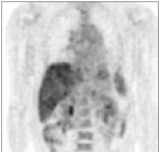
Fig. 3. Positron emission tomography showing increased
18F-FDG uptake in the lower common bile duct and no distant
metastasis.
Fig. 4. Endoscopic retrograde cholangioscopy showing the
swollen papilla of Vater with a erosive mucosa.
from the distal extrahepatic bile duct showed no malig-nancy due to insufficient material. The endoscopic retro-grade cholangiography revealed the protruded and en-gorged ampulla of Vater. The common bile duct and in-trahepatic bile duct were markedly dilated. There was complete obstruction at the distal common bile duct. To decompress the biliary system, an endoscopic retrograde biliary drainage tube was inserted (Fig. 5).
Esophagogastroduodenoscope and colonoscope for eval-uation of the common primary site of signet ring cell car-cinoma did not show any abnormality.
The patient underwent a pylorus preserving pancreati-coduodenectomy (PPPD) with extended lymph node dis-section including paraarotic lymph nodes. On gross
evalu-ation of the resected specimen, the wall of the common bile duct was thickened predominantly at the distal part measuring up to 1 cm. The cut surface of the thickened common bile duct wall showed concentric fibrosis and in-tervening gray tan areas (Fig. 6). The lesion was con-nected to the duodenal muscle and pancreatic paren-chyma. A few gray colored myxoid nodules were noted in the perimuscular connective tissue of the common bile duct. The ampulla of Vater was not prominant, grossly. The proximal part of common bile duct was slightly dilated. The cystic duct, gallbladder, and duodenal mucosa
Fig. 5. Endoscopic retrograde cholangioscopy showing the
plastic biliary stent inserted in the distal common bile duct.
Fig. 6. Surgical specimen showing the thickened wall of the
common bile duct measuring up to 1 cm. P, pancreatic duct; T, tumor mass; PC, pancreas.
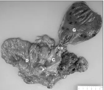
Fig. 7. The ampullar of Vater showing normal feature grossly
and slight dilatation of the proximal common bile duct. D, duodenum; C, common bile duct; P, pancreatic duct; G, gallbladder; CD, cystit duct; T, tumor mass; PC, pancreas.
Fig. 9. Signet ring cell distal common bile duct cancer (H&E
stain, ×100).
Fig. 8. Gastric metaplasia in distal common bile duct mucosa
(H&E stain, ×40).
were grossly unremarkable (Fig. 7).
The histology of the main tumor mass revealed SRCC (Figs. 8 and 9) invading perimuscular soft tissue and
in-volving pancreatic parenchyma, ampulla of Vater, duode-nal submucosa, and myenteric plexus. Extensive neural invasion and mucin pool formation were also detected. The tumor cells were found in a ruptured peripancreatic node. And tumor cells were noted on the proximal re-section margin of common bile duct. With these histo-logic finidings, the patient was confirmed to T3N1M0, stage IIB signet ring cell carcinoma of the distal common bile duct with R1 resection status.
He was taken postoperative concurrent chemoradio-therapy with total radiation dose of 50.4 Gray on the tu-mor bed. The combination of gemcitabine and cisplatin was used for chemotherapeutic regimen of concurrent chemoradiotherapy. After that, he underwent additional 4 cycles of gemcitabine and cisplatin chemotherapy until now without any evidence of recurrence.
DISCUSSION
The incidence of bile duct tumors in large autopsy studies varies from 0.01% to 0.2%, and may constitute about 2% of all reported cancer.1,3 Tumors of the distal
extrahepatic bile duct represent approximately 20% to 30% of all bile duct cancers and 5% to 10% of all peri-ampullary tumors.1,5 The majority of patients are over 65
years of age, and the peak incidence occurs in the seventh decade of life.5
SRCC of the biliary system is extremely rare and most SRCCs of the biliary system are originated from gallbladder. Currently, only one case of SRCC from the lower bile duct has been reported in the English literature.4 And this case is the second case of primary
signet ring cell carcinoma of the distal extrahepatic bile duct reported in literature.
Commonly, bile duct cancer spreads longitudinally along the bile duct wall and connective tissue. It can go through the submucosa in spite of intact epithelial lining. Because the extent of ductal tumor spread is difficult to know preoperatively, the intraopertive frozen section anal-ysis is important to determine the degree of submucosal spreading and to spare tumor-free resection margin.3,6,7
Wakai et al.8 suggested that ductal margin status was a
strong independent prognostic factor and invasive ductal carcinoma at resection margin appeared to have a strong adverse effect on patient survival. In this case, resection margin was clear in intraoperative frozen section analysis, but proximal resection margin was invaded by tumor in final pathologic report. It has long been suggested that extrahepatic bile duct tumors progress slowly.9 However,
progression can be rapid in some patients. In general, SRCCs of other digestive organs progress rapidly and have a poor prognosis.10-12 Hiraki et al.4 reported one case
of extrahepatic SRCC of rapid growth and aggressive nature. It is unclear whether the progression is more rap-id in patients with SRCCs than patients with other type tumors because of extreme rarity. In this case, the patient is still alive without visible remnant lesion after surgery and chemoradiotherapy.
The origin of SRCCs in periampullary area is not known well. There are two possible explanations for this histologic variation. One explanation is that the tumors may arise from ectopic gastric mucosa. Indeed, there are some reported cases of SRCCs with ectopic gastric muco-sa in ampullary tumors. Another explanation suggests that SRCCs may develop from gastric-type epithelial metaplasia. The presence of duodenal ulcer and elevated intraluminal acidity may irritate epithelial lining and can
induce gastric-type metaplasia. It can be etiology of the periampullary SRCCs.12-14 At the present case, no ectopic
gastric mucosa and epithelial metaplasia was found in surgical specimen of the duodenum. The patients also did not have a previous history of peptic ulcer disease. In this case, we could make differential diagnosis between the distal common bile duct cancer and the ampulla of Vater cancer, as there were transitional zone from mural gland to signet ring cell carcinoma in distal portion of the com-mon bile duct.
In conclusion, curative therapy may be possible with surgery and adjuvant therapy in SRCC of distal extra-hepatic bile duct. But due to extreme rarity of this dis-ease, additional case reports are warranted to decide the nature and optimal management of the disease.
REFERENCES
1. Jarnagin WR, Shoup M. Surgical management of cholangio-carcinoma. Semin Liver Dis 2004;24:189-199.
2. Slattery JM, Sahani DV. What is the current state-of- the-art imaging for detection and staging of cholangiocar-cinoma? Oncologist 2006;11:913-922.
3. Akoad M, Jenkins R. Proximal biliary malignancy. Surg Clin North Am 2008;88:1409-1428, x-xi.
4. Hiraki M, Yakushiji H, Hashiguchil K, et al. Signet ring cell carcinoma of the lower bile duct with rapid growth: report of a case. Hepatogastroenterology 2007;54:1922- 1924.
5. Mosconi S, Beretta GD, Labianca R, Zampino MG, Gatta G, Heinemann V. Cholangiocarcinoma. Crit Rev Oncol Hematol 2009;69:259-270.
6. Yamaguchi K, Chijiiwa K, Saiki S, Shimizu S, Takashima M, Tanaka M. Carcinoma of the extrahepatic bile duct: mode of spread and its prognostic implications. Hepatogas-troenterology 1997;44:1256-1261.
7. Shimada H, Niimoto S, Matsuba A, Nakagawara G, Koba-yashi M, Tsuchiya S. The infiltration of bile duct carcino-ma along the bile duct wall. Int Surg 1988;73:87-90. 8. Wakai T, Shirai Y, Moroda T, Yokoyama N, Hatakeyama
K. Impact of ductal resection margin status on long-term survival in patients undergoing resection for extrahepatic cholangiocarcinoma. Cancer 2005;103:1210-1216.
9. Ustundag Y, Bayraktar Y. Cholangiocarcinoma: a compact review of the literature. World J Gastroenterol 2008;14: 6458-6466.
10. Krstić M, Katić V. Histological, mucinohistochemical and immunohistochemical features of gastric signet ring cell carcinoma. Vojnosanit Pregl 2008;65:835-838.
11. Yokota T, Kunii Y, Teshima S, et al. Signet ring cell carci-noma of the stomach: a clinicopathological comparison with the other histological types. Tohoku J Exp Med 1998;186:121-130.
12. Akatsu T, Aiura K, Takahashi S. Signet-ring cell carcinoma of the ampulla of Vater: report of a case. Surg Today 2007;37:1110-1114.